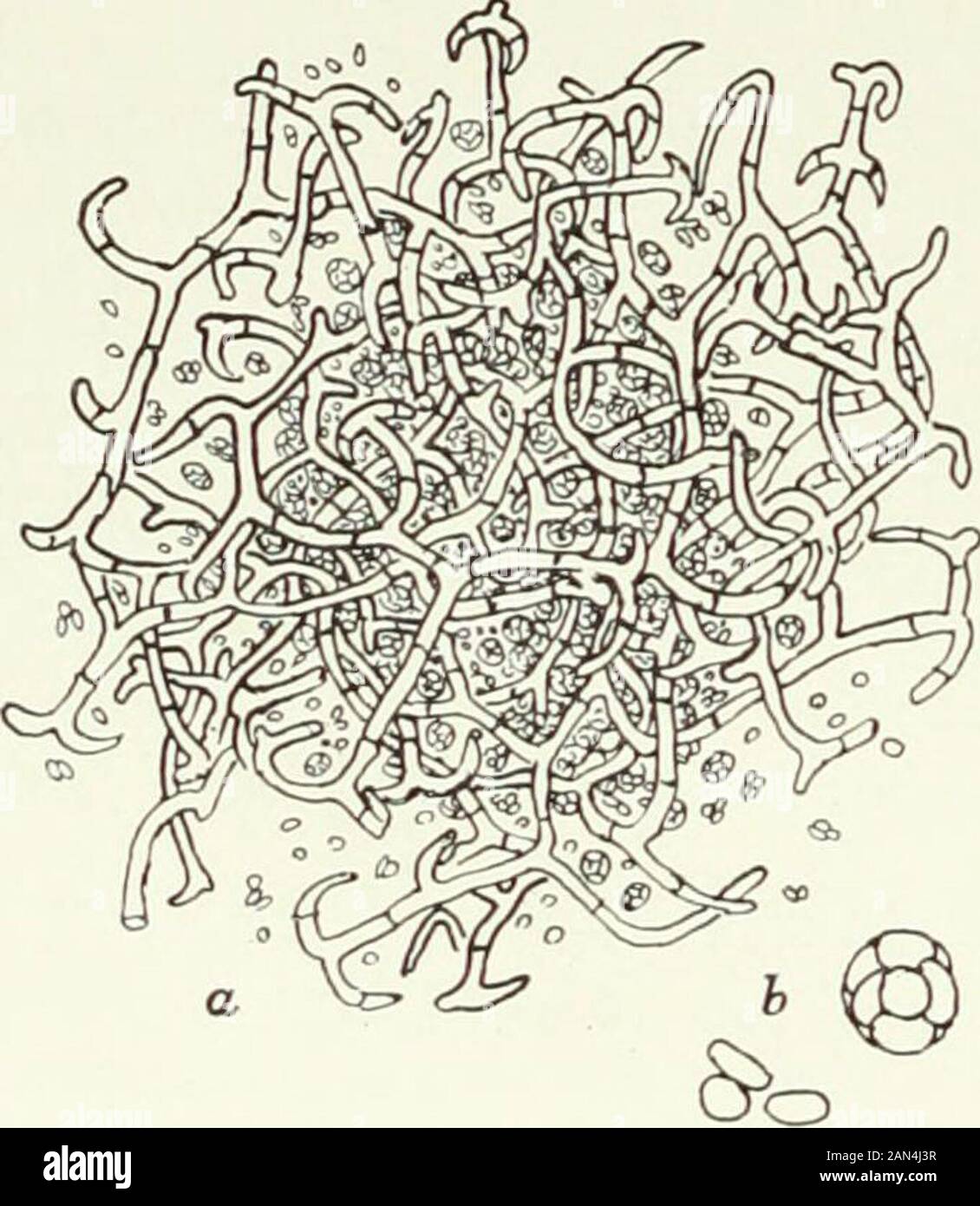
Fungi, Ascomycetes, Ustilaginales, Uredinales . ins eight spores.The species of Gymnoascus occurin various habitats, on dung, beesnests, dead grass, etc. In G. Reesii, according to Dale,two branches grow up from thesame hypha, one on each side ofa septum, and become twistedaround one another. These are theantheridium and oogonium; theirfree ends swell into club-shapedheads which lie in close contact andeach becomes delimited by a transverse septum. The walls between thembreak down, and the contents of the antheridium pass over into theoogonium (fig. 27 a, b). Both cells are at first uninucleat
Image details
Contributor:
The Reading Room / Alamy Stock PhotoImage ID:
2AN4J3RFile size:
7.1 MB (329.6 KB Compressed download)Releases:
Model - no | Property - noDo I need a release?Dimensions:
1474 x 1695 px | 25 x 28.7 cm | 9.8 x 11.3 inches | 150dpiMore information:
This image is a public domain image, which means either that copyright has expired in the image or the copyright holder has waived their copyright. Alamy charges you a fee for access to the high resolution copy of the image.
This image could have imperfections as it’s either historical or reportage.
Fungi, Ascomycetes, Ustilaginales, Uredinales . ins eight spores.The species of Gymnoascus occurin various habitats, on dung, beesnests, dead grass, etc. In G. Reesii, according to Dale, two branches grow up from thesame hypha, one on each side ofa septum, and become twistedaround one another. These are theantheridium and oogonium; theirfree ends swell into club-shapedheads which lie in close contact andeach becomes delimited by a transverse septum. The walls between thembreak down, and the contents of the antheridium pass over into theoogonium (fig. 27 a, b). Both cells are at first uninucleate, but later coeno-cytic, and, though the history of the nuclei has not been traced, it seemsalmost certain that fusion must sooner or later occur. Up to this point the sexual cells are usually quite similar in form, butnow the antheridium grows larger and more spherical, remaining almoststraight, while the oogonium puts out a prolongation which winds aroundit, undergoes septation and soon branches to give rise to ascogenoushyphae (fig. 27 c).. Fi 26. Gymnoascus sp.; a. ascocarp, x 26;b. ascus and free ascospores, x 1040. tn] PLECTASCALES 67 In G. candidus (fig.27 d) the antheridium and oogonium already differin form at the time of their union, and, in the majority of cases, appear to