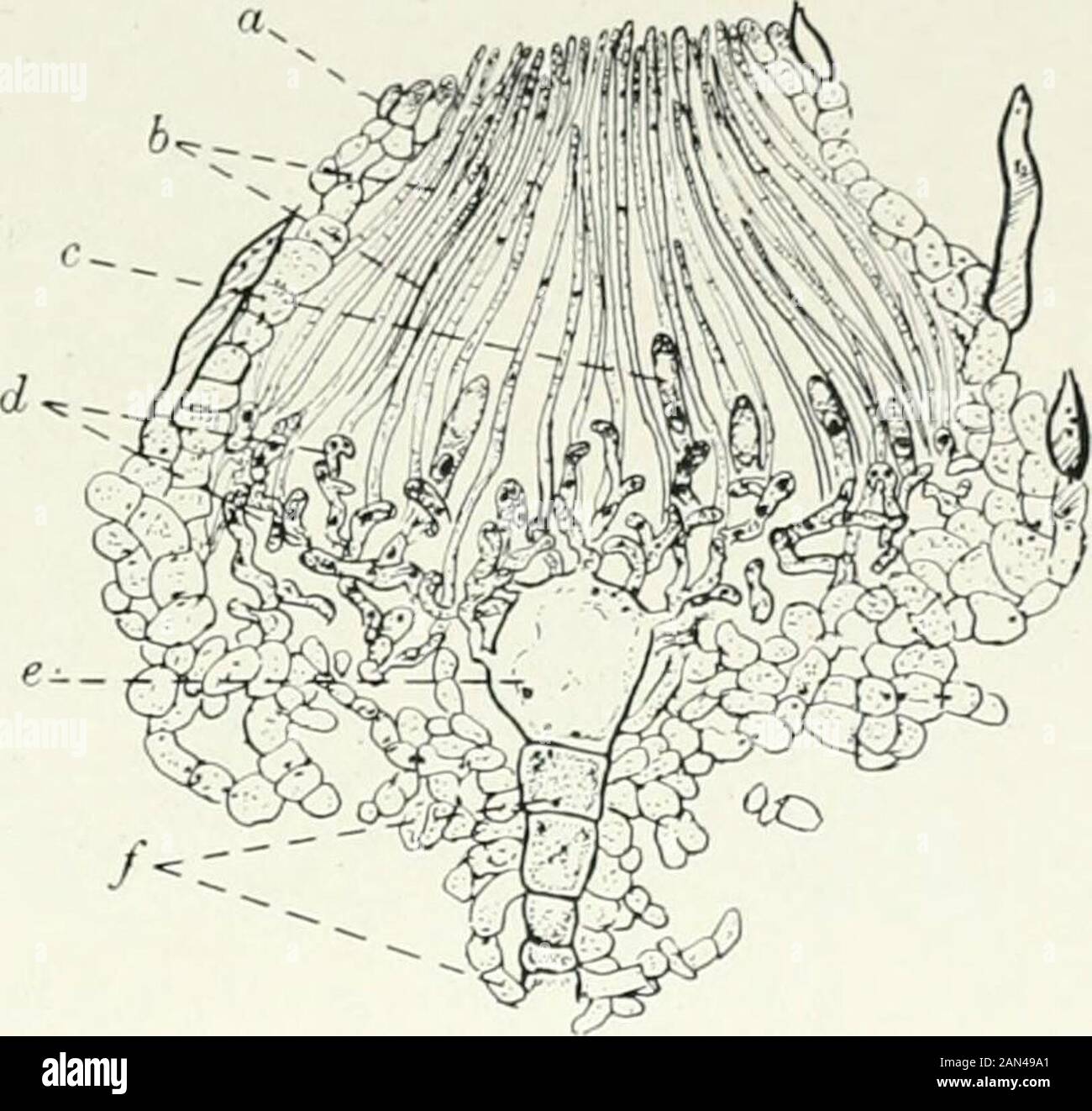
Fungi, Ascomycetes, Ustilaginales, Uredinales . Fig. 51. Otidta aurantia Mass.; apotheci, Fig. 52. Lachn<a stercoral (lers.) Gill.; ascocarp innat. size. longitudinal section showing young asci and para- physes, x 160. a. sheath; b. paraphyses; c. ascus;a. ascogenous hyphae; e. oogonium;/. stalk of archicarp. wall of the cup (fig. 52). The lower part of the cup is filled by the hypo-thecium, a tangle of hyphae, some vegetative, some ascogenous. Thesegive rise to the sub-hymenial layer where the paraphyses have their originand where the young asci are developed. The asci and paraphyses grow
Image details
Contributor:
The Reading Room / Alamy Stock PhotoImage ID:
2AN49A1File size:
7.1 MB (304.7 KB Compressed download)Releases:
Model - no | Property - noDo I need a release?Dimensions:
1623 x 1539 px | 27.5 x 26.1 cm | 10.8 x 10.3 inches | 150dpiMore information:
This image is a public domain image, which means either that copyright has expired in the image or the copyright holder has waived their copyright. Alamy charges you a fee for access to the high resolution copy of the image.
This image could have imperfections as it’s either historical or reportage.
Fungi, Ascomycetes, Ustilaginales, Uredinales . Fig. 51. Otidta aurantia Mass.; apotheci, Fig. 52. Lachn<a stercoral (lers.) Gill.; ascocarp innat. size. longitudinal section showing young asci and para- physes, x 160. a. sheath; b. paraphyses; c. ascus;a. ascogenous hyphae; e. oogonium;/. stalk of archicarp. wall of the cup (fig. 52). The lower part of the cup is filled by the hypo-thecium, a tangle of hyphae, some vegetative, some ascogenous. Thesegive rise to the sub-hymenial layer where the paraphyses have their originand where the young asci are developed. The asci and paraphyses grow uptogether and rise to the surface of the ascocarp forming the hymenium orfertile disc which is spread over the interior of the cup. The asci are moreor less cylindrical and parallel one to another and to the paraphyses (fig. 53).They open either by a lid (fig. 55) or by the ejection of a plui; (fig. 54).They arise in succession so that large numbers may be produced in a singleascocarp. If the hypothecium is well developed the apothecial cup i