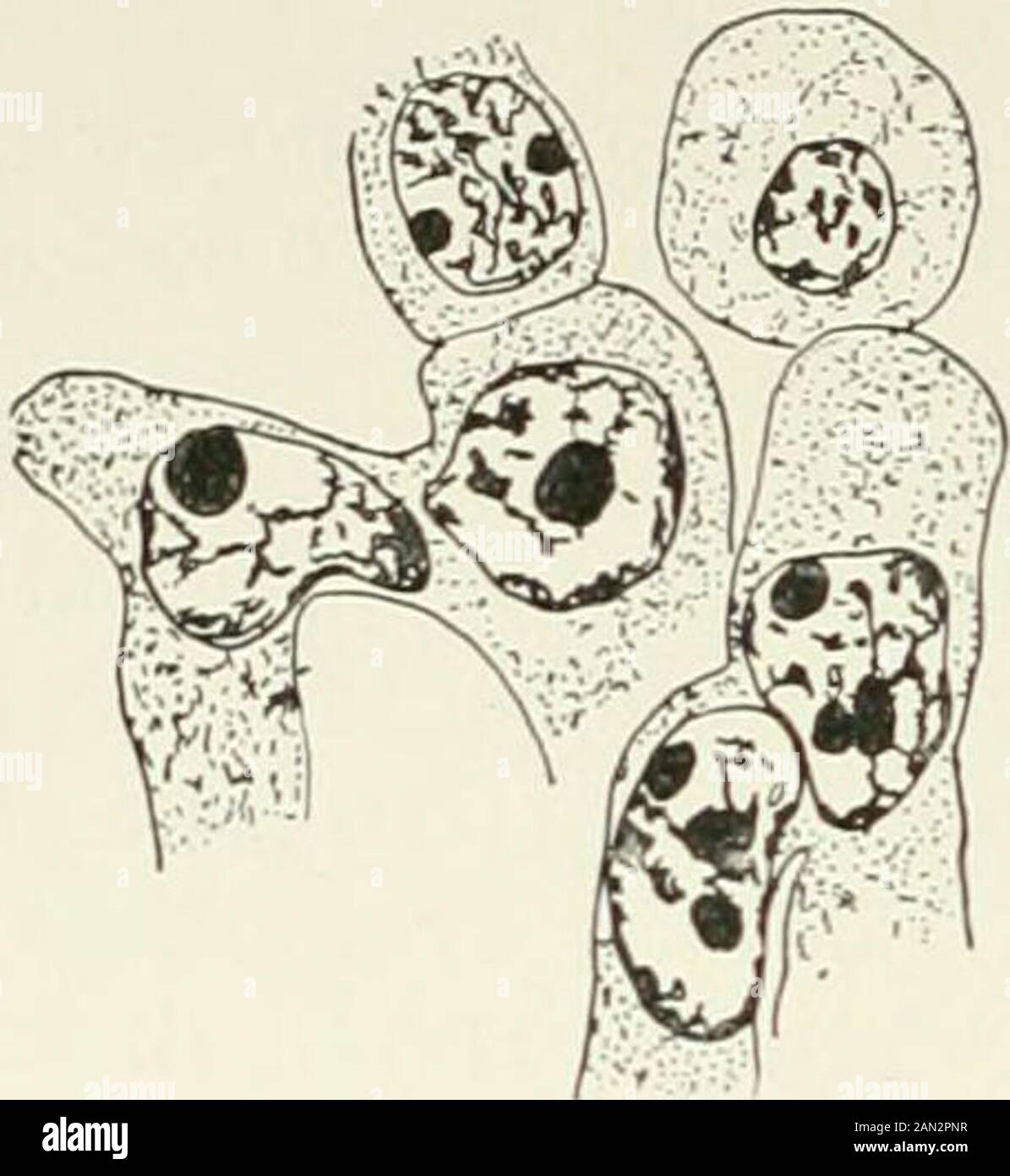
Fungi, Ascomycetes, Ustilaginales, Uredinales . eoma; sterile cell pushing up betweenepidermal cells of host, x 1.500; afterBlackman. Fig-9S- Phragmidium speciosum Fr.;fertile cells after conjugation; aecidio- spore mother-cell above ; after Christ-man. takes place and aecidiospore mother-cells arc cut off so that a single rowof aecidiospores is developed from each pair of gametes. Christman regardsthe fertile cells as isogametes between which conjugation takes place, andthe sterile cells merely as buffers, of which the function is to assist in therupture of the epidermis. His observations on
Image details
Contributor:
The Reading Room / Alamy Stock PhotoImage ID:
2AN2PNRFile size:
7.2 MB (219.6 KB Compressed download)Releases:
Model - no | Property - noDo I need a release?Dimensions:
1516 x 1649 px | 25.7 x 27.9 cm | 10.1 x 11 inches | 150dpiMore information:
This image is a public domain image, which means either that copyright has expired in the image or the copyright holder has waived their copyright. Alamy charges you a fee for access to the high resolution copy of the image.
This image could have imperfections as it’s either historical or reportage.
Fungi, Ascomycetes, Ustilaginales, Uredinales . eoma; sterile cell pushing up betweenepidermal cells of host, x 1.500; afterBlackman. Fig-9S- Phragmidium speciosum Fr.;fertile cells after conjugation; aecidio- spore mother-cell above ; after Christ-man. takes place and aecidiospore mother-cells arc cut off so that a single rowof aecidiospores is developed from each pair of gametes. Christman regardsthe fertile cells as isogametes between which conjugation takes place, andthe sterile cells merely as buffers, of which the function is to assist in therupture of the epidermis. His observations on Phragmidium speciosum were confirmed in 1906 byBlackman and Fraser for Melampsora Rostrupi on Mercurialis (fig. 174);these authors pointed out that the fusion of the fertile cells is a reducedfertilization, strictly comparable to that in Ph. violaceum but achieved by theunion of female cells in pairs instead of by the entrance of a vegetativenucleus into the female cell. This interpretation is confirmed by the fact 216 PROTOBASIDIOMYCETES [CH.. Fig. 196. Triphragmidium Ulmariae(Schum.) Link; primary uredosorus;condition intermediate between migra-tion and conjugation of fertile cell;after Olive. that Moreau found both processes (cell-fusion being considerably morecommon than migration) in the same caeoma in Phragmidium subcorticium.Since 1905 nuclear association by the fusion of fertile cells in pairs hasbeen observed in a number of species, and seems, according to our presentknowledge, to be the usual method. In the primary uredosorus of Triphragmidium Ulmariae and in certainother species, Olive, in 1908, found arrangements of an intermediate type. Here the cells of the young fructificationform a more or less regular layer and cutoff sterile cells in the usual way. Otherhyphae then push up among them, andcell fusion and nuclear association takeplace (fig. 196). Fusion begins througha narrow pore which afterwards broadens, and in many cases the nucleus of theyounger cell migrate