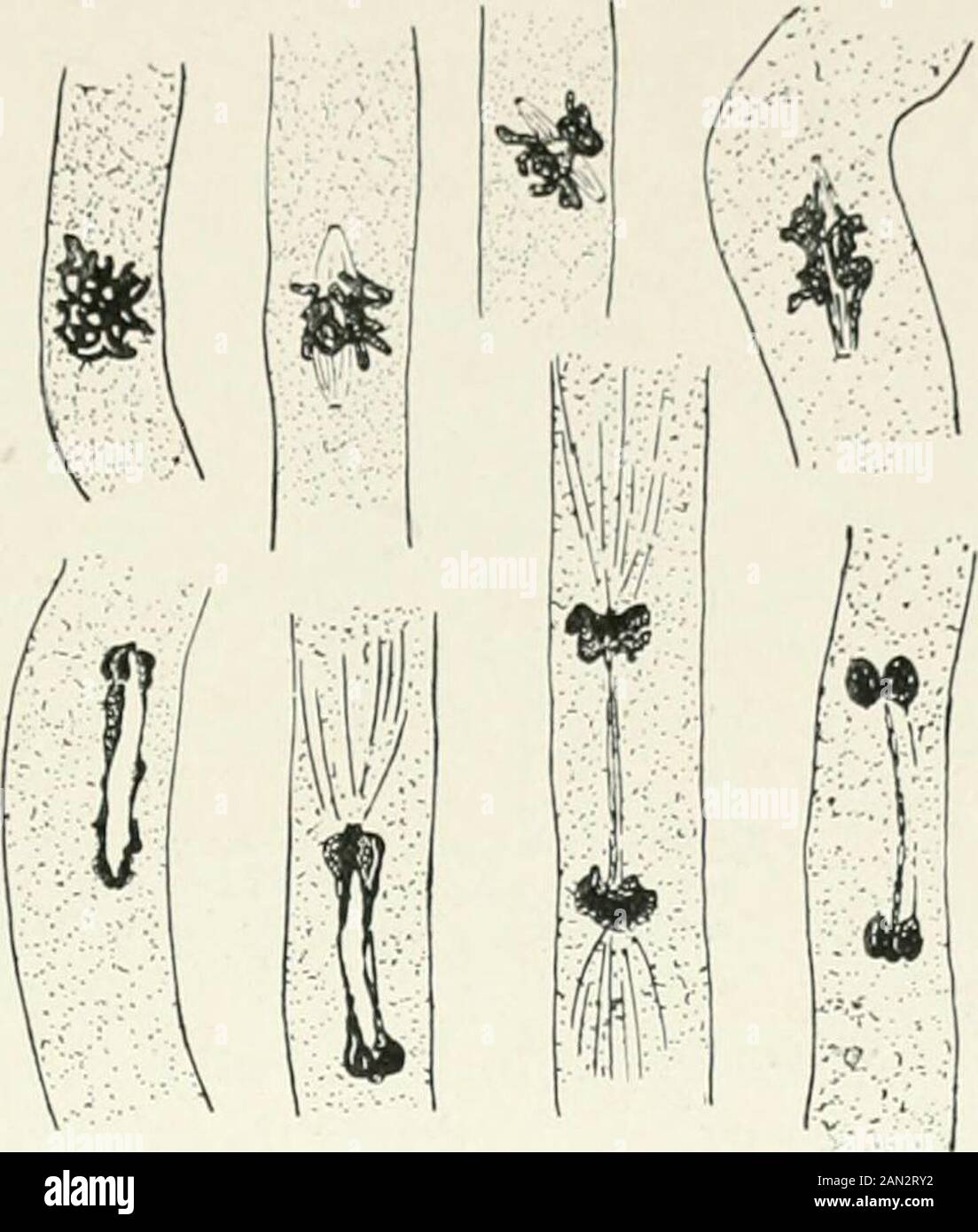
Fungi, Ascomycetes, Ustilaginales, Uredinales . ation. Thecytology of the aecidium wasfirst described in detail in 1904In Blackman, for Phragmidiumvio/aceum, a species occurring onthe bramble. The aecidium here is of the caeoma type, consisting of a groupof fertile cells of indefinite extent and usually bounded at the periphery bya number of thin-walled paraphyses. Its formation begins by the massing of hyphae below the epidermis of the leaf where they form a series of uninucleate cells two or three layers thick. The cells next the epidermis increase in size and each divides by a transverse wa
Image details
Contributor:
The Reading Room / Alamy Stock PhotoImage ID:
2AN2RY2File size:
7.1 MB (243 KB Compressed download)Releases:
Model - no | Property - noDo I need a release?Dimensions:
1456 x 1716 px | 24.7 x 29.1 cm | 9.7 x 11.4 inches | 150dpiMore information:
This image is a public domain image, which means either that copyright has expired in the image or the copyright holder has waived their copyright. Alamy charges you a fee for access to the high resolution copy of the image.
This image could have imperfections as it’s either historical or reportage.
Fungi, Ascomycetes, Ustilaginales, Uredinales . ation. Thecytology of the aecidium wasfirst described in detail in 1904In Blackman, for Phragmidiumvio/aceum, a species occurring onthe bramble. The aecidium here is of the caeoma type, consisting of a groupof fertile cells of indefinite extent and usually bounded at the periphery bya number of thin-walled paraphyses. Its formation begins by the massing of hyphae below the epidermis of the leaf where they form a series of uninucleate cells two or three layers thick. The cells next the epidermis increase in size and each divides by a transverse wall parallel to the surface of the leaf, separating an upper sterile cell from the fertile cell below. The sterile cells remain cubical and ultimately disintegrate; the fertile cells elongate to form a more or less regular layer and paired nuclei appear in them, first at the centre and later towards the periphery of the group (fig. 191 ). „. , , ... Fig. 191. Phragmidium vielaceum Went.; caeoma, The second nucleus in the , , 40: , lMrl Blackman.. Fig. 190. Gymnosporangium clavariaeforme Reesfirst division in basidium, x [460; after Blackman.