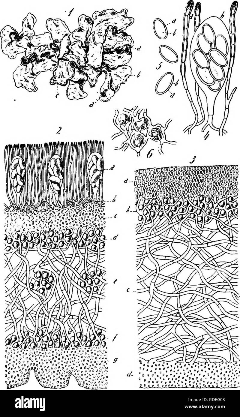
. A text-book of mycology and plant pathology . Plant diseases; Fungi in agriculture; Plant diseases; Fungi. 8o MYCOtOGY. Fig. 26.—A foliaceous lichen, Parmelia perlata. i, Plant slightly reduced in size; a, apothecia; b, lobe of thallus; c, patches of soredia; 2, longitudinal section of apothecium and cross-section of thallus; o, ascus; b, c, hypothecium; d. upper gonidial (upper algal) layer; e, medullary layer; /, lower gonidial layer; g, lower cortical layer; I, 3, cross-section of vegetative thallus. (From Gager. After Schneider.). Please note that these images are extracted from scanned
Image details
Contributor:
The Book Worm / Alamy Stock PhotoImage ID:
RDEG03File size:
7.1 MB (624 KB Compressed download)Releases:
Model - no | Property - noDo I need a release?Dimensions:
1240 x 2015 px | 21 x 34.1 cm | 8.3 x 13.4 inches | 150dpiMore information:
This image is a public domain image, which means either that copyright has expired in the image or the copyright holder has waived their copyright. Alamy charges you a fee for access to the high resolution copy of the image.
This image could have imperfections as it’s either historical or reportage.
. A text-book of mycology and plant pathology . Plant diseases; Fungi in agriculture; Plant diseases; Fungi. 8o MYCOtOGY. Fig. 26.—A foliaceous lichen, Parmelia perlata. i, Plant slightly reduced in size; a, apothecia; b, lobe of thallus; c, patches of soredia; 2, longitudinal section of apothecium and cross-section of thallus; o, ascus; b, c, hypothecium; d. upper gonidial (upper algal) layer; e, medullary layer; /, lower gonidial layer; g, lower cortical layer; I, 3, cross-section of vegetative thallus. (From Gager. After Schneider.). Please note that these images are extracted from scanned page images that may have been digitally enhanced for readability - coloration and appearance of these illustrations may not perfectly resemble the original work.. Harshberger, John W. (John William), 1869-1929. Philadelphia : P. Blakiston's Son & Co