. The surgical anatomy of the horse ... Horses. Plate XXVIII.—Metatarsal Region, showing Arteries, Tendons, Ligaments, Bones, etc. A.—INNER aspect I. Cunean tendon. 2. Cuneiform parvum. 3. Scaphoid. 4. Head of inner small metatarsal bone. 5. Cuneiform magnum. 6. Perforatus tendon. 7. Large metatarsal bone. 8. Perforans tendon. 9. Internal plantar interosseous artery. 10. Internal plantar nerve. 11. Suspensory ligament. 12. Large metatarsal artery. 13. Anastomosis of large metatarsal and internal plantar interosseous arteries. 14. Division of large metatarsal into the two digital arteries. B.—O
Image details
Contributor:
Central Historic Books / Alamy Stock PhotoImage ID:
PG1H6BFile size:
7.1 MB (410.4 KB Compressed download)Releases:
Model - no | Property - noDo I need a release?Dimensions:
1337 x 1869 px | 22.6 x 31.6 cm | 8.9 x 12.5 inches | 150dpiMore information:
This image is a public domain image, which means either that copyright has expired in the image or the copyright holder has waived their copyright. Alamy charges you a fee for access to the high resolution copy of the image.
This image could have imperfections as it’s either historical or reportage.
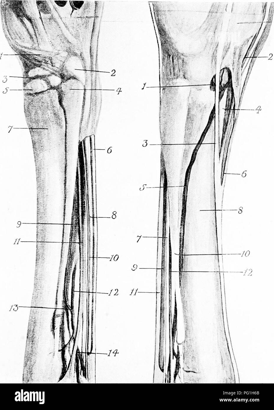
. The surgical anatomy of the horse ... Horses. Plate XXVIII.—Metatarsal Region, showing Arteries, Tendons, Ligaments, Bones, etc. A.—INNER aspect I. Cunean tendon. 2. Cuneiform parvum. 3. Scaphoid. 4. Head of inner small metatarsal bone. 5. Cuneiform magnum. 6. Perforatus tendon. 7. Large metatarsal bone. 8. Perforans tendon. 9. Internal plantar interosseous artery. 10. Internal plantar nerve. 11. Suspensory ligament. 12. Large metatarsal artery. 13. Anastomosis of large metatarsal and internal plantar interosseous arteries. 14. Division of large metatarsal into the two digital arteries. B.—OUTER ASPECT I. Perforating metatarsal artery. 2. Anterior tibial artery. 3. Peroneal tendon. 4. Extensor brevis. 5. Large metatarsal artery. 6. Tendon of extensor pedis. 7. External plantar nerve. 8. Large metatarsal bone. g. Perforans tendon. 10 External small metatarsal bone. 11. Per- foratus tendon. 12. Suspensory ligament. The flexor tendons and suspensory ligament have been displaced slightly backwards to display more fully the arteries. Part of the extensor brevis muscle has also been removed to show the division of the anterior tibial artery.. Please note that these images are extracted from scanned page images that may have been digitally enhanced for readability - coloration and appearance of these illustrations may not perfectly resemble the original work.. Share-Jones, John T. London, Williams and Norgate