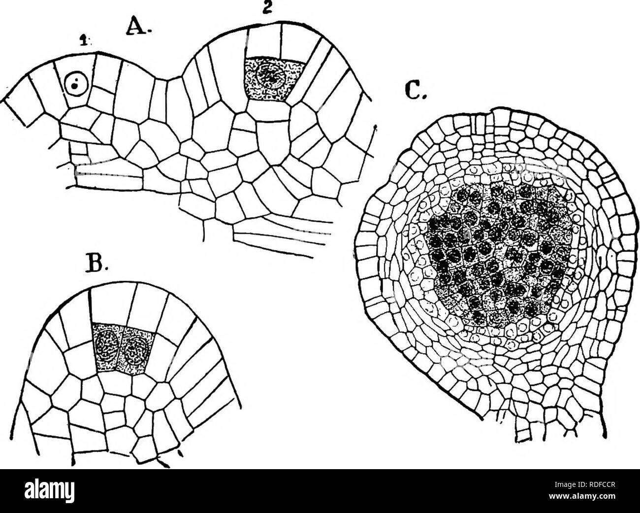
. The structure and development of mosses and ferns (Archegoniatae). Plant morphology; Mosses; Ferns. VII PTERIDOPHYTA—FILICINEM—OPHIOGLOSSACEM 269 point, but the youngest stages found by me in which the sporangial nature of the outgrowths was unmistaiiable, would not forbid such an interpretation, although there was no doubt that the basal part of the sporangium is derived in part from the surrounding tissue. From the central cell, by a periclinal wall, an inner cell, the archesporium, is separated from an outer one. The outer cell divides next by cross walls, and this is followed by similar
Image details
Contributor:
The Book Worm / Alamy Stock PhotoImage ID:
RDFCCRFile size:
7.1 MB (357.2 KB Compressed download)Releases:
Model - no | Property - noDo I need a release?Dimensions:
1847 x 1352 px | 31.3 x 22.9 cm | 12.3 x 9 inches | 150dpiMore information:
This image is a public domain image, which means either that copyright has expired in the image or the copyright holder has waived their copyright. Alamy charges you a fee for access to the high resolution copy of the image.
This image could have imperfections as it’s either historical or reportage.
. The structure and development of mosses and ferns (Archegoniatae). Plant morphology; Mosses; Ferns. VII PTERIDOPHYTA—FILICINEM—OPHIOGLOSSACEM 269 point, but the youngest stages found by me in which the sporangial nature of the outgrowths was unmistaiiable, would not forbid such an interpretation, although there was no doubt that the basal part of the sporangium is derived in part from the surrounding tissue. From the central cell, by a periclinal wall, an inner cell, the archesporium, is separated from an outer one. The outer cell divides next by cross walls, and this is followed by similar divisions in the inner cells (Fig. 148). The succeeding divi-. Development of the sporangia. A, X240; all median longitudinal sections, Fig. 148.—Botrychium Virginianum. x^cvciupuicni. ui luc ispuiangia. j», », *, Very young sporangia; B, a somewhat older one, X480; C, older sporangium, '* ^ r^-j;_-i —*: tjjg sporogenous cells are shaded. sions in the outer cells are now mainly periclinal, and transform the four cells lying immediately above the archesporium into as many rows of tabular cells. Growth is active in the mean- time in the basal part of the sporangium, which projects more and more until it becomes almost spherical. To judge from the account given by Goebel (3) and Bower (16) of 5. lunaria, this species corresponds closely in its early stages to B. Vir- ginianum. The later divisions in the archesporium do not apparently follow any definite rule, but divisions take place in all directions until a very large number of cells is formed.. Please note that these images are extracted from scanned page images that may have been digitally enhanced for readability - coloration and appearance of these illustrations may not perfectly resemble the original work.. Campbell, Douglas Houghton, 1859-1953. New York, The Macmillan Company;