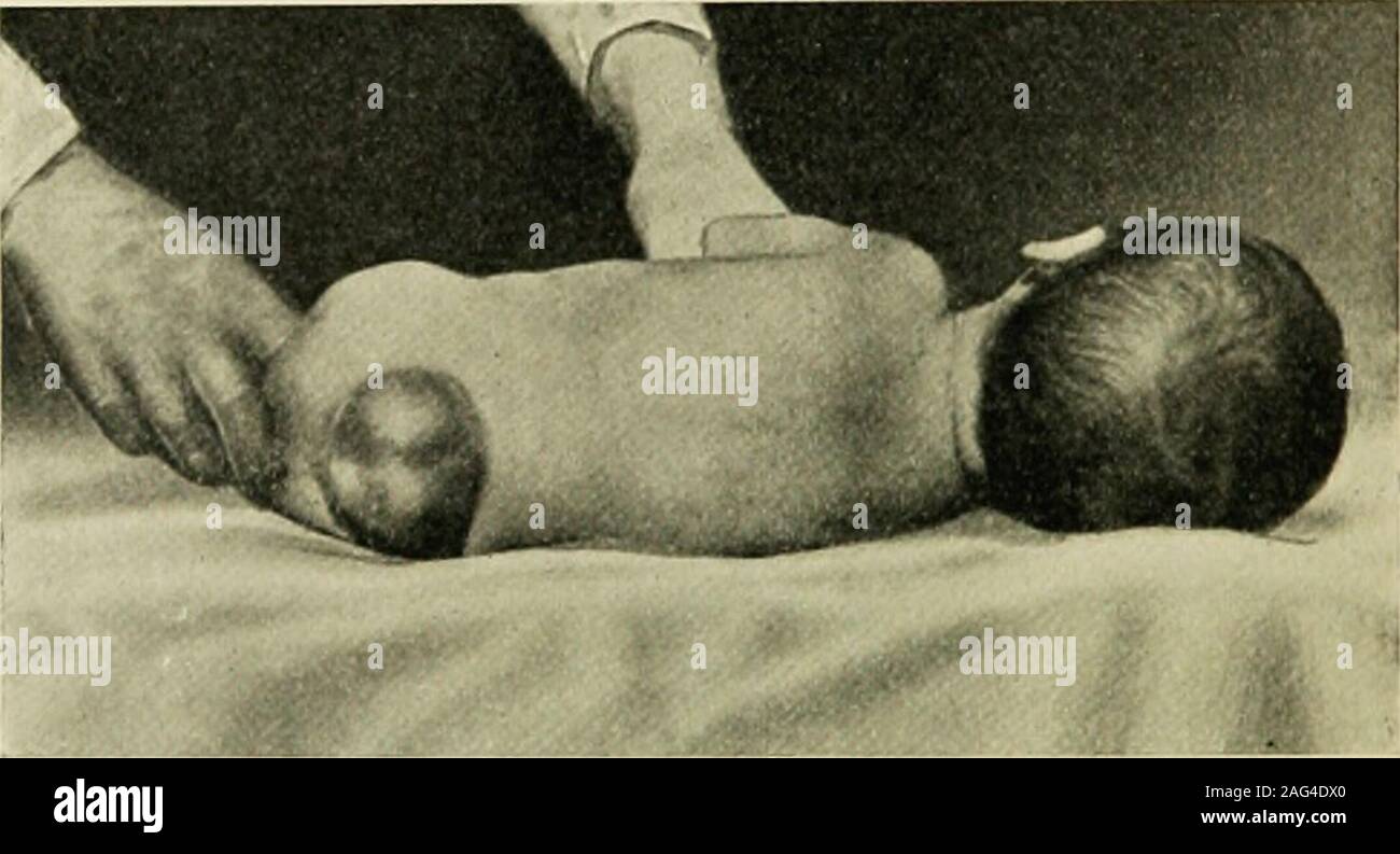
. The practice of pediatrics. Fig. 62.—Microcephalic idiot. 484 THE PRACTICE OF PEDIATRICS neurology, rather than to general pediatrics. The terms themselvesroughly define the respective conditions. Spina Bifida.—Spina bifida is the term applied to a congenital cleftin the vertebral column which permits of a hernia of part of the con-tents of the canal. The defect is found most frequently in the cervicalor lower dorsal vertebra?. In meningocele of the cord the membranes alone constitute the hernialsac. Myelomeningocele is a protrusion of a portion of the spinal cord andits attached nerve-roots
Image details
Contributor:
The Reading Room / Alamy Stock PhotoImage ID:
2AG4DX0File size:
7.2 MB (230.5 KB Compressed download)Releases:
Model - no | Property - noDo I need a release?Dimensions:
2149 x 1163 px | 36.4 x 19.7 cm | 14.3 x 7.8 inches | 150dpiMore information:
This image is a public domain image, which means either that copyright has expired in the image or the copyright holder has waived their copyright. Alamy charges you a fee for access to the high resolution copy of the image.
This image could have imperfections as it’s either historical or reportage.
. The practice of pediatrics. Fig. 62.—Microcephalic idiot. 484 THE PRACTICE OF PEDIATRICS neurology, rather than to general pediatrics. The terms themselvesroughly define the respective conditions. Spina Bifida.—Spina bifida is the term applied to a congenital cleftin the vertebral column which permits of a hernia of part of the con-tents of the canal. The defect is found most frequently in the cervicalor lower dorsal vertebra?. In meningocele of the cord the membranes alone constitute the hernialsac. Myelomeningocele is a protrusion of a portion of the spinal cord andits attached nerve-roots, together with an accumulation of fluid, whichusually has its origin in the anterior subarachnoid space. In syringomyelocele, hydro?nyelocele, or myelocystocele the centralcanal of the cord is dilated with fluid, and the cord substance itselfforms the lining of the sac. The malformations just described are frequently accompanied byother abnormalities in the same subject, such as hydrocephalus, club-. Fig. 63.—Spina bifida. foot, sensory and trophic disturbances, and exstrophy of the bladder.With myelomeningocele and syringomyelocele, paralysis of the extremi-ties, bladder, and rectum may exist. Diagnosis of the type of spina bifida present in a given case is notalways easy. Simple spinal meningocele is frequently found in the sacral region.This tumor is often translucent. It protrudes through a small cleftin the canal and is pedunculated. It is seldom associated with symp-toms of paralysis. In myelomeningocele and syringomyelocele the swelling is ordinarilyless transparent and has a broader base. Pressure on the tumor maycause distention of the fontanel. These forms commonly occur in thelumbosacral region, but may exist in any region of the spine. Paralyticsymptoms are much more common than in cases of meningocele. Of the three forms, syringomyelocele is far the most frequentlyassociated with a hydrocephalus. Prognosis.—Simple meningocele offers a fair prognosis under