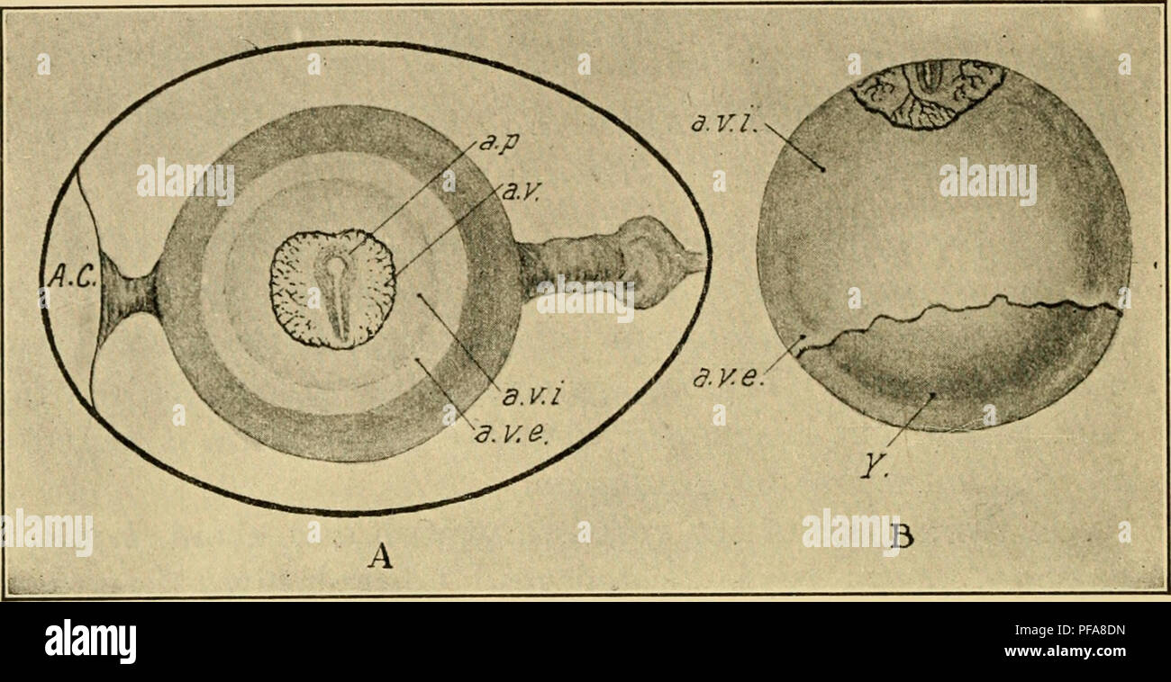
. The development of the chick : an introduction to embryology. Embryology; Chickens -- Embryos. 62 THE DEVELOPMENT OF THE CHICK vitellina, which may be divided into inner and outer zones (Figs, 32 and 33). The development of the embryo during the same period is indicated in the same figures.. Fig. 32. — A. Hen's egg at about the twenty-sixth hour of incubation, to show the zones of the blastoderm and the orientation of the embryo with reference to the axis of the shell. (After Duval.) B. Yolk of hen's egg incubated about 50 hours to show the extent of overgrowth of the blastoderm. (After Duva
Image details
Contributor:
Central Historic Books / Alamy Stock PhotoImage ID:
PFA8DNFile size:
7.2 MB (403.4 KB Compressed download)Releases:
Model - no | Property - noDo I need a release?Dimensions:
2204 x 1134 px | 37.3 x 19.2 cm | 14.7 x 7.6 inches | 150dpiMore information:
This image is a public domain image, which means either that copyright has expired in the image or the copyright holder has waived their copyright. Alamy charges you a fee for access to the high resolution copy of the image.
This image could have imperfections as it’s either historical or reportage.
. The development of the chick : an introduction to embryology. Embryology; Chickens -- Embryos. 62 THE DEVELOPMENT OF THE CHICK vitellina, which may be divided into inner and outer zones (Figs, 32 and 33). The development of the embryo during the same period is indicated in the same figures.. Fig. 32. — A. Hen's egg at about the twenty-sixth hour of incubation, to show the zones of the blastoderm and the orientation of the embryo with reference to the axis of the shell. (After Duval.) B. Yolk of hen's egg incubated about 50 hours to show the extent of overgrowth of the blastoderm. (After Duval.) A. C, Air chamber, a. p., Area pellucida. a. v., Area vasculosa. a. v. e., Area vitellina externa, a. v. i., Area vitellina interna. Y., Uncovered portion of yolk. The blastoderm early becomes divided in two layers as far as the margin of the vascular area. The outer layer, known as the somatopleure, is continuous with the body-wall, which is open ventrally in the young embryo. The inner one, known as the splanchnopleure, is continuous with the wall of the intestine which is likewise open ventrally. The space between these two membranes, the extra-embryonic body-cavity, is continuous with the body-cavity of the embryo. Ultimately, the splitting of the blastoderm is carried around the entire yolk, so that the latter is enclosed in a separate sac of the splanchnopleure, the yolk-sac, which is connected by a stalk, the yoik-stalk, to the intestine of the embryo. This stalk runs through an opening in the ventral body-wall, the umbilicus, where the amnion, which has developed from the extra-embryonic somatopleure, joins the body-wall (Fig. 33 B). About the nineteenth day of incubation the yolk-sac is drawn. Please note that these images are extracted from scanned page images that may have been digitally enhanced for readability - coloration and appearance of these illustrations may not perfectly resemble the original work.. Lillie, Frank Rattray, 1870-1947. New York : Henry Ho