The anatomy of the nervous system, from the standpoint of development and function . the dorsal cochlearnucleus run medially upon the floor of the fourth ventricle in the stria? medullares(Fig. 89), and sinking into the tegmentum join the fibers from the ventral coch-lear nucleus in the trapezoid body. IS© THE NERVOUS SYSTEM The trapezoid body (corpus trapezoideum), which in most mammals appearson the surface of the medulla near the border of the pons (Fig. 83), is coveredin man by the enlarged pars basalis pontis. In sections through the more caudalportions of the pons the trapezoid body form
Image details
Contributor:
The Reading Room / Alamy Stock PhotoImage ID:
2AWGNBRFile size:
7.1 MB (428.1 KB Compressed download)Releases:
Model - no | Property - noDo I need a release?Dimensions:
1742 x 1434 px | 29.5 x 24.3 cm | 11.6 x 9.6 inches | 150dpiMore information:
This image is a public domain image, which means either that copyright has expired in the image or the copyright holder has waived their copyright. Alamy charges you a fee for access to the high resolution copy of the image.
This image could have imperfections as it’s either historical or reportage.
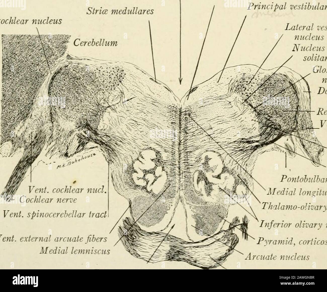
The anatomy of the nervous system, from the standpoint of development and function . the dorsal cochlearnucleus run medially upon the floor of the fourth ventricle in the stria? medullares(Fig. 89), and sinking into the tegmentum join the fibers from the ventral coch-lear nucleus in the trapezoid body. IS© THE NERVOUS SYSTEM The trapezoid body (corpus trapezoideum), which in most mammals appearson the surface of the medulla near the border of the pons (Fig. 83), is coveredin man by the enlarged pars basalis pontis. In sections through the more caudalportions of the pons the trapezoid body forms a conspicuous bundle of trans-verse fibers in the ventral portion of the reticular formation (Fig. 108). Thefibers are associated with the terminal nuclei of the cochlear nerve, especiallythe ventral one, and with the superior olivary nucleus, around the ventral borderof which they swing in such a way as to form a bay for its reception. Farthermedialward they pass through the medial lemniscus at right angles to its con- Dorsal cochlear nucleus Fourth ventricleStrice medullares. Vent, spinocerebellar tract Vent, external arcuate fibersMedial lemniscus Nucleus of emineniia teres Principal vestibular nucleus Lateral vestibularnucleus Nucleus of tractussolitarius Glossopharyngeal*C nerve, }X Dorsal cochlear[ || nucleus stiform bodyentral cochlearnucleus Spinal trad andnucleus N. V Trapezoid bodyPontobulbar bodyMedial longitudinal fasciculusThalamo-olivary tractInferior olivary nucleusPyramid, corticospinal tractArcuate nucleus Foramen caecum Pons Fig. 107.—Section through caudal border of the pons and the cochlear nuclei of a child. Pal- Weigert method. (X 4.) stituent fibers and decussate in the median raphe. The trapezoid body de-scribes a curve with convexity directed rostrally as well as ventrally, and as aresult its lateral portions are seen best in sections through the lower borderof the pons (Fig. 107), while the rest of it is in evidence in sections at a higherlevel (F