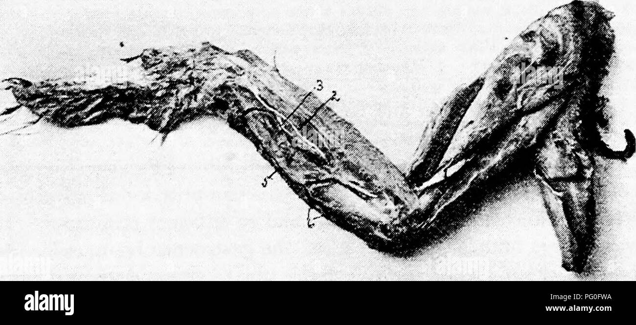
. The anatomy of the domestic fowl . Domestic animals; Veterinary medicine; Poultry. Fig. 66.—Blood-vessels and nerves of the posterior extremity. Outside view. i. Anterior tibial artery. 2, Metatarsal artery. 3, Digital arteries. 4, Vena cutaneous crurus. 5, Ischiadic artery. 6, Vena cruralis. 7, Lateral cutaneous branch of the ischiadic nerve. between two of the main lobes of the kidney. The continuation of the posterior aorta is called the sacralis media' (Fig. 64, No. 32). The ischiadic artery gives off a recurrent renalis on the. Fig. 67.—Blood-vessels and nerves of the fore limb. Outside
Image details
Contributor:
Central Historic Books / Alamy Stock PhotoImage ID:
PG0FWAFile size:
7.1 MB (411.4 KB Compressed download)Releases:
Model - no | Property - noDo I need a release?Dimensions:
2376 x 1051 px | 40.2 x 17.8 cm | 15.8 x 7 inches | 150dpiMore information:
This image is a public domain image, which means either that copyright has expired in the image or the copyright holder has waived their copyright. Alamy charges you a fee for access to the high resolution copy of the image.
This image could have imperfections as it’s either historical or reportage.
. The anatomy of the domestic fowl . Domestic animals; Veterinary medicine; Poultry. Fig. 66.—Blood-vessels and nerves of the posterior extremity. Outside view. i. Anterior tibial artery. 2, Metatarsal artery. 3, Digital arteries. 4, Vena cutaneous crurus. 5, Ischiadic artery. 6, Vena cruralis. 7, Lateral cutaneous branch of the ischiadic nerve. between two of the main lobes of the kidney. The continuation of the posterior aorta is called the sacralis media' (Fig. 64, No. 32). The ischiadic artery gives off a recurrent renalis on the. Fig. 67.—Blood-vessels and nerves of the fore limb. Outside view, i. Median nerve. 3, Ulnar nerve. 4, Radial artery. 5, Recurrent ulnaris artery. 6, Twigs of ulnar artery to wing feathers. posterior lobe of the kidney (Fig. 64, No. 33). On the left side it gives off a branch to the oviduct and to the hgament of the oviduct. The main trunk leaves the cavity with the ischiadic nerve (Fig. 65, . Please note that these images are extracted from scanned page images that may have been digitally enhanced for readability - coloration and appearance of these illustrations may not perfectly resemble the original work.. Kaupp, Benjamin Franklyn, 1874-. Philadelphia ; London : W. B. Saunders Company