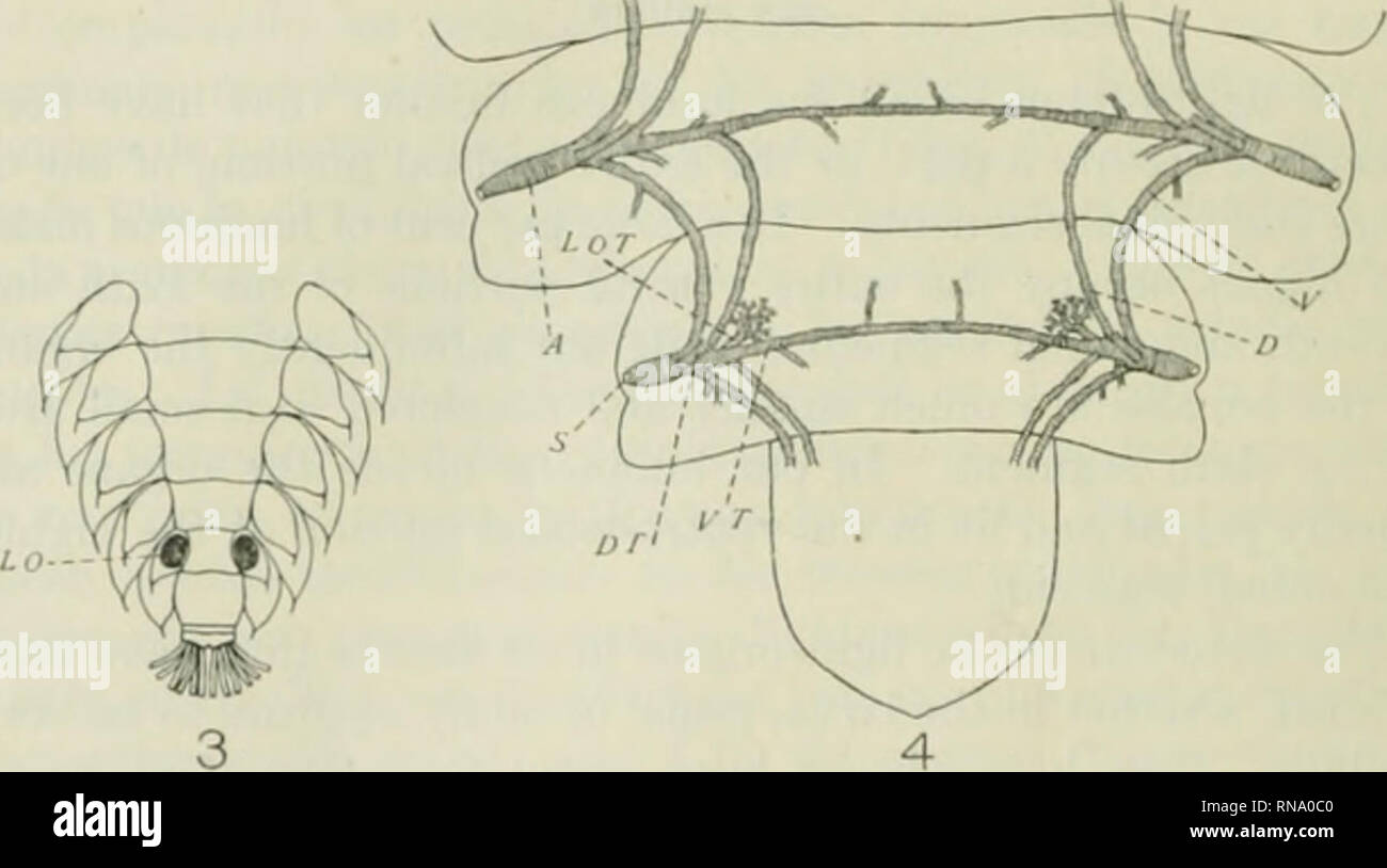
. The anatomical record. Anatomy; Anatomy. Fig. 1 Pliotinus scintillans, malo, (liiiKrammatic drawing to represent the two layers of flie light-organ and the general arrangement of the larger trachea. A, ampulla; D T, dorsal tracheal trunk; P, photogenic layer; R, reflector layer; S, spiracle; V T, ventral tracheal trunk. Fig. 2 Photurus pennsylvanica, larva, diagrammatic drawing to show the two layers of the light-organ, and the arrangement of the trachea. A, ampulla; D T, dorsal tracheal trunk; /', jihotogenic layer; l{, reflector layer; N, spiracle; V T, ventral tracheal trunk. Fig. 3 Photu
Image details
Contributor:
Library Book Collection / Alamy Stock PhotoImage ID:
RNA0C0File size:
7.2 MB (143.9 KB Compressed download)Releases:
Model - no | Property - noDo I need a release?Dimensions:
2118 x 1180 px | 35.9 x 20 cm | 14.1 x 7.9 inches | 150dpiMore information:
This image is a public domain image, which means either that copyright has expired in the image or the copyright holder has waived their copyright. Alamy charges you a fee for access to the high resolution copy of the image.
This image could have imperfections as it’s either historical or reportage.
. The anatomical record. Anatomy; Anatomy. Fig. 1 Pliotinus scintillans, malo, (liiiKrammatic drawing to represent the two layers of flie light-organ and the general arrangement of the larger trachea. A, ampulla; D T, dorsal tracheal trunk; P, photogenic layer; R, reflector layer; S, spiracle; V T, ventral tracheal trunk. Fig. 2 Photurus pennsylvanica, larva, diagrammatic drawing to show the two layers of the light-organ, and the arrangement of the trachea. A, ampulla; D T, dorsal tracheal trunk; /', jihotogenic layer; l{, reflector layer; N, spiracle; V T, ventral tracheal trunk. Fig. 3 Photurus pennsylvanica, larva, ventral view of abdomen. The dark elliptic.il areas which arc located on the eighth abdominal segment represent the larval light-organs, L f>. Fig. 4 Photurus pennsylvanica, larva, dorsal view of the princi|^al trachea of the seventh and eighth abdominal'segments. .4, ampulla; I), dorsal longi- tudinal tracheal connective; D T, dorsal tracheal trunk; LOT, light-organ tra- chea; .S', spiracle; 1', ventral longitudinal tracheal connective. .Ml tracheae shown with small branches supply the light-organ.. Please note that these images are extracted from scanned page images that may have been digitally enhanced for readability - coloration and appearance of these illustrations may not perfectly resemble the original work.. Bardeen, Charles Russell, 1871-1935, ed; Boyden, Edward A. (Edward Allen), 1886-1976; Bremer, John Lewis, 1874- ed; Hardesty, Irving, b. 1866, ed; American Association of Anatomists; American Society of Zoologists; Wistar Institute of Anatomy and Biology. [New York, etc. ] A. R. Liss [etc. ]