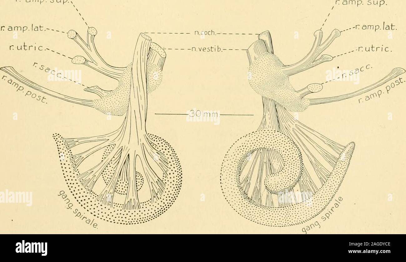
. The American journal of anatomy. ,r amp. sup./ ,-r amp. I at.r. utnc. qanqspirale a m p. 5 u p.,. MEDIAN VIEW LATERAL VIEW Fig. 5. Stages in the differentiation of the acoustic nerve complex. Vesti-bular ganglion shown by fine dots, and spiral ganglion by large dots. 158 Development of Ear and YII-VIII Cranial Nerves ganglion cells connecting the anlage of the spiral ganglion with thebrain, and the conversion of this column into fibroblasts produces theearly fibers of the trunk; this would explain the abrupt appearance ofthe nerve trunk in all parts of its course at once. Proceeding to embry
Image details
Contributor:
The Reading Room / Alamy Stock PhotoImage ID:
2AGDYCEFile size:
7.2 MB (272.9 KB Compressed download)Releases:
Model - no | Property - noDo I need a release?Dimensions:
2082 x 1201 px | 35.3 x 20.3 cm | 13.9 x 8 inches | 150dpiMore information:
This image is a public domain image, which means either that copyright has expired in the image or the copyright holder has waived their copyright. Alamy charges you a fee for access to the high resolution copy of the image.
This image could have imperfections as it’s either historical or reportage.
. The American journal of anatomy. , r amp. sup./ , -r amp. I at.r. utnc. qanqspirale a m p. 5 u p., . MEDIAN VIEW LATERAL VIEW Fig. 5. Stages in the differentiation of the acoustic nerve complex. Vesti-bular ganglion shown by fine dots, and spiral ganglion by large dots. 158 Development of Ear and YII-VIII Cranial Nerves ganglion cells connecting the anlage of the spiral ganglion with thebrain, and the conversion of this column into fibroblasts produces theearly fibers of the trunk; this would explain the abrupt appearance ofthe nerve trunk in all parts of its course at once. Proceeding to embryos 30 mm. long, the same as seen in Figs. a. h, c, Plate I, we meet with conditions which are practically those found inthe adult. There is the vestibular nerve, on whose trunk is situated itsganglion mass, consisting of an upper and lower division. The upperdivision is connected with the labyrinth by the branches supplying theanterior and lateral ampullae, and the utricle; the lower division givesoff branches to the sacculus and posterior ampulla. In the adult thedivision between the pars super