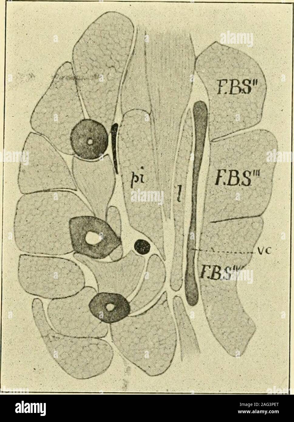
. The American journal of anatomy. flexores brevesmedii reach a much greater devel-opment than in the lower groupand are arranged in two distinctlayers, the superficial one (Fig.11, I) lying immediately beneaththe volar cartilage, from which ittakes origin, while the deeperone (pi) is in relation with theunderlying metacarpal bones. This latter layer does not concern us at present and will be left for con-sideration on another occasion. The superficial layer when traced distallydivides into four portions which pass to the II-V digits, there being noportion for the pollex. Each portion lies ben
Image details
Contributor:
The Reading Room / Alamy Stock PhotoImage ID:
2AG3PETFile size:
7.1 MB (387.3 KB Compressed download)Releases:
Model - no | Property - noDo I need a release?Dimensions:
1365 x 1830 px | 23.1 x 31 cm | 9.1 x 12.2 inches | 150dpiMore information:
This image is a public domain image, which means either that copyright has expired in the image or the copyright holder has waived their copyright. Alamy charges you a fee for access to the high resolution copy of the image.
This image could have imperfections as it’s either historical or reportage.
. The American journal of anatomy. flexores brevesmedii reach a much greater devel-opment than in the lower groupand are arranged in two distinctlayers, the superficial one (Fig.11, I) lying immediately beneaththe volar cartilage, from which ittakes origin, while the deeperone (pi) is in relation with theunderlying metacarpal bones. This latter layer does not concern us at present and will be left for con-sideration on another occasion. The superficial layer when traced distallydivides into four portions which pass to the II-V digits, there being noportion for the pollex. Each portion lies beneath the correspondingportion of the flexor superficialis, being separated from it by the strongtendon derived from the volar cartilage. More distally each of the por-tions corresponding to digits II-IV divides into two slips which come tolie on either side of the corresponding strong tendon ap-d are finally in-serted into opposite sides of the base of the metacarpo-phalangeal fibro-cartilage of the digit to which they belong-. 14. Fig. 11. Transverse section through thepalm of Liolepisma laterale. F. B. S., flexorbrevis dig-itorura superflcialis; I, lumbriealis;i. e., superficial layer of the flexor brevismedius; pi, palmar adductors; i. t., deep layerof the flexor brevis medius; vc, volar cartilage. 20G The Phylogeiiy of the Forcann Flexors In the case of the fifth digit the conditions are slightly different, inthat the superficial sheet of the flexor medius does not extend laterallybeyond its radial border, and hence, when the division of the sheet intoseparate slips takes place, that for the minimus lies upon the radial side of its digit and does not divide intotwo terminal slips as do the others, but inserts entirely into the radialside of the arthrodial filnocartilage.The arrangement of these muscle-slips is shown diagrammatically inFig. 12. If now we proceed to comparethese arrangements with those seenin the amphibia we arrive at thefollowing conclusions. The por-tion of