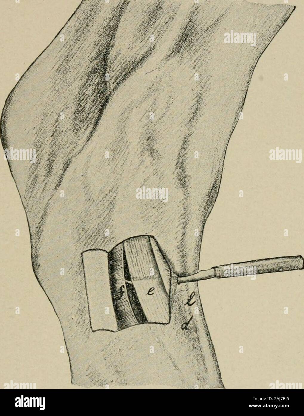
Surgical and obstetrical operations . has beensevered. By keeping as close to the anterior border of thetendon as possible we can avoid injury to the plantar nerve,the common digital artery, the internal cutaneous, and theinternal and external interosseous veins which run betweenthe flexor pedis and the suspensory ligament. After the removal of the knife and seeing that there is awide space between the ends of the tendon, the foot is un-bound from the splint and a bandage applied to the meta-carpus, which rests upon the fetlock joint and remains inposition for eight days. Healing of the cutane
Image details
Contributor:
The Reading Room / Alamy Stock PhotoImage ID:
2AJ7BJ5File size:
7.1 MB (371.6 KB Compressed download)Releases:
Model - no | Property - noDo I need a release?Dimensions:
1400 x 1785 px | 23.7 x 30.2 cm | 9.3 x 11.9 inches | 150dpiMore information:
This image is a public domain image, which means either that copyright has expired in the image or the copyright holder has waived their copyright. Alamy charges you a fee for access to the high resolution copy of the image.
This image could have imperfections as it’s either historical or reportage.
Surgical and obstetrical operations . has beensevered. By keeping as close to the anterior border of thetendon as possible we can avoid injury to the plantar nerve, the common digital artery, the internal cutaneous, and theinternal and external interosseous veins which run betweenthe flexor pedis and the suspensory ligament. After the removal of the knife and seeing that there is awide space between the ends of the tendon, the foot is un-bound from the splint and a bandage applied to the meta-carpus, which rests upon the fetlock joint and remains inposition for eight days. Healing of the cutaneous woundby primary union. 35. PERONEAL TENOTOMY.PI.ATK XXIII. Object. The relief of Stringhalt. Instruments. Razor, scissors, sharp tenotome. Technic. On the lateral side of the metatarsus a triangle, d, opening toward the tarsus is formed by the tendons of theextensor pedis longus muscle, /, and the lateral extensor ofthe foot, e, which unite on the anterior surface of the middleof the metatarsus. The synovial sheath of the extensor. Pirate XXIII. Peroneai, Tenotomy for Stringhai^t. Right hind foot seen from the external side.The skin covering the lateral extensor of thefoot is laid back in the form of a flap, the cruralfascia divided. e. Peroneal tendon ; /, cruralfascia ; /, tendon of the anterior extensor pedismusale ; d, the triangle formed by / and <?. lO 145 146 CUNEAN TENOTOMY. pedis longus muscle extends inferiorly to near the point ofjuncture of the two tendons ; the sheath of the lateral ex-tensor ends below 3 to 4 cm. above the point of union. Inthe middle of this space without a sheath, which is 3 to 4cm. long, and below the annular ligament of the hock theoperation is carried out. After the skin hos been shavedand disinfected, confine in the stocks or operate upon thestanding horse, with the aid of locil anaesthesia, a twitchbeing applied to the nose and the opposite hind foot held upwith the side-line. The tendon of the lateral extensor iseasily felt under the