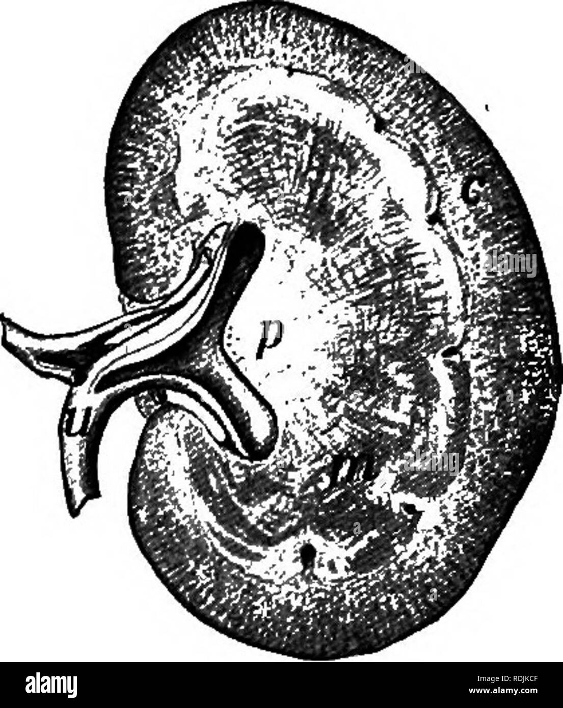
. Practical anatomy of the rabbit : an elementary laboratory textbook in mammalian anatomy . Rabbits; Anatomy, Comparative. The Urinogenital System. 93 THE KIDNEYS. urinogenital sinus. This development reaches its extreme in the higher mammalia, where the urinogenital. sinus is completely separated from the digestive tube, and where the urinary ducts are also transferred from a posterior or hypocystic position on the wall of the. urinogenital sinus to an anterior or epicystic position on the dorsal wall of the bladder. The chief organs of the urinary system are the kidneys. They are paired org
Image details
Contributor:
The Book Worm / Alamy Stock PhotoImage ID:
RDJKCFFile size:
7.1 MB (362.9 KB Compressed download)Releases:
Model - no | Property - noDo I need a release?Dimensions:
1457 x 1714 px | 24.7 x 29 cm | 9.7 x 11.4 inches | 150dpiMore information:
This image is a public domain image, which means either that copyright has expired in the image or the copyright holder has waived their copyright. Alamy charges you a fee for access to the high resolution copy of the image.
This image could have imperfections as it’s either historical or reportage.
. Practical anatomy of the rabbit : an elementary laboratory textbook in mammalian anatomy . Rabbits; Anatomy, Comparative. The Urinogenital System. 93 THE KIDNEYS. urinogenital sinus. This development reaches its extreme in the higher mammalia, where the urinogenital. sinus is completely separated from the digestive tube, and where the urinary ducts are also transferred from a posterior or hypocystic position on the wall of the. urinogenital sinus to an anterior or epicystic position on the dorsal wall of the bladder. The chief organs of the urinary system are the kidneys. They are paired organs, lying against the dorsal abdominal wall, approxi- mately in the position of the embryonic inter- mediate cell mass from which they are formed. That of the left side is displaced backward, out of the position of symmetry, oa account of the posterior development of the greater curvature of the stomach. The kidneys appear as solid organs, brownish in colour and bean-like in general shape, enclosed by a fibrous coat, and connected medially with the expanded end of the ureter. In the rabbit the kidney appears as an almost continuous mass, in which, how- ever, slight traces of lobulation can be distinguished. In many mammals, such as sheep and bear, the organ is composed of distinct and separable lobules. This condition is clearly shown in the human kidney in foetal life, and though much more concentrated in the adult, the lobulated con- dition appears internally in the division of the ureter into several renal calyces, each of them connected with a corresponding renal papilla. When horizontally divided (Fig. 50), the kidney is seen to be made up of a more vascular and granular external layer, termed the cortex, and of a somewhat radially striated, central mass, termed the medulla. In the rabbit, there is a single renal papilla, and the expended end or pelvis of the ureter is undivided. Notwith- standing the solid appearance of cortex and medulla, the kidney is made up of a syst