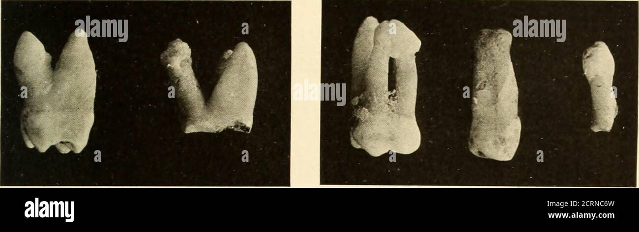
. Oral Roentgenology : a Roentgen study of the anatomy and pathology of the oral cavity . tion. Figure 141.Patient: Mrs. T. M. G. Roentgen Examination: Shows irregular outline of mesial root of first molar,a condition which indicates absorption due to chronic inflammation. Figure 142.Patient: Mrs. H. G. B. Roentgen Examination: Granulomata at the ends of the roots of the first molarare indicated by the radiolucent areas. The mesial root shows marked exostosis. Figures 143 and 145.Roentgen Examination: Shows in Figure 143 absorption of the palatal root ofthe upper first molar and in Figure 145
Image details
Contributor:
Reading Room 2020 / Alamy Stock PhotoImage ID:
2CRNC6WFile size:
7.1 MB (204.4 KB Compressed download)Releases:
Model - no | Property - noDo I need a release?Dimensions:
2911 x 858 px | 24.6 x 7.3 cm | 9.7 x 2.9 inches | 300dpiMore information:
This image could have imperfections as it’s either historical or reportage.
. Oral Roentgenology : a Roentgen study of the anatomy and pathology of the oral cavity . tion. Figure 141.Patient: Mrs. T. M. G. Roentgen Examination: Shows irregular outline of mesial root of first molar, a condition which indicates absorption due to chronic inflammation. Figure 142.Patient: Mrs. H. G. B. Roentgen Examination: Granulomata at the ends of the roots of the first molarare indicated by the radiolucent areas. The mesial root shows marked exostosis. Figures 143 and 145.Roentgen Examination: Shows in Figure 143 absorption of the palatal root ofthe upper first molar and in Figure 145 absorption of the end of the lateral incisor. Figure 144. Patient: Mrs. I. O. H. History: Patient says she had periostitis 30 years ago in Norway. A piece ofnecrosed bone was removed by her dentist. Tooth is firm and reacts on the heat test. Roentgen Examination: Shows one root very much shortened. An irregularradiopaque area indicates scar bone and there is a radiolucent area at the distal sideunder the filling, which shows decay. ROENTGEXOGRAPHIC STUDY OF PATHOLOGICAL CONDITIONS 105. Figure 135 Figure 136.