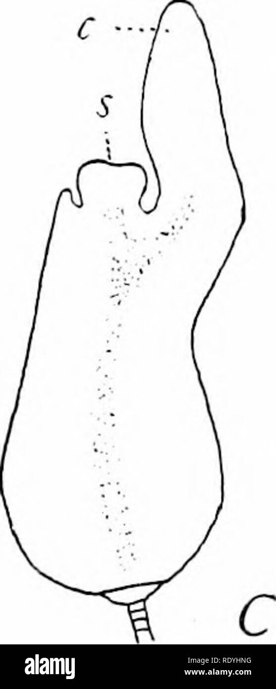
. Morphology of angiosperms (Morphology of spermatophytes. Part II). Angiosperms; Plant morphology. B Fig. 88.—Zannichellia palustris. Development of embryo. A, young embryo; x 320; B, later stage, showing beginning of differentiation into stem-tip (*) and cotyledon (c), both coming from the cells derived from terminal cell of proembryo ; x- 160 ; C stem- tip (s) and cotyledon (c) clearly differentiated; x 60.—After Campbell.41 the others, not merely because it characterizes the primitive hydrophytic forms, but also because it is the simplest type, and the others may well be modifications of
Image details
Contributor:
The Book Worm / Alamy Stock PhotoImage ID:
RDYHNGFile size:
7.1 MB (105.7 KB Compressed download)Releases:
Model - no | Property - noDo I need a release?Dimensions:
1035 x 2413 px | 17.5 x 40.9 cm | 6.9 x 16.1 inches | 150dpiMore information:
This image is a public domain image, which means either that copyright has expired in the image or the copyright holder has waived their copyright. Alamy charges you a fee for access to the high resolution copy of the image.
This image could have imperfections as it’s either historical or reportage.
. Morphology of angiosperms (Morphology of spermatophytes. Part II). Angiosperms; Plant morphology. B Fig. 88.—Zannichellia palustris. Development of embryo. A, young embryo; x 320; B, later stage, showing beginning of differentiation into stem-tip (*) and cotyledon (c), both coming from the cells derived from terminal cell of proembryo ; x- 160 ; C stem- tip (s) and cotyledon (c) clearly differentiated; x 60.—After Campbell.41 the others, not merely because it characterizes the primitive hydrophytic forms, but also because it is the simplest type, and the others may well be modifications of it. In the Pistia type the suspensor is suppressed; in the Lilium type it becomes massive and meristematic; in the Orchid type it is developed as a special haustorium that passes out of the ovule on account of the lack of endosperm, and perhaps for the same reason the embryo does not reach the stage of differentiating organs. There have been observed certain departures from the mon- ocotyledonous type of embryo that deserve special mention.. Please note that these images are extracted from scanned page images that may have been digitally enhanced for readability - coloration and appearance of these illustrations may not perfectly resemble the original work.. Coulter, John Merle, 1851-1928; Chamberlain, Charles Joseph, b. 1863. New York, D. Appleton