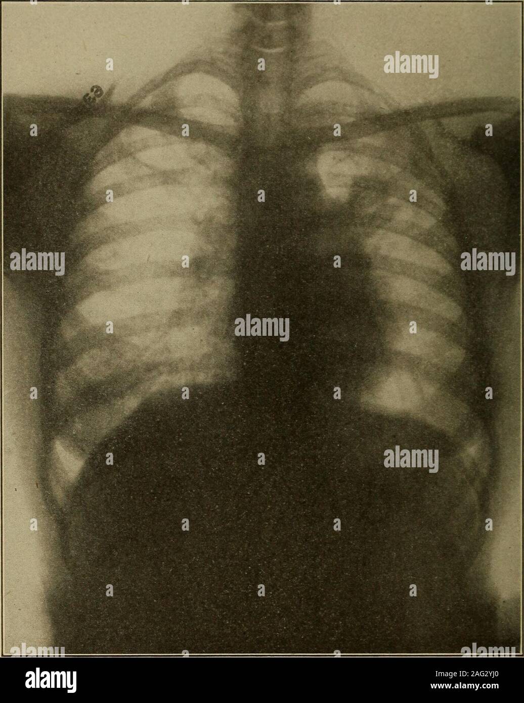
. Medical diagnosis for the student and practitioner. Fig. 127.—Apical Tuberculosis—Healed or Latent. Note that lesions are closely cir-cumscribed by apparently normal lung. {Dr. Frank S. Bissell.) This is true because the more characteristic changes tend to becomemasked by the effects of fibrosis and mixed infections. Usually, however,the distribution of the lesions points the way to a correct diagnosis or somearea of slight involvement is found where the changes are more typical. Compensatory emphysema frequently exists in some degree, manefesting Emphysema. 3i8 MKDICAL I)IA(;OSIS itself ch
Image details
Contributor:
The Reading Room / Alamy Stock PhotoImage ID:
2AG2YJ0File size:
7.1 MB (430.2 KB Compressed download)Releases:
Model - no | Property - noDo I need a release?Dimensions:
1411 x 1770 px | 23.9 x 30 cm | 9.4 x 11.8 inches | 150dpiMore information:
This image is a public domain image, which means either that copyright has expired in the image or the copyright holder has waived their copyright. Alamy charges you a fee for access to the high resolution copy of the image.
This image could have imperfections as it’s either historical or reportage.
. Medical diagnosis for the student and practitioner. Fig. 127.—Apical Tuberculosis—Healed or Latent. Note that lesions are closely cir-cumscribed by apparently normal lung. {Dr. Frank S. Bissell.) This is true because the more characteristic changes tend to becomemasked by the effects of fibrosis and mixed infections. Usually, however, the distribution of the lesions points the way to a correct diagnosis or somearea of slight involvement is found where the changes are more typical. Compensatory emphysema frequently exists in some degree, manefesting Emphysema. 3i8 MKDICAL I)IA(;OSIS itself chiefly by increased translucency of the area so affected. This trans-lucency, when the emphysema is extensive, aids materially in the studyand recognition of the tuberculous foci by lending sharper contrast to them.Thus the clinicians handicap becomes the advantage of the roentgenologist.In advanced tuberculosis the greatest value of the roentgen examination. Fig. 128.—Pulmonary Tuberculosis with Cavitation. {Dr. Frank S. Bissell.) lies in the accuracy with which may be demonstrated the extent of thedisease, the presence and nature of complications, as well as other factors inprognosis. The extent of involvement of a lung is usually found greater thanphysical signs or symptoms would lead one to suspect because so many of thelesions lie in the deeper portions of the lung where they escape the detection ofthe keenest clinician. ROENTGENOGRAPHS EXAMINATION OF LUNGS AND PLEURA 310 Cavities.- -Cavitalion is easily recognized although one must be carefulnot to interpret all ring-like shadows in the lung field as cavities. Theremust be a total absence of normal lung structure within the ring and specialcare must be used to differentiate from bronchiectasis or partial pneumo-thorax. Areas of lung consolidation surrounding a cavity filled with exudate