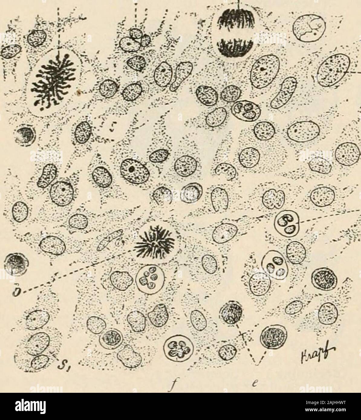
Manual of pathology : including bacteriology, the technic of postmortems, and methods of pathologic research . iscalled the fibroblast. Ap-parently it is the productof proliferative changes inthe fixed connective-tissuecells of the part. The rela-tion of the newly formed fibrous tissue to these cells renders it clear thatthe fibrils are in some way developed from them, but exactly how theelaboration of fibrils is accomplished is less certain. Of the many methodsthat have been suggested, ohree are deserving of consideration: (i) Theolder view that the fibrils resulted from elongation and attenu
Image details
Contributor:
The Reading Room / Alamy Stock PhotoImage ID:
2AJHHWTFile size:
7.1 MB (409.4 KB Compressed download)Releases:
Model - no | Property - noDo I need a release?Dimensions:
1515 x 1649 px | 25.7 x 27.9 cm | 10.1 x 11 inches | 150dpiMore information:
This image is a public domain image, which means either that copyright has expired in the image or the copyright holder has waived their copyright. Alamy charges you a fee for access to the high resolution copy of the image.
This image could have imperfections as it’s either historical or reportage.
Manual of pathology : including bacteriology, the technic of postmortems, and methods of pathologic research . iscalled the fibroblast. Ap-parently it is the productof proliferative changes inthe fixed connective-tissuecells of the part. The rela-tion of the newly formed fibrous tissue to these cells renders it clear thatthe fibrils are in some way developed from them, but exactly how theelaboration of fibrils is accomplished is less certain. Of the many methodsthat have been suggested, ohree are deserving of consideration: (i) Theolder view that the fibrils resulted from elongation and attenuation of thecell protoplasm followed by disappearance of the nucleus is not in accordwith the best information as to the method by which cells ordinarilyaccomplish the specific purposes toward which their activity is directed.(2) According to this view, the fibrils are formed at the periphery ofthe cell from which they are shaled off or shed, new fibrils arising asrapidly as the older become an integral part of the developing cicatrix.Eventually when the fibrous tissue formation is complete the nuclei of. -/ Fig. 160.—Cellular Elements of Formative Tissue.—(Schmaus.) X 500 diameters. a. a. Polyblasts. b. Mother star. c. Daughter star. d. Skeinst.-ige. e Round or lymphoid cells. /, /. Polymorphonu-clear leukocytes. INK LAM MAT ION ANU KIvlAIK. 303 many ol the fibroblasts disappear, otliers remaining as the fixed connec-tive-tissue cells of the scar. (3) It is known that the matrix of boneand cartilage is formed through the secretory activity f cells (osteo-blasts or chondroblasts) and tliat most other cells accomjjlish functionby a process called secretion. In order to coordinate fibrogenesis withosteogenesis and chondrogenesis it is necessary to assume that the fibro-blasts in a way secrete the fibrils forming the cicatrix; in other words, that the cicatricial tissue is laid down by these cells exactly as thematrix of bone or of cartilage is formed. It is possible to see in