. Local and regional anesthesia; with chapters on spinal, epidural, paravertebral, and parasacral analgesia, and other applications of local and regional anesthesia to the surgery of the eye, ear, nose and throat, and to dental practice. Lachrymal tier. Fig. 172.—Scheme of the ophthalmic nerve after Corning. (Braun.) recurrent branch from the lacrimal artery to the dura, and theophthalmic vein. The relative position of these structures is seen in Fig. 170. 542 LOCAL ANESTHESLA. In making deep orbital injections for the purpose of blocking thosebranches of the trigeminus which pass through this
Image details
Contributor:
The Reading Room / Alamy Stock PhotoImage ID:
2AGDHDWFile size:
7.1 MB (298.8 KB Compressed download)Releases:
Model - no | Property - noDo I need a release?Dimensions:
1445 x 1729 px | 24.5 x 29.3 cm | 9.6 x 11.5 inches | 150dpiMore information:
This image is a public domain image, which means either that copyright has expired in the image or the copyright holder has waived their copyright. Alamy charges you a fee for access to the high resolution copy of the image.
This image could have imperfections as it’s either historical or reportage.
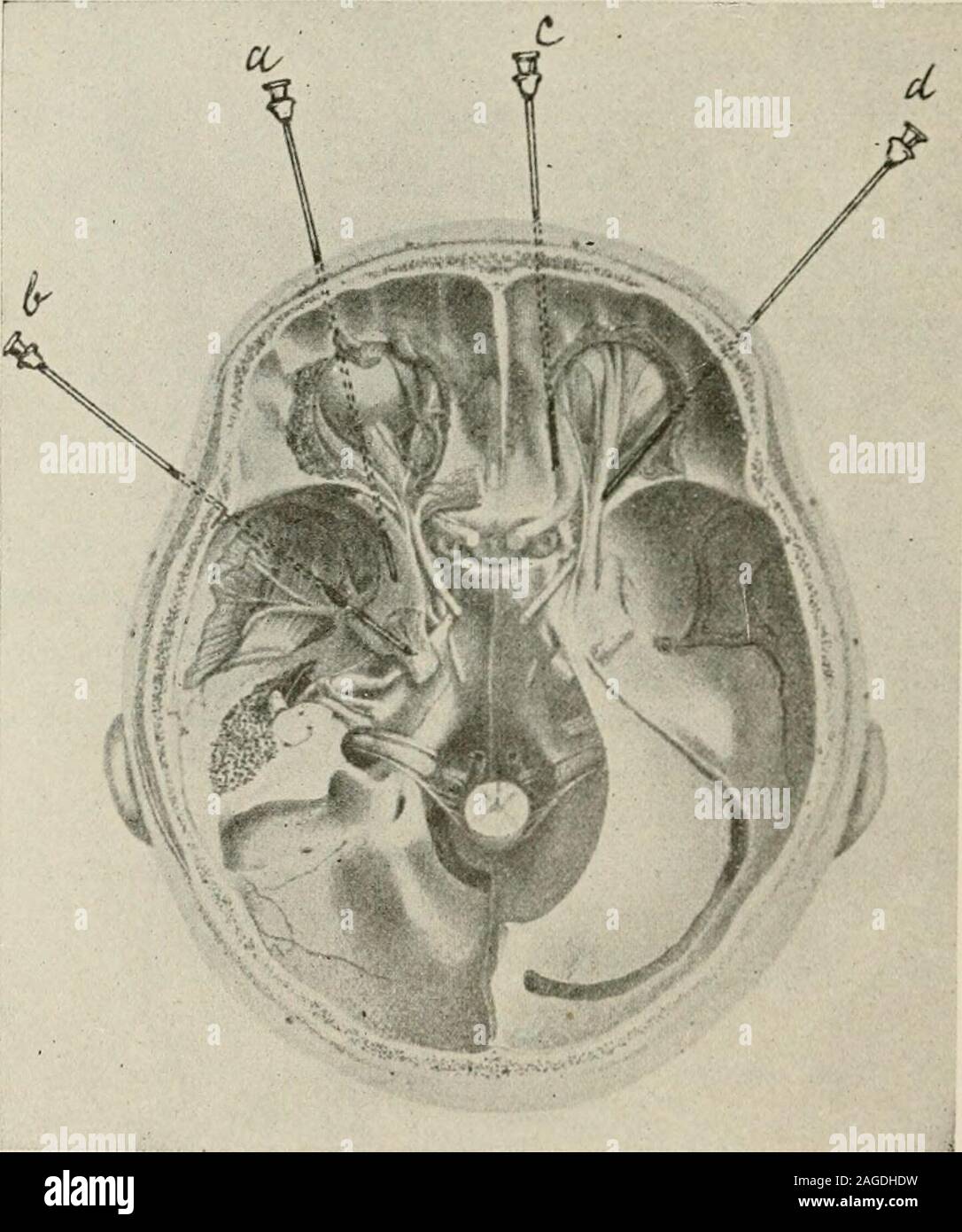
. Local and regional anesthesia; with chapters on spinal, epidural, paravertebral, and parasacral analgesia, and other applications of local and regional anesthesia to the surgery of the eye, ear, nose and throat, and to dental practice. Lachrymal tier. Fig. 172.—Scheme of the ophthalmic nerve after Corning. (Braun.) recurrent branch from the lacrimal artery to the dura, and theophthalmic vein. The relative position of these structures is seen in Fig. 170. 542 LOCAL ANESTHESLA. In making deep orbital injections for the purpose of blocking thosebranches of the trigeminus which pass through this fossa on their wayto other parts, we should try to select such routes of puncture as liealong smooth and regular bony surfaces, using these surfaces as aguide in approaching the deeper parts, and always keeping the needle-point in close contact with the bone; in this way, by keeping welltoward the peripheral limits of the orbit, we are in the zone outside. Fig. 173.—Base of skull with cranial nerves, from Arnold. Needles a and b same as Fig.217, c, to nasal nerve, d to frontal and lacrimal nerves. (Hjirtel). of the eye and its attached muscles. This idea of utilizing the orbitas a means of approach to the intra- and retro-orbital nerve-trunksmay appear to the inexperienced as a hazardous procedure; this, however, is a misconception, as the i)uncture under proper technicshould be a perfectly innocent undertaking, except in the knowndangerous region of the orbit—in its axis or at its apex. In extensiveoperations upon the eye, as in enucleation, these regions are inten-tionally invaded. (Figs. 171, 172 show the arrangement of thenerves within the orbit.) THE HEAD, SCALP, CRANIUM, BRAIN, AND FACE 543 The recognized routes of orbital puncture are: 1. Medial orbital route, first described by Peuckart for reachingthe nasal nerve (Figs. 173-181). 2. Lateral orbital route of Braun for reaching the frontal andlacrimal branches of the ophthalmi