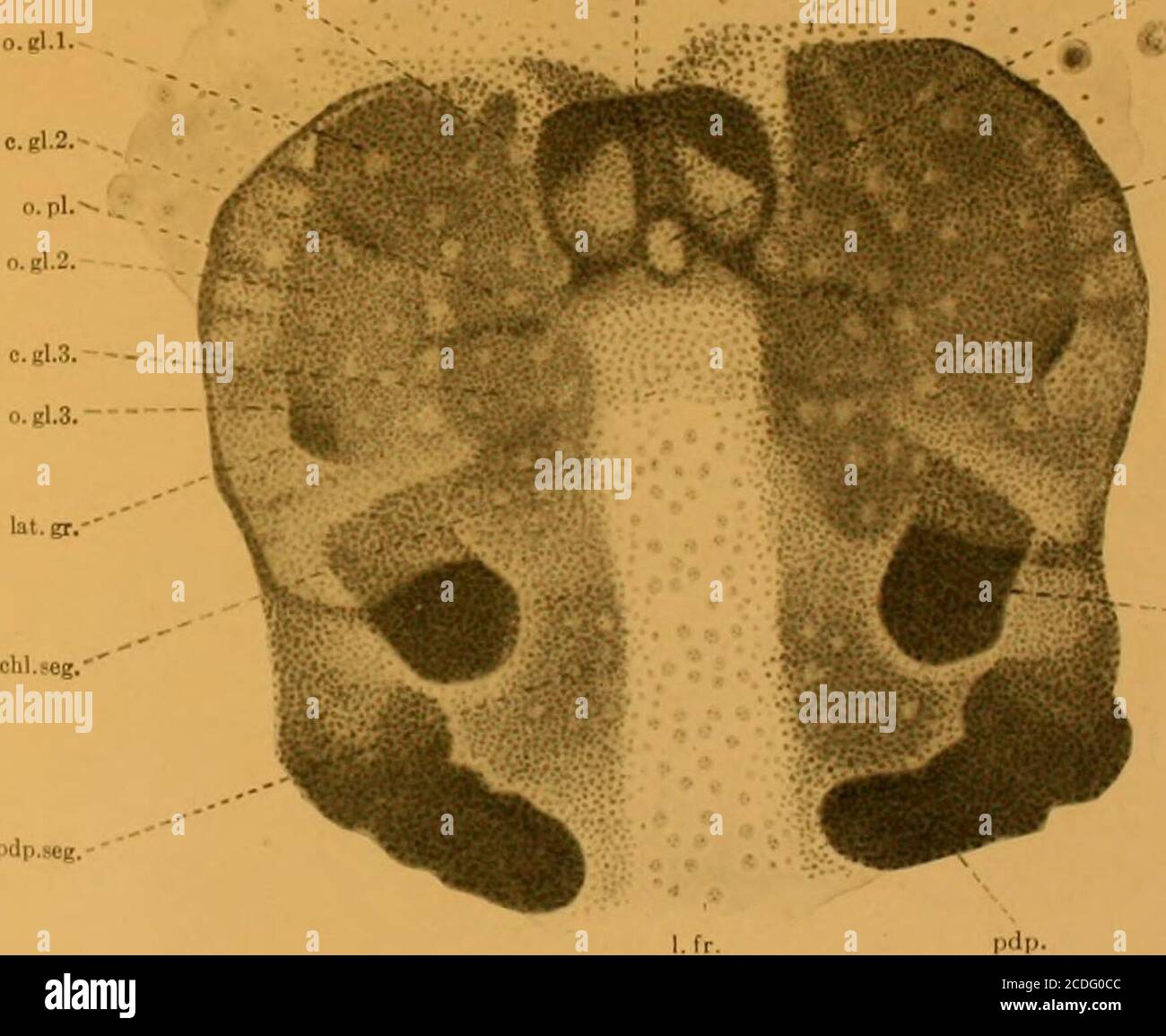
. Journal of morphology . fe. ^i^>-i> «r r)dp.»eg. nt.gr. f.e.l.. l.fr. pdp.soK. pdp- Fio. 21. Tub .TouKNAi of Moupiiolouv.—Vol. XX, No. 3. Tlate VII.Fig. 23, Stage IV. Showing the partial coalescence of the two parts of therostral rudiment, the extension of the optic plate, o. ?>/., the formation ofa distinct coxal portion to the pedipalps, pdp. c, which forms the rudimentof the mandibles. The increase in the number of neuroblasts in the cerebraland optic areas, and their extension to the neiiromeres of the chelicer?e,pedipalps, and the thoracic appendages, is apparent. Fig. 24. Stag
Image details
Contributor:
Reading Room 2020 / Alamy Stock PhotoImage ID:
2CDG0CCFile size:
7.1 MB (253.4 KB Compressed download)Releases:
Model - no | Property - noDo I need a release?Dimensions:
1746 x 1431 px | 29.6 x 24.2 cm | 11.6 x 9.5 inches | 150dpiMore information:
This image is a public domain image, which means either that copyright has expired in the image or the copyright holder has waived their copyright. Alamy charges you a fee for access to the high resolution copy of the image.
This image could have imperfections as it’s either historical or reportage.
. Journal of morphology . fe. ^i^>-i> «r r)dp.»eg. nt.gr. f.e.l.. l.fr. pdp.soK. pdp- Fio. 21. Tub .TouKNAi of Moupiiolouv.—Vol. XX, No. 3. Tlate VII.Fig. 23, Stage IV. Showing the partial coalescence of the two parts of therostral rudiment, the extension of the optic plate, o. ?>/., the formation ofa distinct coxal portion to the pedipalps, pdp. c, which forms the rudimentof the mandibles. The increase in the number of neuroblasts in the cerebraland optic areas, and their extension to the neiiromeres of the chelicer?e, pedipalps, and the thoracic appendages, is apparent. Fig. 24. Stage V. The advances made in the development of the cephalicplate are shown in the deepening of the anterior groove, ant. gr., and ofthe lateral grooves, hit. gr., the shifting of the rostrum from the anteriormargin of the plate to a point between the cerebral ganglia, the more com-plete union of the parts of the rostrum, and the beginning of the foldwhich forms the roof of the cerebral vesicle, o. f. PBOCEPHALIC LOBES OF EPEIRA CINEREA.AVERT E. LAMBERT.