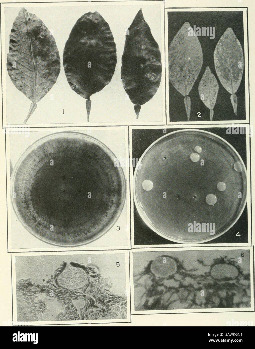
Journal of agricultural research . Journal of Agricultural Research Vol. VI, No.2 Citrus Canker Plate XI. Journal of Agricultural Research Vol. VI, No. 2 PLATE XI Fig. I.—Cankers on old grapefruit leaves which have enlarged during the secondgrowing season. Fig. 2.—Citrus canker resulting from immersion of leaves in a bacterial suspension.Lesions involving a large part of the lower leaf surface are thus formed. Fig. 3.—Culture of Phoma socia showing pycnidial formation in concentric rings. Fig. 4.—Dilution poured plate of Pseudomonas citri on green-bean agar. The spotson the colonies are the re
Image details
Contributor:
The Reading Room / Alamy Stock PhotoImage ID:
2AWKGN1File size:
7.1 MB (449.2 KB Compressed download)Releases:
Model - no | Property - noDo I need a release?Dimensions:
1398 x 1787 px | 23.7 x 30.3 cm | 9.3 x 11.9 inches | 150dpiMore information:
This image is a public domain image, which means either that copyright has expired in the image or the copyright holder has waived their copyright. Alamy charges you a fee for access to the high resolution copy of the image.
This image could have imperfections as it’s either historical or reportage.
Journal of agricultural research . Journal of Agricultural Research Vol. VI, No.2 Citrus Canker Plate XI. Journal of Agricultural Research Vol. VI, No. 2 PLATE XI Fig. I.—Cankers on old grapefruit leaves which have enlarged during the secondgrowing season. Fig. 2.—Citrus canker resulting from immersion of leaves in a bacterial suspension.Lesions involving a large part of the lower leaf surface are thus formed. Fig. 3.—Culture of Phoma socia showing pycnidial formation in concentric rings. Fig. 4.—Dilution poured plate of Pseudomonas citri on green-bean agar. The spotson the colonies are the reflection of the windows of the room in which the exposurewas made. Colonies 14 days old, the last 5 of which days the plates were kept in anice chest at a temperature of about 55°. Fig. 5.—Photomicrograph of pycnidium of Phoma socia taken in reflected simlight. Fig. 6.—Photomicrograph of pycnidia of Phoma socia taken in diffuse light. o ADDITIONAL COPIES OF TmS PUBLICATION MAT BE PROCUEED FROM THE SUPERINTENDENT OF DOCUMENTS GOVERNMENT PRINTING OFFICE ?WASHINGTON, D. C. AT 15 CENTS PER