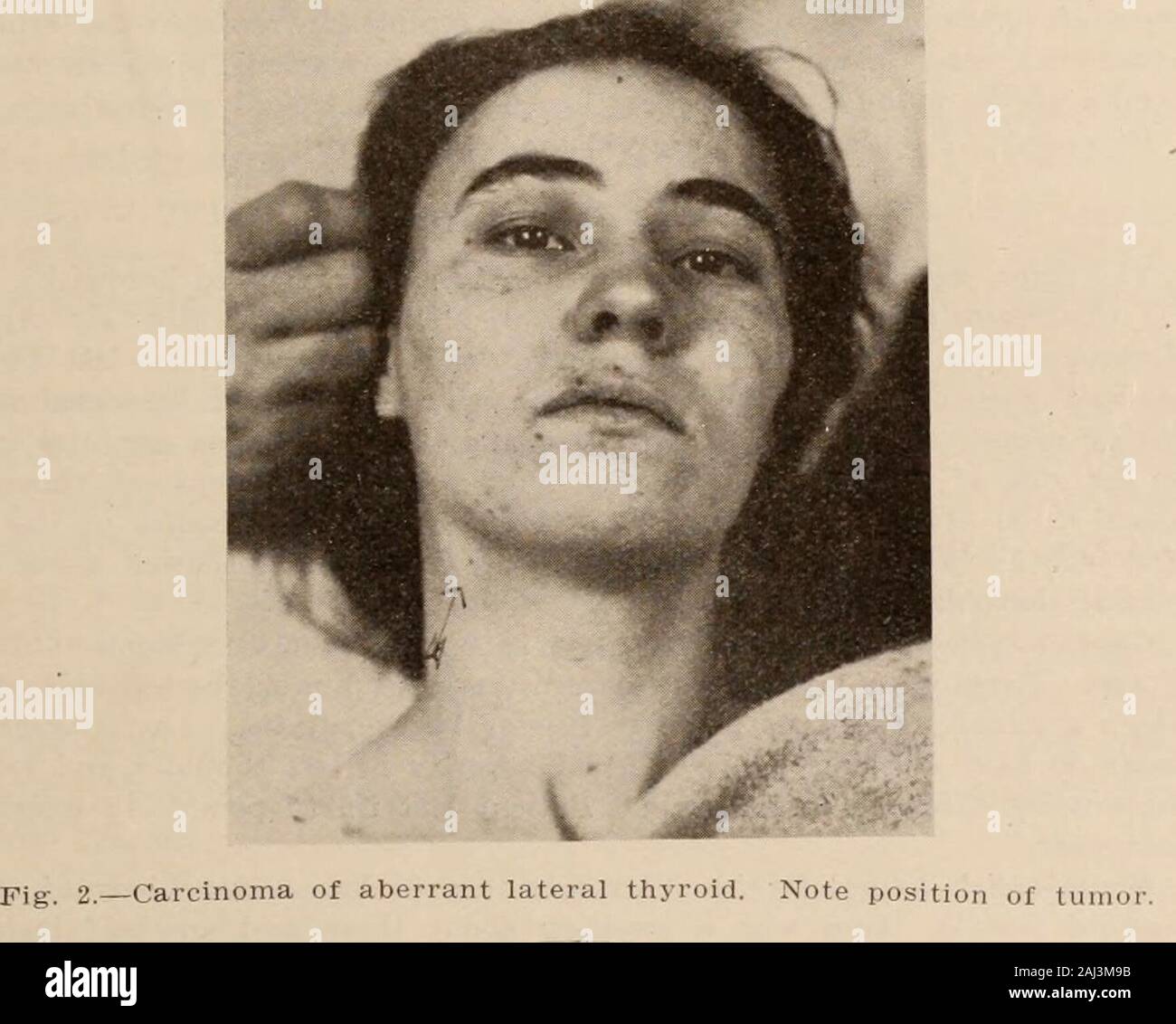
Interstate medical journal . peration.—By Dr. R. P. Condon. An incision along the anterior borderof right sternomastoid. Beneath the edge of the muscle was a tumor whichlooked like a lymph node. It was easily shelled out. It was the size of alarge pecan. About two centimeters below it, and lying deep under thesternomastoid, was another lymph node-like body about one-fourth the sizeof the first and attached to it. There were three other small lymph nodes,the size of small peas, and were connected with the one just described. Theywere of a reddish color and had the gross appearance of a large gi
Image details
Contributor:
The Reading Room / Alamy Stock PhotoImage ID:
2AJ3M9BFile size:
7.1 MB (239.3 KB Compressed download)Releases:
Model - no | Property - noDo I need a release?Dimensions:
1765 x 1415 px | 29.9 x 24 cm | 11.8 x 9.4 inches | 150dpiMore information:
This image is a public domain image, which means either that copyright has expired in the image or the copyright holder has waived their copyright. Alamy charges you a fee for access to the high resolution copy of the image.
This image could have imperfections as it’s either historical or reportage.
Interstate medical journal . peration.—By Dr. R. P. Condon. An incision along the anterior borderof right sternomastoid. Beneath the edge of the muscle was a tumor whichlooked like a lymph node. It was easily shelled out. It was the size of alarge pecan. About two centimeters below it, and lying deep under thesternomastoid, was another lymph node-like body about one-fourth the sizeof the first and attached to it. There were three other small lymph nodes, the size of small peas, and were connected with the one just described. Theywere of a reddish color and had the gross appearance of a large giant cell sar-coma found in maxillary antrum. They were not in contact with the great ves- Wohl: Carcinoma of Lateral Aberrant Thyroid 1047 sels of the neck and no other enlargements were noticed. The tumor wasremoved without drainage. Intensive x-ray treatment was used as a post-operative measure. Patient made an uneventful recovery and at the timeof her dismissal from the hospital no evidence of metastasis could be found.. Fig. 3.—Photograph of tumor removed. Note appearance after longitudinalsection. Pathological Report.—The specimen consists of five nodules varying in sizefrom a pecan to a pea. They are encapsulated and interconnected by con-nective tissue bands. Upon section the larger nodules, (A) and (B), presenta gray white appearance, and the smaller nodules are of a more reddish, and 1048 INTERSTATE MEDICAL JOURNAL in places, gray color. (Macroscopically the tumor has no resemblance tothyroid tissue.) Microscopical.—Section from larger two nodules shows follicles filled withan albuminous material which has an orange color with Van Giesons stain.The follicles are lined with cylindrical cells, which in places fill the luminaof the follicles. In other places there is a papillary formation with invasionof the stroma. The connective tissue stroma is increased in amount and isvascular. Section from the smaller nodules reveals lymphoid structure withnests of epithelia