. Fig. 36.—Flower of Radish hypertrophied by Cystopv.i cundidv.s. The white .«;wollen conidial cushions occupy the enlarged petals, sepals and ovaries. (Dr. Bruns' phot.) develop to intercellular mycelia, tine short lateral twigs of which pierce the wall of the host-cells and become little spherical haustoria. The oogonia arise as thick-walled spherical swellings on the mycelium. The antheridium, after applying itself to the oogonium, widens and projects a fine fertilization-tube through the wall to the egg-cell. After fertilization is effected, the egg-cell is enclosed in a firm uneven membra
Image details
Contributor:
The Bookworm Collection / Alamy Stock PhotoImage ID:
MCK6JYFile size:
14.3 MB (378.9 KB Compressed download)Releases:
Model - no | Property - noDo I need a release?Dimensions:
2236 x 2236 px | 37.9 x 37.9 cm | 14.9 x 14.9 inches | 150dpiMore information:
This image is a public domain image, which means either that copyright has expired in the image or the copyright holder has waived their copyright. Alamy charges you a fee for access to the high resolution copy of the image.
This image could have imperfections as it’s either historical or reportage.
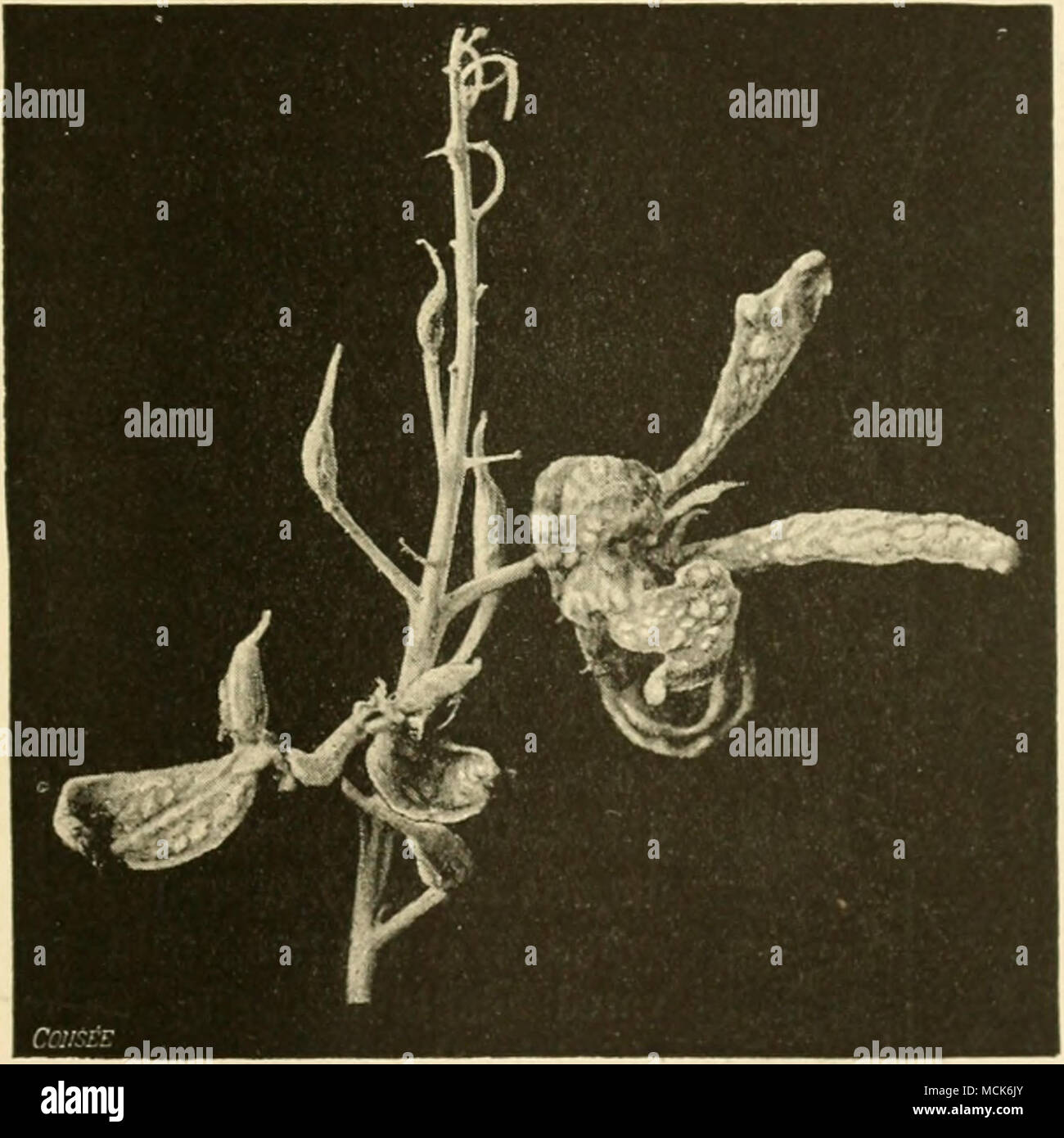
. Fig. 36.—Flower of Radish hypertrophied by Cystopv.i cundidv.s. The white .«;wollen conidial cushions occupy the enlarged petals, sepals and ovaries. (Dr. Bruns' phot.) develop to intercellular mycelia, tine short lateral twigs of which pierce the wall of the host-cells and become little spherical haustoria. The oogonia arise as thick-walled spherical swellings on the mycelium. The antheridium, after applying itself to the oogonium, widens and projects a fine fertilization-tube through the wall to the egg-cell. After fertilization is effected, the egg-cell is enclosed in a firm uneven membrane, and hibernates inside the oogonium. In spring the plasma of the oospore forms numerous biciliate