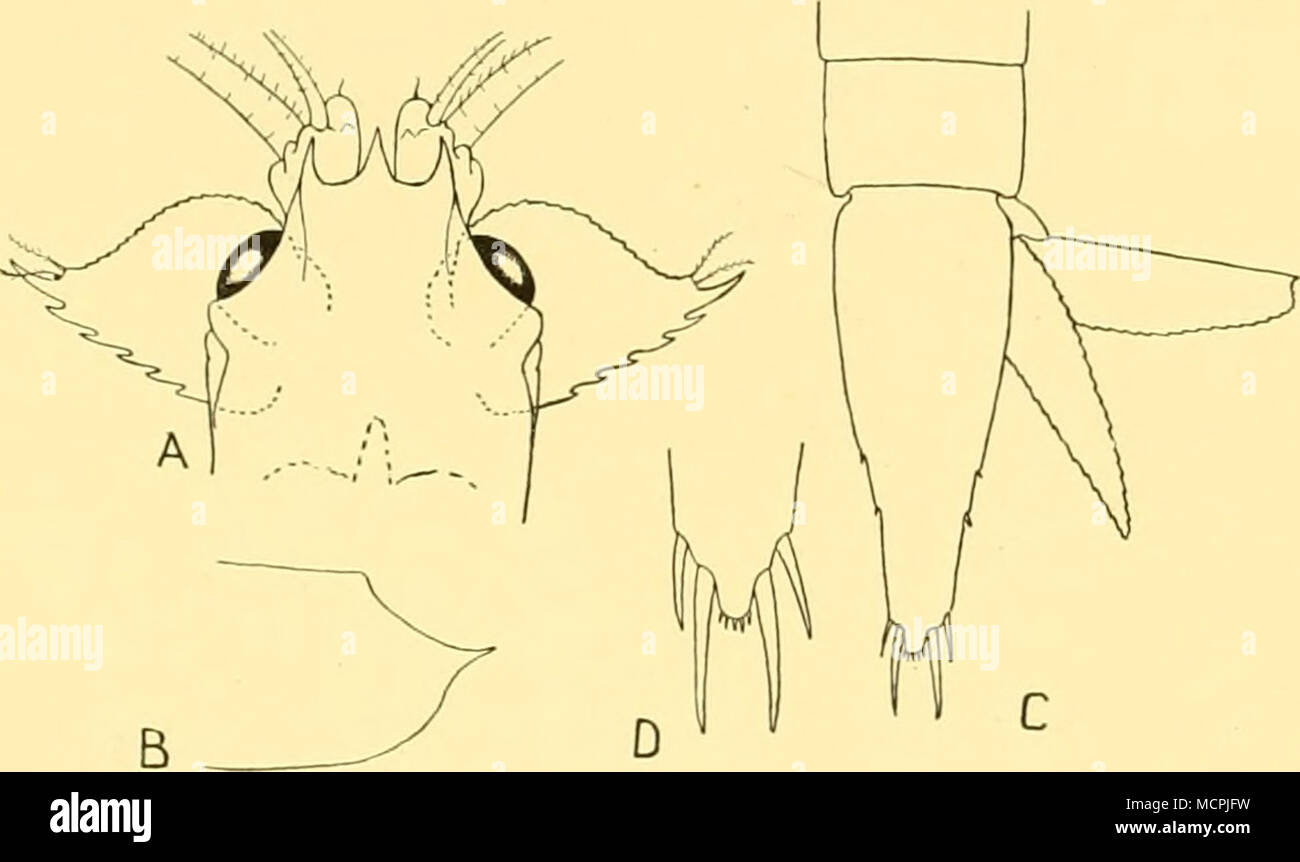
. Fig. 2. Lophogaster challengeri Fage. A, anterior end in dorsal view, x 10; B, lateral view of postero-lateral region of carapace ('wing'); C, telson and right uropod in dorsal view, x 10; D, distal end of telson (enlarged), x 19. Iww*.. Sh«-. 9 The integument of the carapace, especially in the mid-dorsal area, is covered with more or less well-developed tubercles in both typicus and challengeri. This condition is much more pronounced in juveniles and as growth proceeds the tubercles tend to disappear. In the Discovery specimens these tubercles are comparatively few in number and very large.
Image details
Contributor:
The Bookworm Collection / Alamy Stock PhotoImage ID:
MCPJFWFile size:
14.3 MB (207.4 KB Compressed download)Releases:
Model - no | Property - noDo I need a release?Dimensions:
2900 x 1723 px | 24.6 x 14.6 cm | 9.7 x 5.7 inches | 300dpiMore information:
This image is a public domain image, which means either that copyright has expired in the image or the copyright holder has waived their copyright. Alamy charges you a fee for access to the high resolution copy of the image.
This image could have imperfections as it’s either historical or reportage.
. Fig. 2. Lophogaster challengeri Fage. A, anterior end in dorsal view, x 10; B, lateral view of postero-lateral region of carapace ('wing'); C, telson and right uropod in dorsal view, x 10; D, distal end of telson (enlarged), x 19. Iww*.. Sh«-. 9 The integument of the carapace, especially in the mid-dorsal area, is covered with more or less well-developed tubercles in both typicus and challengeri. This condition is much more pronounced in juveniles and as growth proceeds the tubercles tend to disappear. In the Discovery specimens these tubercles are comparatively few in number and very large. The animals are all small and very immature and since neither Sars nor Fage mentioned the tuberculation of the integument of the carapace in their larger specimens of challengeri, I assume that, as in typicus, the tubercles become less pronounced with growth. In small juveniles of both species the margin of the carapace bordering the eye is fringed with a close row of fine teeth which disappear completely with growth. In L. typicus this pectination can still be seen in animals of 12 mm. in length but in L. challengeri it disappears much earlier. At station 277 (haul 1) all the specimens of less than 5 mm. in length had marked pectination but in those of 5-5-5 mm. only very faint traces of it remained. I could not see any sign of the pectination of the margins of the epimeral plates such as occurs in juveniles of L. typicus. G. O. Sars (18856, p. 14) neither mentioned nor figured a post-orbital spine in his Challenger specimens, although M. Sars made a special point of it in his original description of L. typicus. Examination of the Challenger specimens confirms that they have no such spine and I am unable to find one in the Discovery specimens, which I here refer to challengeri. In all the European specimens