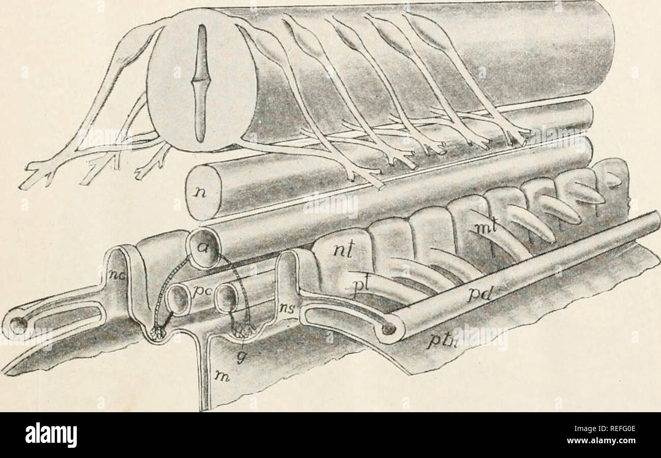
. Comparative anatomy of vertebrates. Anatomy, Comparative; Vertebrates -- Anatomy. UROGENITAL SYSTEM. 313 so that the whole resembles a renal corpuscle, but is different in origin. In either case the exuding fluid passes into the metaccele from which it is drawn by the cilia of the nephrostomes and passed into the tubules. The blood is brought to the glomus or glomeruli by short segmental arteries arising from the dorsal aorta (fig. 318) and, after passing through the capillaries, it is carried away by the postcardinal veins of the corresponding side to the heart, these veins keeping pace in
Image details
Contributor:
The Book Worm / Alamy Stock PhotoImage ID:
REFG0EFile size:
7.1 MB (341.5 KB Compressed download)Releases:
Model - no | Property - noDo I need a release?Dimensions:
2002 x 1248 px | 33.9 x 21.1 cm | 13.3 x 8.3 inches | 150dpiMore information:
This image is a public domain image, which means either that copyright has expired in the image or the copyright holder has waived their copyright. Alamy charges you a fee for access to the high resolution copy of the image.
This image could have imperfections as it’s either historical or reportage.
. Comparative anatomy of vertebrates. Anatomy, Comparative; Vertebrates -- Anatomy. UROGENITAL SYSTEM. 313 so that the whole resembles a renal corpuscle, but is different in origin. In either case the exuding fluid passes into the metaccele from which it is drawn by the cilia of the nephrostomes and passed into the tubules. The blood is brought to the glomus or glomeruli by short segmental arteries arising from the dorsal aorta (fig. 318) and, after passing through the capillaries, it is carried away by the postcardinal veins of the corresponding side to the heart, these veins keeping pace in their backward development with the development of the nephridial tubules.. FIG. 318.—Stereogram of developing pro- and mesonephros. a, aorta; g, glomus or glomerulus; in, mesentery; mt, mesonephric tubule; n, notochord; nc, cavity of («/) nephro- tome; ns, nephrostome; pc, postcardinal vein; pd, pronephric duct; pt, pronephric tubule; ptm, peritoneal membrane. There is much that goes to show that the pronephros formerly had a much greater extension than at present, including a larger number of somites. It has, however, been replaced in the adults of all vertebrates (with the possible exception of Bdellostoma) by the mesonephros, and later, in the amniotes, by the metanephros as described below. Mesonephros.—The mesonephros or Wolffian body is the second excretory organ to arise. It arises after the pronephros and its duct are formed, by the development of a series of mesonephric tubules, which grow out from the nephrotomes behind those concerned in the formation of the pronephros. These tubules extend laterally until they meet and fuse with the pronephric duct, which now acts as the excretory canal of the new gland. In some cases the point of origin. Please note that these images are extracted from scanned page images that may have been digitally enhanced for readability - coloration and appearance of these illustrations may not perfectly resemble the original work.. King