. Chordate morphology. Morphology (Animals); Chordata. Sharpey's fibers osGl canal acellular lamellar bony base Figure 8-31. Section through the scale of a ray, Dosybotus sp?. (After 0rvig, 1951) In the holocephalans, denticles occur only on the claspers, and in the young there may be a double row of small den- ticles along the back. The denticles and spine structure are like those of the shark. General observations The placoid scale of the shark has been described as a basic skeletal unit, and it is apparent that denticles of one shape or another are characteristic of the bone and scales of v
Image details
Contributor:
Library Book Collection / Alamy Stock PhotoImage ID:
RJ6XT0File size:
7.1 MB (155.6 KB Compressed download)Releases:
Model - no | Property - noDo I need a release?Dimensions:
1575 x 1586 px | 26.7 x 26.9 cm | 10.5 x 10.6 inches | 150dpiMore information:
This image is a public domain image, which means either that copyright has expired in the image or the copyright holder has waived their copyright. Alamy charges you a fee for access to the high resolution copy of the image.
This image could have imperfections as it’s either historical or reportage.
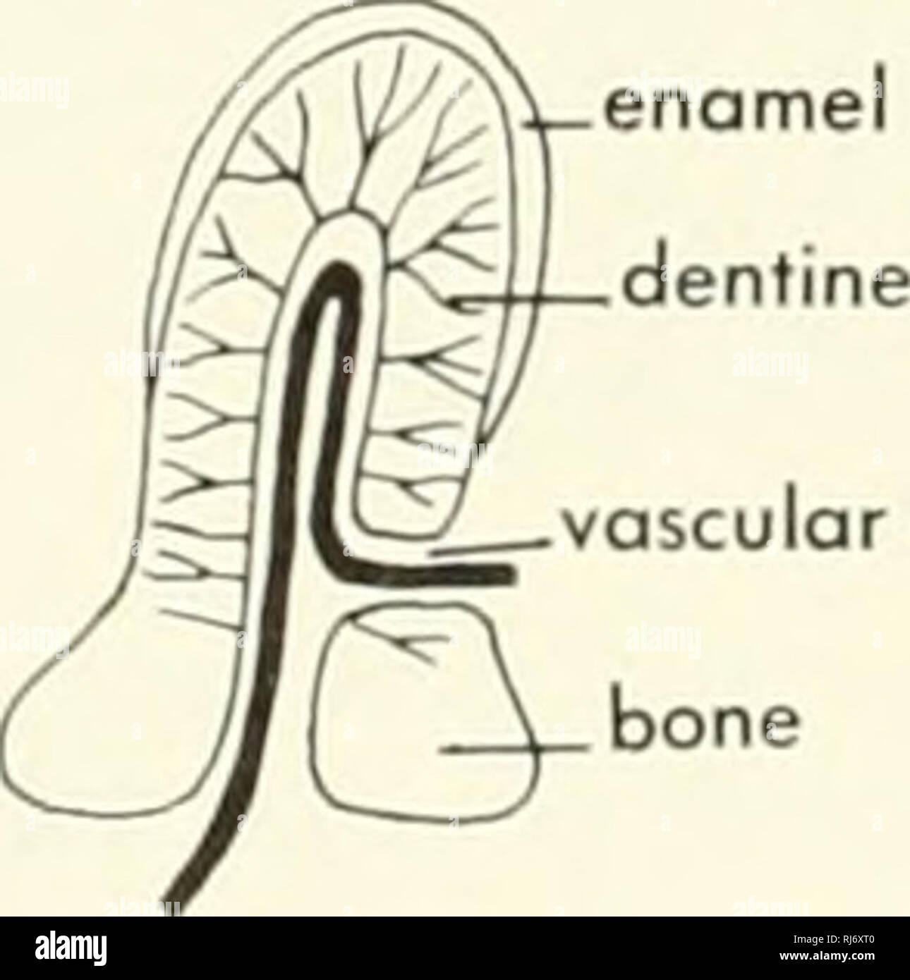
. Chordate morphology. Morphology (Animals); Chordata. Sharpey's fibers osGl canal acellular lamellar bony base Figure 8-31. Section through the scale of a ray, Dosybotus sp?. (After 0rvig, 1951) In the holocephalans, denticles occur only on the claspers, and in the young there may be a double row of small den- ticles along the back. The denticles and spine structure are like those of the shark. General observations The placoid scale of the shark has been described as a basic skeletal unit, and it is apparent that denticles of one shape or another are characteristic of the bone and scales of vertebrates. In very primitive sharks, small units, called lepidomoria, are found (Figure 8-32). In more advanced sharks, the simple tooth-like lepidomoria tend to fuse together to form larger compound units. It has been assumed that the history of vertebrate scales and plates has been one of marginal aggregation of these lepidomorial units around a central element—this growth was accom- panied by acquisition and thickening of the base. The shark placoid scale is peculiar in its irregular shape as contrasted with the regular-shaped scales of other groups. Actinopterygian fishes Ganoid scale The scales and the dermal bone of primitive actinopterygian fishes are described as ganoid because of the "shiny, " enamel-like material covering their surface. As already pointed out, the fact that ganoin may be produced by the dermis, and not by the overlying epidermis does not mean that, in essence, it is not phylogenetically the same material. If the material occurs on the tooth of the fish, it is described as enamel; if it occurs on the scale, it is described as ganoin. This distinction does not seem quite proper. In the primitive ganoid scale, the layers of enamel overlie a zone of dentine. The dentine has a network of vascular canals below it; these open at the neck of the scale as well as through the base and the outer surface by one or a few channels. The base of the scale