. Chordate morphology. Morphology (Animals); Chordata. Figure 8-3. Development of the human epidermis and hair follicle. A, epidermis of 2.1 -mm embryo; B, epidermis of 16-mm embryo; C, epidermis of 32-mm embryo; D, epidermis of 85-mm embryo; E to H, progressive stages in the development of the follicle and hair. (After Patten, 1946) of the snout. It is not strengthened by a bony core as in the cow but is seated on a bony knob of the skull. The pointed tip is produced and maintained by wear. Bmbryological development The ectoderm is at first a simple cuboidal epithelium which gradually becomes
Image details
Contributor:
Library Book Collection / Alamy Stock PhotoImage ID:
RJ6YK7File size:
7.2 MB (411.4 KB Compressed download)Releases:
Model - no | Property - noDo I need a release?Dimensions:
1868 x 1338 px | 31.6 x 22.7 cm | 12.5 x 8.9 inches | 150dpiMore information:
This image is a public domain image, which means either that copyright has expired in the image or the copyright holder has waived their copyright. Alamy charges you a fee for access to the high resolution copy of the image.
This image could have imperfections as it’s either historical or reportage.
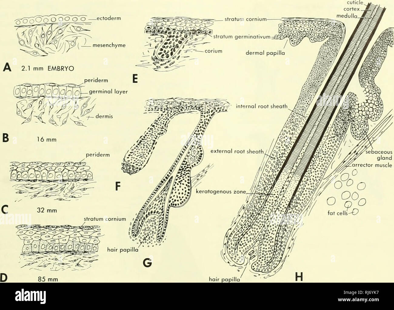
. Chordate morphology. Morphology (Animals); Chordata. Figure 8-3. Development of the human epidermis and hair follicle. A, epidermis of 2.1 -mm embryo; B, epidermis of 16-mm embryo; C, epidermis of 32-mm embryo; D, epidermis of 85-mm embryo; E to H, progressive stages in the development of the follicle and hair. (After Patten, 1946) of the snout. It is not strengthened by a bony core as in the cow but is seated on a bony knob of the skull. The pointed tip is produced and maintained by wear. Bmbryological development The ectoderm is at first a simple cuboidal epithelium which gradually becomes strati- fied (Figure 8-3). Hair begins its development as thicken- ings of the epidermis. Each such thickening grows down into the dermis as a strand which carries the stratum germina- tivum before it. A dermal papilla now forms which projects upward into the strand. The cells overlying the papilla form the matrix from which the hair grows upward through the strand. The strand is thus converted into a follicle. Seba- ceous glands form from the wall of the follicle. The sweat glands form as ingrowths from the epidermis. The soft keratin of the stratum corneum is formed by ac- cumulation of granules within the cells which pass through a number of steps. Being cellular, the stratum corneum des- quamates at its surface. The mammary glands are generally considered to be modi- fied sweat glands, although they might just as easily be viewed as sebaceous glands. Pertinent to this question is the obser- vation that in man the apocrine sweat gland of the arm pit and the nipple undergo cyclic changes which can be cor- related with the menstrual cycle. Unlike the sweat glands, the mammary glands are compound and alveolar, and they are related to mammary hairs. In man about eight hair fol- scale (hord keratin). Please note that these images are extracted from scanned page images that may have been digitally enhanced for readability - coloration and appearance of these illustrations may not