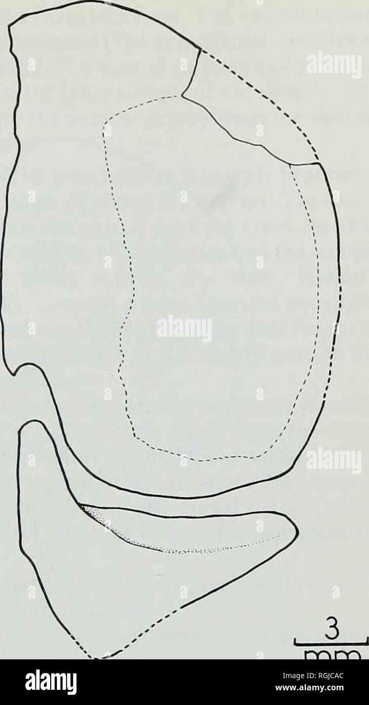
. Bulletin of the British Museum (Natural History), Geology. . mm Fig. 10 Macrosemius rostratus Agassiz. Lateral views of proximal parts of A, last branchiostegal and B, penultimate branchiostegal in 37094. C, opercular and subopercular of AS. 1.640 in lateral view. The sinuous suture between angular and retroarticular is interrupted by a canal which presumably transmitted the external mandibular ramus of the facial nerve. Amia has a similar foramen (Allis 1897 : pi. 20, fig. 6). The dentary bears about 10 large teeth. Each tooth is laterally compressed and tapers to a point. The teeth are mos
Image details
Contributor:
Book Worm / Alamy Stock PhotoImage ID:
RGJCACFile size:
7.1 MB (128.6 KB Compressed download)Releases:
Model - no | Property - noDo I need a release?Dimensions:
1183 x 2112 px | 20 x 35.8 cm | 7.9 x 14.1 inches | 150dpiMore information:
This image is a public domain image, which means either that copyright has expired in the image or the copyright holder has waived their copyright. Alamy charges you a fee for access to the high resolution copy of the image.
This image could have imperfections as it’s either historical or reportage.
. Bulletin of the British Museum (Natural History), Geology. . mm Fig. 10 Macrosemius rostratus Agassiz. Lateral views of proximal parts of A, last branchiostegal and B, penultimate branchiostegal in 37094. C, opercular and subopercular of AS. 1.640 in lateral view. The sinuous suture between angular and retroarticular is interrupted by a canal which presumably transmitted the external mandibular ramus of the facial nerve. Amia has a similar foramen (Allis 1897 : pi. 20, fig. 6). The dentary bears about 10 large teeth. Each tooth is laterally compressed and tapers to a point. The teeth are most closely set in the anterior region where the dentary curves sharply towards the mental symphysis. The lateral surface of the dentary is exposed in 37051 (Fig. 4) and 37094. The oral border of the bone rises steeply and forms the greater part of the dermal coronoid process. The open trough for the mandibular sensory canal is very wide and occupies about half of the depth of the dentary below the tooth-row. Three small indentations on the dorsal and ventral margins of the trough may be the remnants of resorbed arches of bone which spanned the canal at an earlier stage of development. The small surangular occupies the upper posterior part of the coronoid process. The mandibular sensory canal continued along a large trough in the ventral part of the angular before turning dorsally beneath the quadrate articulation past the remains of another arch of bone. The angular forms a long tapering extension above the sensory canal in the dentary and forms an interdigitating suture with this bone in the medial wall of the canal trough. The coronoid and prearticular are preserved displaced from the remainder of the mandible in AS. 1.639 and AS. 1.770 (Fig. 9). The coronoid is short and bears six mammiliform teeth each with a nipple of ganoine. The surfaces of several of these teeth have been worn flat. The two medial teeth, in opposition to the pair of large vomerine teeth, are larger than