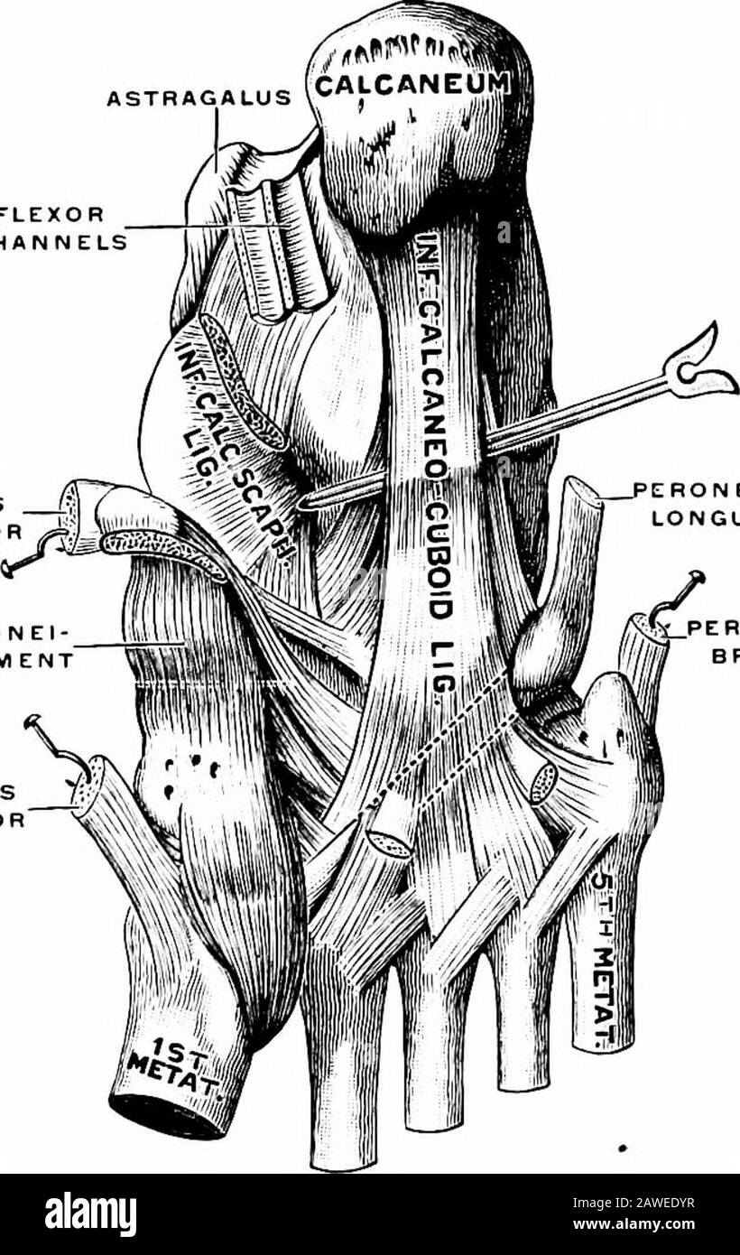
Applied anatomy and kinesiology, the mechanism of muscular movement . 109.—Bones of the foot. (Gray.) kept from spreading by ligaments and muscles, forming an effi-cient shock-absorbing mechanism to lessen the jar that wouldotherwise result in walking, running, and jumping. The bones areas follows: Seven tarsal bones: astragalus, calcaneum, scaphoid or navicu-lar, cuboid, and three cuneiform bones numbered from withinoutward; 1SS MOVEMENTS OF THE FOOT Five metatarsal bones, numbered from within outward, andFourteen phalanges, three for each toe except the first, which hastwo. The principal arc

Image details
Contributor:
The Reading Room / Alamy Stock PhotoImage ID:
2AWEDYRFile size:
7.1 MB (435.6 KB Compressed download)Releases:
Model - no | Property - noDo I need a release?Dimensions:
1256 x 1989 px | 21.3 x 33.7 cm | 8.4 x 13.3 inches | 150dpiMore information:
This image is a public domain image, which means either that copyright has expired in the image or the copyright holder has waived their copyright. Alamy charges you a fee for access to the high resolution copy of the image.
This image could have imperfections as it’s either historical or reportage.
Applied anatomy and kinesiology, the mechanism of muscular movement . 109.—Bones of the foot. (Gray.) kept from spreading by ligaments and muscles, forming an effi-cient shock-absorbing mechanism to lessen the jar that wouldotherwise result in walking, running, and jumping. The bones areas follows: Seven tarsal bones: astragalus, calcaneum, scaphoid or navicu-lar, cuboid, and three cuneiform bones numbered from withinoutward; 1SS MOVEMENTS OF THE FOOT Five metatarsal bones, numbered from within outward, andFourteen phalanges, three for each toe except the first, which hastwo. The principal arch passes transversely beneath the foot, as seenin Fig. 108, the calcaneum forming its rear base and the minorarch forming its front base. The minor arch is formed by the meta-tarsal bones, the anterior ends of the first and fifth resting uponthe ground and the intervening three supported between them.The weight of the body is transmitted through the tibia to the ASTRAGALUS FLEXORCH ANN ELS TIBIALISPOSTERIOR SCAPHO-CU N EFORM LIGAM ENT Tl BIALI S 7v ANTERIOR N«Xj. ERON EUSLONGUS PERONEUSBREVIS Fig. 110.—The plantar ligaments. (Gerrish.J astragalus, which serves as the keystone of the main arch. Sogreat a weight pressing down on such flat arches tends to flattenthem out, requiring three strong supports to tie together the threebases; one from the heel to the first metatarsal, one from the heelto the fifth metatarsal, and one between the inner and outer meta-tarsals. Of these three supports, acting like bow-strings to keepthe arches intact, one is ligamentous and the other two muscular.The two calcaneocuboid or plantar ligaments, which are next tothe patellar ligaments the strongest in the body, bind the calcaneumto the cuboid and the last three metatarsals beneath the outer side MOVEMENTS OF THE FOOT 189 of the main arch, as seen in Fig. 110, so firmly that this side of thefoot acts almost as one solid piece. The inner side of the mainarch, which is better suited in some res