. Anatomy, descriptive and applied. Anatomy. THE CHOROID, CILIARY BODY, AND IRIS 1095 Structure.—The ciliary processes are similar in structure to the choroid, but the vessels are larger, and have chiefly a longitudinal direction. They constitute the most vascular portion of the eyeball. The processes are covered on their inner surface by two strata of black pigment cells, which are continued forward from the retina, and are named the pars ciliaris retinae (Fig. 814). In the stroma of the ciliary processes there are also stellate pigment cells, which, how- ever, are not so numerous as in the c
Image details
Contributor:
Library Book Collection / Alamy Stock PhotoImage ID:
RN5N5MFile size:
7.1 MB (376.8 KB Compressed download)Releases:
Model - no | Property - noDo I need a release?Dimensions:
1218 x 2051 px | 20.6 x 34.7 cm | 8.1 x 13.7 inches | 150dpiMore information:
This image is a public domain image, which means either that copyright has expired in the image or the copyright holder has waived their copyright. Alamy charges you a fee for access to the high resolution copy of the image.
This image could have imperfections as it’s either historical or reportage.
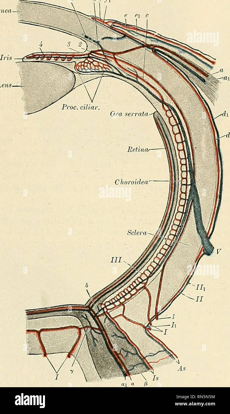
. Anatomy, descriptive and applied. Anatomy. THE CHOROID, CILIARY BODY, AND IRIS 1095 Structure.—The ciliary processes are similar in structure to the choroid, but the vessels are larger, and have chiefly a longitudinal direction. They constitute the most vascular portion of the eyeball. The processes are covered on their inner surface by two strata of black pigment cells, which are continued forward from the retina, and are named the pars ciliaris retinae (Fig. 814). In the stroma of the ciliary processes there are also stellate pigment cells, which, how- ever, are not so numerous as in the choroid itself. ''//i. Fig. 810.—Diagram of the bloodvessels of the eye, as seen in a horizontal section. (Leber, after Stohr.) Course of vasa centralia retinae: a. Al-teria. ai. Vena centralis retinae. /^. Anastomosis with vessels of outer coats 7. Anastomosis with branches of short posterior ciliary arteries, ri. Anastomosis with chorioideal vessels. Course of vasa ciliar. postic. brev.: I. Arteriae, and Ii. Venae ciliar. postic. brev. II. Episcleral artery. IIi. Episcleral vein. III. Capillaries of lamina choriocapiilaris. Course of vasa ciliar. postic. long.: 1. a. ciliar. post, longa. 2. Circulus iridis major cut across. 3. Branches to ciliary body. 4. Branches to iris. ' Course of vasa ciliar. ant.: a. Arteria. ai. Vena ciliar. ant. b. Junction with the circulus iridis major. c. Junction with lamina choriocapill. d. Arterial, and d. Venous episcleral branches, e. Arterial, and ei. Venous branches to conjunctiva sclerae. f. Arterial, and fi. Venous branches to corneal border. V. Vena vorticosa. S. Transverse section of sinus venosus sclerae. The Ciliary muscle (Bowman's muscle) (m. ciliaris) (Figs. 814 and 816) con- sists of unstriped fibres; it forms a grayish, semitransparent, circular band, about 3 mm. (one-eighth of an inch) broad, on the outer surface of the fore part of the choroid, between the choroid and the iris and back of the sclerocorneal junction. It is th