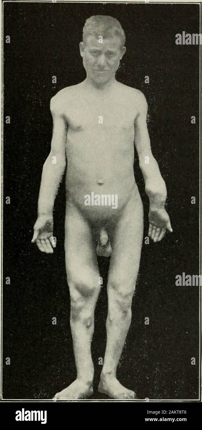
American practice of surgery : a complete system of the science and art of surgery . f the forearmand lower leg and the upper end ofthe tibia usually showing the great-est change. The bones of the handsand feet, however, are rarely involved.The femur and humerus also shownumerous growths, and they fre-quently occur on the pelvis, scapula,and ribs, while the vertebra^ and ster-num are rarely affected. The exos-toses arise at or near the epiphysealline and grow backward over theshaft as in the single form. Theyare best developed in the lower endof the femur or in the upper endof the tibia, where
Image details
Contributor:
The Reading Room / Alamy Stock PhotoImage ID:
2AKT8T6File size:
7.2 MB (267.7 KB Compressed download)Releases:
Model - no | Property - noDo I need a release?Dimensions:
1122 x 2228 px | 19 x 37.7 cm | 7.5 x 14.9 inches | 150dpiMore information:
This image is a public domain image, which means either that copyright has expired in the image or the copyright holder has waived their copyright. Alamy charges you a fee for access to the high resolution copy of the image.
This image could have imperfections as it’s either historical or reportage.
American practice of surgery : a complete system of the science and art of surgery . f the forearmand lower leg and the upper end ofthe tibia usually showing the great-est change. The bones of the handsand feet, however, are rarely involved.The femur and humerus also shownumerous growths, and they fre-quently occur on the pelvis, scapula, and ribs, while the vertebra^ and ster-num are rarely affected. The exos-toses arise at or near the epiphysealline and grow backward over theshaft as in the single form. Theyare best developed in the lower endof the femur or in the upper endof the tibia, where they may be long and branching. In other localities—as, for example, the lower end of the radius—they are seen as irregular masses, of varying size, surrounding the end of the bone and extending for a consider-able distance up the shaft. Although by far the greater number of these casesshow the exostoses at or near the epiphyses, in certain others they arise nearthe centre of the shaft, but are tipped with cartilage and are identical with the *Rev. dorthopedic, 1899, x.. 466.. Fig. 185.—Multiple Cartilaginous Exostoses ina Male Twenty-one Years Old. Note the shorteningof the upper arms and the symmetry of the exostosesof the tibiae. (Massachusetts General Hospital.) 416 AMERICAN PRACTICE OF SURGERY. common form. The point of origin of these is disputed. They may arise atan early stage of foetal development from the cartilaginous centre of ossifi-cation of the shaft (Fischer; Lippert) or from a bit of epiphyseal carti-lage which became isolated very early m life (Bessel-Hagen). The deformity, which is a marked feature of the disease, is usually greatest in theforearms, and is caused, not only by the presence of the multiple growths, bul also by arelative lack of development of the hones. Bessel-Hagens theory was thatthere was enough epiphyseal cartilage to form a norma] adult hone, hut that ifsome of this was utilized in forming exostoses there would he a degree of short-eni