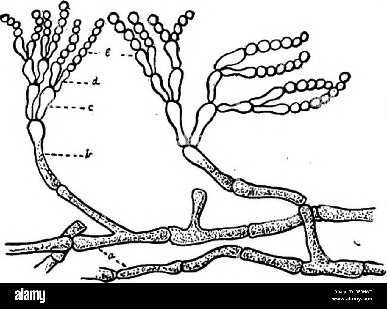
. A text-book upon the pathogenic Bacteria and Protozoa for students of medicine and physicians. Bacteriology; Pathogenic bacteria; Protozoa. Fig. 15.—Aspergillus glaucus: A, A portion of the mycelium m, with a con- idiaphore c, and a young perithrecium F, magnified 190 diameters; B and B', conidiaphore with conidia; B, individual sterigma greatly magnified; C, early stage of the development of the fructifying organ; D, young perithrecium in longitudinal section; w, the future wall of the contents; as, the screw, magnified 250 diameters; E, an aseus with spores from a perithrecium, magnified 6
Image details
Contributor:
The Book Worm / Alamy Stock PhotoImage ID:
RE0HWTFile size:
7.2 MB (237.6 KB Compressed download)Releases:
Model - no | Property - noDo I need a release?Dimensions:
1849 x 1352 px | 31.3 x 22.9 cm | 12.3 x 9 inches | 150dpiMore information:
This image is a public domain image, which means either that copyright has expired in the image or the copyright holder has waived their copyright. Alamy charges you a fee for access to the high resolution copy of the image.
This image could have imperfections as it’s either historical or reportage.
. A text-book upon the pathogenic Bacteria and Protozoa for students of medicine and physicians. Bacteriology; Pathogenic bacteria; Protozoa. Fig. 15.—Aspergillus glaucus: A, A portion of the mycelium m, with a con- idiaphore c, and a young perithrecium F, magnified 190 diameters; B and B', conidiaphore with conidia; B, individual sterigma greatly magnified; C, early stage of the development of the fructifying organ; D, young perithrecium in longitudinal section; w, the future wall of the contents; as, the screw, magnified 250 diameters; E, an aseus with spores from a perithrecium, magnified 600 diameters (duBary). whole germinal organ thus comes to resemble a whisk-broom or, as Hiss describes it, a skeleton hand, in which the conidiaphore cor-. Fig. 16.—Penicillium: a, Mycelium; b, conidiaphores; c, d, sterigmata; e, spores (Eyre). responds to the wrist; the sterigrhata, to the metacarpal bones; the chains of spores, to the phalanges. None of the penicillia is know to be pathogenic either for man or animals.. Please note that these images are extracted from scanned page images that may have been digitally enhanced for readability - coloration and appearance of these illustrations may not perfectly resemble the original work.. McFarland, Joseph, 1868-. Philadelphia and London, W. B. Saunders Company