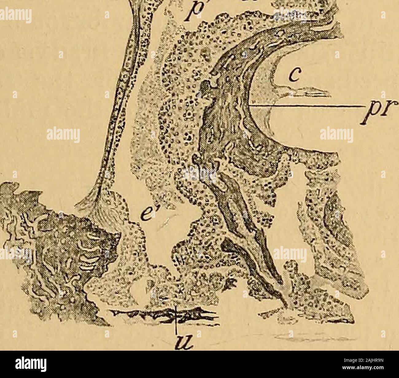
A text-book of the diseases of the ear for students and practitioners . Fig. 158.—Frontal Section through the Tympanic Cavity or a Childwho died of Measles. 0, Superior ; u, Inferior wall of the tympanic cavity; t, Membrana tympani withsection of handle of malleus; n, Recess of fenestra vestibuli; pr, Promontory;c, Cochlea; /, Facial nerve ; st, Base of stapes ; p, Inflammatory infiltrated mucousmembrane of the attic, adherent to the inner wall ; p, Inflamed and swollenmucous membrane of the promontory ; e, e, Exudate in the tympanic cavity.(From a preparation in the authors collection.) vascu
Image details
Contributor:
The Reading Room / Alamy Stock PhotoImage ID:
2AJHR9NFile size:
7.1 MB (377.6 KB Compressed download)Releases:
Model - no | Property - noDo I need a release?Dimensions:
1686 x 1482 px | 28.5 x 25.1 cm | 11.2 x 9.9 inches | 150dpiMore information:
This image is a public domain image, which means either that copyright has expired in the image or the copyright holder has waived their copyright. Alamy charges you a fee for access to the high resolution copy of the image.
This image could have imperfections as it’s either historical or reportage.
A text-book of the diseases of the ear for students and practitioners . Fig. 158.—Frontal Section through the Tympanic Cavity or a Childwho died of Measles. 0, Superior ; u, Inferior wall of the tympanic cavity; t, Membrana tympani withsection of handle of malleus; n, Recess of fenestra vestibuli; pr, Promontory;c, Cochlea; /, Facial nerve ; st, Base of stapes ; p, Inflammatory infiltrated mucousmembrane of the attic, adherent to the inner wall ; p, Inflamed and swollenmucous membrane of the promontory ; e, e, Exudate in the tympanic cavity.(From a preparation in the authors collection.) vascular layers were infiltrated with numerous mononuclear and polynuclearleucocytes. This cell-infiltration diminished to such an extent in the deeperlayers of the mucous membrane, that the structures lying in contact withthe bone appeared almost normal (Siegfried Weiss). This layer of mucousmembrane, which was free from infiltration, presented a structure, as SiegfriedWeiss showed, similar to that of the embryonic mucous membrane. Thebone itself showed no changes, wit