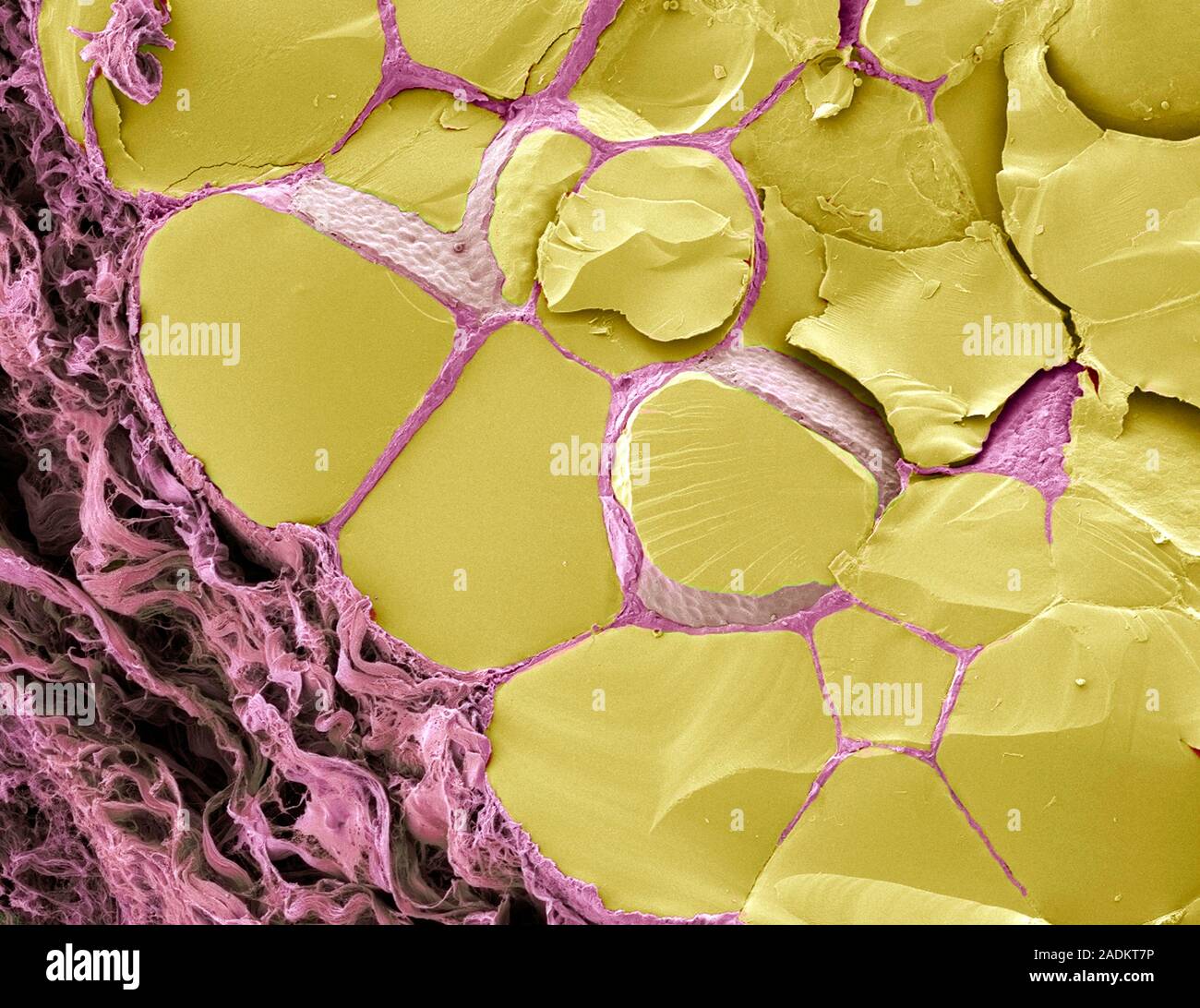
Thyroid gland. Coloured scanning electron micrograph (SEM) of a fracture through the thyroid gland revealing several follicles (yellow). Between the f
RMID:Image ID:2ADKT7P
Image details
Contributor:
Science Photo Library / Alamy Stock PhotoImage ID:
2ADKT7PFile size:
28.6 MB (1.3 MB Compressed download)Releases:
Model - no | Property - noDo I need a release?Dimensions:
3600 x 2776 px | 30.5 x 23.5 cm | 12 x 9.3 inches | 300dpiDate taken:
31 May 2007Photographer:
STEVE GSCHMEISSNER/SCIENCE PHOTO LIBRARYMore information:
Thyroid gland. Coloured scanning electron micrograph (SEM) of a fracture through the thyroid gland revealing several follicles (yellow). Between the follicles is connective tissue (purple). A follicle consists of a layer of epithelial cells (grey) around a central storage chamber. The cells produce the thyroid hormones tri-iodothyronin (T3) and thyroxine (T4) and secrete them into the central chamber where they are stored as the glycoprotein thyroid colloid (yellow). Thyroid hormones regulate the body's metabolism.