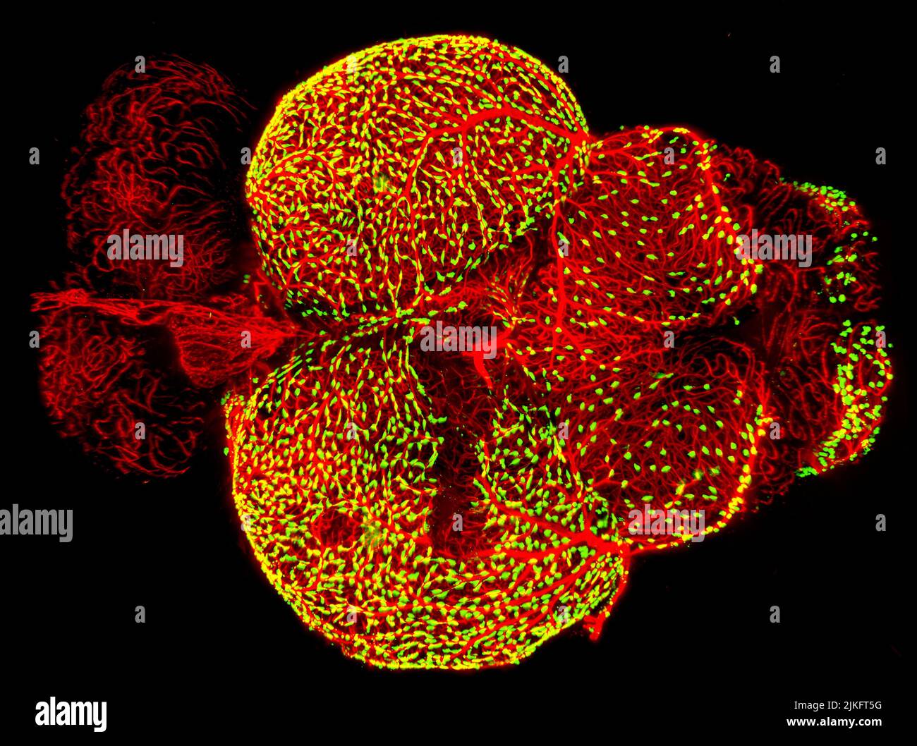
This image, winner of the Federation of American Societies for Experimental Biology's 2017 BioArt competition, shows the brain of an adult zebrafish, a popular organism for studying brain function. It captures dense networks of blood vessels (red) lining the outer surface of the brain. Next to many of these vessels are previously little-studied cells called fluorescent (yellowish-green) granular perithelial cells.
RMID:Image ID:2JKFT5G
Image details
Contributor:
BSIP SA / Alamy Stock PhotoImage ID:
2JKFT5GFile size:
35.8 MB (1.3 MB Compressed download)Releases:
Model - no | Property - noDo I need a release?Dimensions:
4096 x 3052 px | 34.7 x 25.8 cm | 13.7 x 10.2 inches | 300dpiDate taken:
14 July 2022Photographer:
NIH / IMAGE POINT FR / BSIP