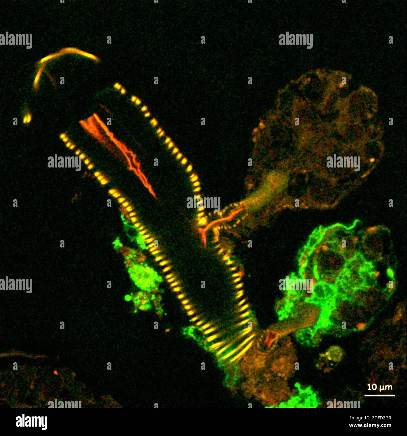
This confocal microscope image shows a cross section of a tick salivary gland infected with Langat virus (green). Two rounded structures on the right,
RFID:Image ID:2DFD2GR
Image details
Contributor:
BSIP SA / Alamy Stock PhotoImage ID:
2DFD2GRFile size:
27.5 MB (1.2 MB Compressed download)Releases:
Model - no | Property - noDo I need a release?Dimensions:
3100 x 3100 px | 26.2 x 26.2 cm | 10.3 x 10.3 inches | 300dpiDate taken:
31 October 2020Photographer:
IMAGE POINT FR / NIH / NIAIDMore information:
This confocal microscope image shows a cross section of a tick salivary gland infected with Langat virus (green). Two rounded structures on the right, called acini, are shown attached to a duct (yellow). The lower acinus is infected, as denoted by the green fluorescent signal. Credit: NIAID.