The frog: an introduction to anatomy, histology, and embryology . els, but incomplete, is present in thefourth branchial arch : and vessels formed on the same plan, butstill less complete, and showing signs of degenerative changes,are present in the hyoid and mandibular arches. There are thus six sets of branchial vessels on each side ofthe pharynx: of these, three, in the first, second, and thirdbranchial arches, are complete; one, in the fourth branchialarch, is incomplete ; and two, in the hyoid and mandibulararches, are rudimentary. 2. The Circulation during the time the tadpole is breathi
Image details
Contributor:
The Reading Room / Alamy Stock PhotoImage ID:
2AM6F81File size:
7.2 MB (302.7 KB Compressed download)Releases:
Model - no | Property - noDo I need a release?Dimensions:
1274 x 1962 px | 21.6 x 33.2 cm | 8.5 x 13.1 inches | 150dpiMore information:
This image is a public domain image, which means either that copyright has expired in the image or the copyright holder has waived their copyright. Alamy charges you a fee for access to the high resolution copy of the image.
This image could have imperfections as it’s either historical or reportage.
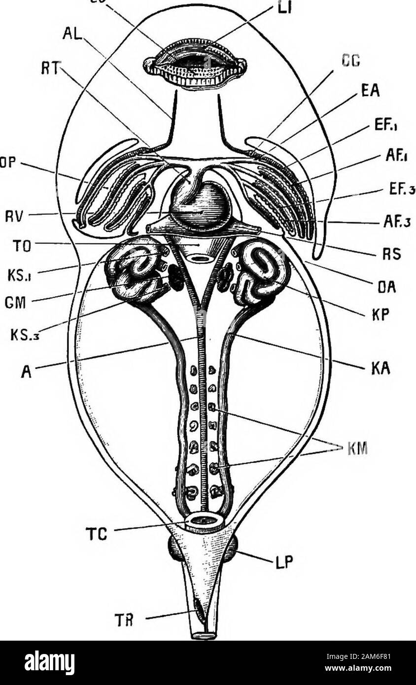
The frog: an introduction to anatomy, histology, and embryology . els, but incomplete, is present in thefourth branchial arch : and vessels formed on the same plan, butstill less complete, and showing signs of degenerative changes, are present in the hyoid and mandibular arches. There are thus six sets of branchial vessels on each side ofthe pharynx: of these, three, in the first, second, and thirdbranchial arches, are complete; one, in the fourth branchialarch, is incomplete ; and two, in the hyoid and mandibulararches, are rudimentary. 2. The Circulation during the time the tadpole is breathingby internal gills. On the formation of the internal gills, additional loops ofcommunication are formed in the gill tufts between the afferentand efferent vessels of the first, second, and third branchialarches, and also a series of simUar loops between the afferentand efferent vessels of the fourth branchial arch. The vesselsin the hyoid and mandibular arches undergo further retrogradechanges, and need not be described in detail. 132 DEYELOPMENT OF THE FROGLJ. Fig. 33—^A 12 mm. tadpole dissected from the ventral surfaceto show the heart, the internal gills, the branchial vessels, and thehead kidneys and their ducts. The tail, which is about double thelength of the head and body, has been removed. X 22. A, dorsal aorta; AFi, AF3, afferent branchial vessels of first andthiid branchial arches ; AL, lingual artery ; CG, carotid gland ; EA, junction between afferent and efferent branchial vessels of firstbranchial arch; EFi, EF3, efferent branchial vessels of first and thirdbranchial arches; GM, glomerulus; KA, archinephric or segmentalduct; KM, Wolffian tubules ; KP, pronephros or head kidney;KSi, KS3, first and third nephrostomes of head kidney; LI, upperlip ; LJ, lower Up ; LP, hind limb ; OA, aperture of opercular cavity;OP, opercular cavity; RS, sinus venosus; RT, truncus arteriosus;RV, ventricle; TC, cloaca; TO, oesophagus, cut short; TP rectal spout. THE VASCULAR SYSTEM 13