. The cyclopædia of anatomy and physiology. Anatomy; Physiology; Zoology. TONGUE. 1121 terior surface of the epiglottis, the sides of the pharynx, and upwards to the soft palate and posterior parts of the cheeks. As we proceed forwards we find it investing the sides, and gradually more and more of the under surface, reflected thence over the hyoglossi and ge- nioglossi muscles, the sublingual glands, vessels, and nerves, and much areolar tissue, which separate it from the mylohyoid muscle, to the inner surface of the alveoli of the lower jaw, where it becomes continuous with the mucous membran
Image details
Contributor:
The Book Worm / Alamy Stock PhotoImage ID:
RD54W2File size:
7.1 MB (484.4 KB Compressed download)Releases:
Model - no | Property - noDo I need a release?Dimensions:
1171 x 2133 px | 19.8 x 36.1 cm | 7.8 x 14.2 inches | 150dpiMore information:
This image is a public domain image, which means either that copyright has expired in the image or the copyright holder has waived their copyright. Alamy charges you a fee for access to the high resolution copy of the image.
This image could have imperfections as it’s either historical or reportage.
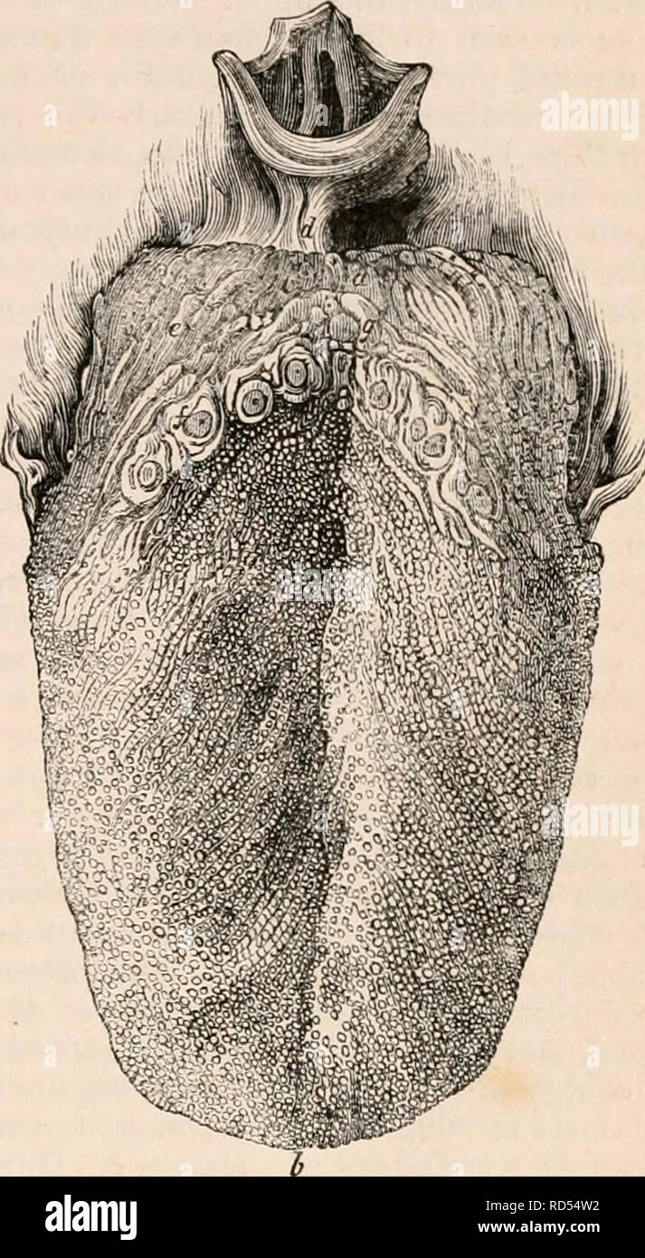
. The cyclopædia of anatomy and physiology. Anatomy; Physiology; Zoology. TONGUE. 1121 terior surface of the epiglottis, the sides of the pharynx, and upwards to the soft palate and posterior parts of the cheeks. As we proceed forwards we find it investing the sides, and gradually more and more of the under surface, reflected thence over the hyoglossi and ge- nioglossi muscles, the sublingual glands, vessels, and nerves, and much areolar tissue, which separate it from the mylohyoid muscle, to the inner surface of the alveoli of the lower jaw, where it becomes continuous with the mucous membrane covering the gums. At certain points where this membrane leaves the tongue it forms distinct folds, which, from their being constant, have received par- ticular names, and which act to a certain ex- tent as ligaments orfrena of the tongue, not so much by virtue of their being folds of mucous membrane, as from their containing within their reduplications a certain amount of a more or less unyielding tissue ; in some this tissue is a mixture of white and yellow fibre, in others it is muscle. Of the first sort are three folds, a middle and two lateral, passing from the base of the tongue to the epiglottis, called the glosso- epiglottid folds, of which the central, which is always present, and has been called the pos- terior frcemim of the tongue, and frccnum epi- glottidis, is much the most considerable (jfig. 745. rf.) : they serve rather to check the move- ments of the epiglottis than as lingual liga- ments. From the sides of the base of the tongue, passing thence to the soft palate, are seen four more folds, two on each side, which, from their position, have been called the pillars of the fauces. They are formed by the raising of the mucous membrane from the general surface by two muscles: the posterior, the least considerable, by the palato-pharyn- geus; the anterior, more marked, by the palatoglossus. The interval between these two is called the amygdaloid fossa, from its