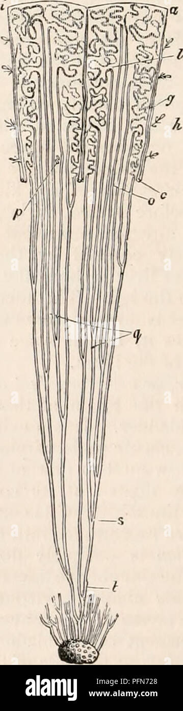
. The cyclopædia of anatomy and physiology. Anatomy; Physiology; Zoology. 236 REN. on the left side is covered by its corresponding vein, and crosses the left psoas muscle. The renal arteries occasionally present some anomalies as to their origin, mode of division, or number. In some instances they arise below the usual situation, from the aorta, or even from the common iliac or hypogastric artery. The two last-mentioned origins are usually associated with an unusual po- sition of the kidney, either in the iliac fossa or in the cavity of the pelvis. Meckel* has observed the two renal arteries
Image details
Contributor:
Central Historic Books / Alamy Stock PhotoImage ID:
PFN728File size:
7.2 MB (278.1 KB Compressed download)Releases:
Model - no | Property - noDo I need a release?Dimensions:
832 x 3005 px | 7 x 25.4 cm | 2.8 x 10 inches | 300dpiMore information:
This image is a public domain image, which means either that copyright has expired in the image or the copyright holder has waived their copyright. Alamy charges you a fee for access to the high resolution copy of the image.
This image could have imperfections as it’s either historical or reportage.
. The cyclopædia of anatomy and physiology. Anatomy; Physiology; Zoology. 236 REN. on the left side is covered by its corresponding vein, and crosses the left psoas muscle. The renal arteries occasionally present some anomalies as to their origin, mode of division, or number. In some instances they arise below the usual situation, from the aorta, or even from the common iliac or hypogastric artery. The two last-mentioned origins are usually associated with an unusual po- sition of the kidney, either in the iliac fossa or in the cavity of the pelvis. Meckel* has observed the two renal arteries arising by a common trunk from the anterior part of the aorta. The artery sometimes divides into two or more branches immediately after its origin, in which case one branch usually leaves the others to enter one of the extremities of the kidney. This irregularity forms an approach to another, which consists in an increase in the number of renal arteries, each kidney re- ceiving two, three, or four branches having a separate origin from the aorta. The emiilgent or renal vein commences in the substance of the kidney by numerous mi- nute branches, which unite into four or five trunks, and these again unite to form a single trunk, either in the fissure of the kidney, or at a short distance from this point. The vein passes almost transversely inwards to the vena cava, the right vein being shorter than the left, on account of the position of the vena cava to the right of the spine; the junction of the right vein with the cava is also somewhat higher than that of the left. The vena cava presents a marked increase of size immediately after receiving the renal veins. Each renal vein is placed in front of the corresponding artery ; the vein on the left side crosses over the aorta. The renal veins receive some small branches from the supra-renal capsule, and from the reticular and adipose tissue sur- rounding the kidney, and the left renal vein is usually joined by the spermatic of the s