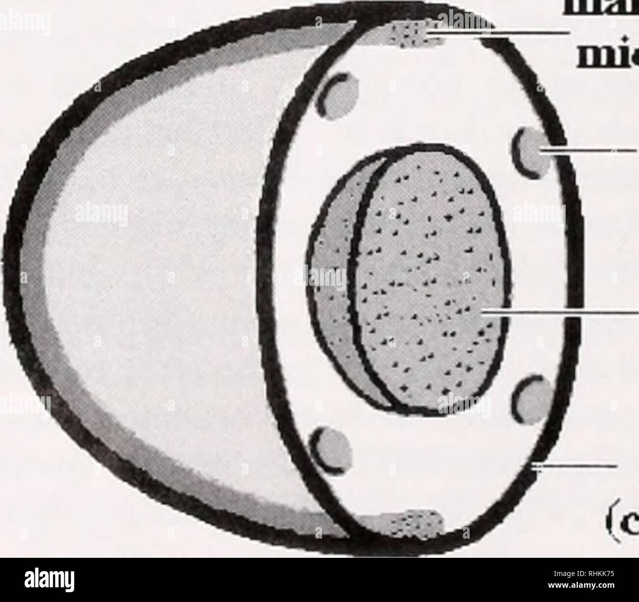
. The Biological bulletin. Biology; Zoology; Biology; Marine Biology. Figure 8. Cytoskeletal and nuclear structure in lipopolysaccharide- activated cells (TEM). (a) —5 min post-activation; low-magnification view of deformed nucleus, (b) Higher magnification view of area delimited in (a), showing bundles of microtubules adjacent to nucleus (arrows), (c) —10 min post-activation; exocytosis is complete, with no intact granules re- maining. Centrioles (arrow, and inset) remain intact. Bars: (a. c) = 1 /xm); (b) = 0.25 fim; (c inset) = 0.2 ju,m. (Nemhauser el <//.. 1980; Cohen and Nemhauxer. !9X

Image details
Contributor:
Library Book Collection / Alamy Stock PhotoImage ID:
RHKK75File size:
7.2 MB (204.2 KB Compressed download)Releases:
Model - no | Property - noDo I need a release?Dimensions:
1691 x 1478 px | 28.6 x 25 cm | 11.3 x 9.9 inches | 150dpiMore information:
This image is a public domain image, which means either that copyright has expired in the image or the copyright holder has waived their copyright. Alamy charges you a fee for access to the high resolution copy of the image.
This image could have imperfections as it’s either historical or reportage.
. The Biological bulletin. Biology; Zoology; Biology; Marine Biology. Figure 8. Cytoskeletal and nuclear structure in lipopolysaccharide- activated cells (TEM). (a) —5 min post-activation; low-magnification view of deformed nucleus, (b) Higher magnification view of area delimited in (a), showing bundles of microtubules adjacent to nucleus (arrows), (c) —10 min post-activation; exocytosis is complete, with no intact granules re- maining. Centrioles (arrow, and inset) remain intact. Bars: (a. c) = 1 /xm); (b) = 0.25 fim; (c inset) = 0.2 ju, m. (Nemhauser el <//.. 1980; Cohen and Nemhauxer. !9Xv Tahlin and Levin. 1988). The same mechanism applies to platelets and to non-mammalian thromhoeytes (Lee <•/ <//.. 2004). and it is similar to that proposed previously tor the MB-eontaining nucleated erythrocytes of all non-mamma- lian vertebrates (Joseph-Silverstein and Cohen. 19X4. 1985). There is, however, one fundamental difference between the MB-containing cytoskeleton of unactivated nucleated erythrocytes and that of invertebrate or vertebrate clotting cells. The erythrocyte system is designed for long-term maintenance of circulating cell shape, with the MB inter- acting with a filamentous network—the actin-spectrin mem- brane skeleton—that is highly specialized for stability. In contrast, the cortical layer of the clotting cell—through interaction with the MB—must maintain the unactivated circulating shape for long periods, while remaining at all times responsive to the signals that induce the rapid shape transformations associated with clotting. As shown in the current work with Liinulus amebocytes, as well as in pre- vious work on platelets and non-mammalian vertebrate thrombocytes, the ultimate effector targeted by such signal- ing appears to be F-actin. Unactivated cytoskelelul variants The presence of minor numbers of discoidal cells con- taining discoid MBs and pointed cells containing a pointed microtubular cytoskeleton (Fig. 3) raises the pos