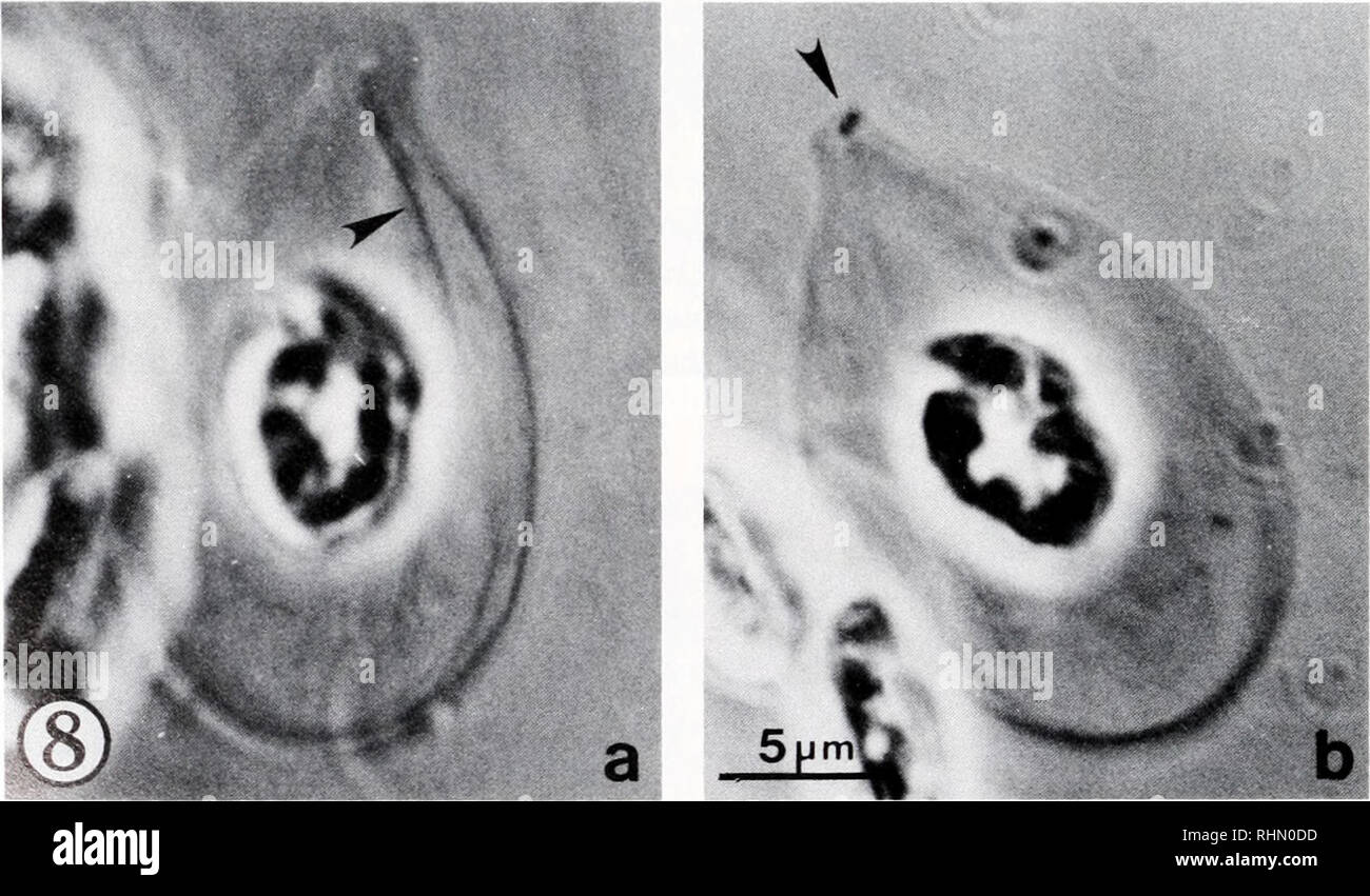
. The Biological bulletin. Biology; Zoology; Biology; Marine Biology. FIGURE 7. Skate erythrocyte cytoskeleton whole mount, uranyl acetate staining, TEM. Example in which the centriole pair is located closer to nucleus than to MB. (a) Survey view; centrioles at arrow; N = nucleus, (b) Higher magnification view of centrioles; few, if any, radiating microtubules are present. The centrioles are enmeshed between the two surface-associated cytoskeleton layers, which form a surrounding network (SAC), (c) Underexposed print of the centriole pair.. FIGURE 8. A rarely observed pointed skate erythrocyte

Image details
Contributor:
Library Book Collection / Alamy Stock PhotoImage ID:
RHN0DDFile size:
7.2 MB (436.3 KB Compressed download)Releases:
Model - no | Property - noDo I need a release?Dimensions:
2066 x 1210 px | 35 x 20.5 cm | 13.8 x 8.1 inches | 150dpiMore information:
This image is a public domain image, which means either that copyright has expired in the image or the copyright holder has waived their copyright. Alamy charges you a fee for access to the high resolution copy of the image.
This image could have imperfections as it’s either historical or reportage.
. The Biological bulletin. Biology; Zoology; Biology; Marine Biology. FIGURE 7. Skate erythrocyte cytoskeleton whole mount, uranyl acetate staining, TEM. Example in which the centriole pair is located closer to nucleus than to MB. (a) Survey view; centrioles at arrow; N = nucleus, (b) Higher magnification view of centrioles; few, if any, radiating microtubules are present. The centrioles are enmeshed between the two surface-associated cytoskeleton layers, which form a surrounding network (SAC), (c) Underexposed print of the centriole pair.. FIGURE 8. A rarely observed pointed skate erythrocyte cytoskeleton, as viewed in phase contrast under oil immersion, (a) One of several fibers (arrowhead) emanating from pointed (upper) region of cytoskeleton. These fibers radiate toward the distant closed end of the MB. (b) Different optical section of same c eleton, showing the pair of centrioles present at the apex of this pointed region (arrowhead). (Note: material at left in photos is part of adjacent clump of cytoskeletons, and different orientation of cytoskeietoi: in b is due to its movement under coverslip between photographs.). Please note that these images are extracted from scanned page images that may have been digitally enhanced for readability - coloration and appearance of these illustrations may not perfectly resemble the original work.. Marine Biological Laboratory (Woods Hole, Mass. ); Marine Biological Laboratory (Woods Hole, Mass. ). Annual report 1907/08-1952; Lillie, Frank Rattray, 1870-1947; Moore, Carl Richard, 1892-; Redfield, Alfred Clarence, 1890-1983. Woods Hole, Mass. : Marine Biological Laboratory