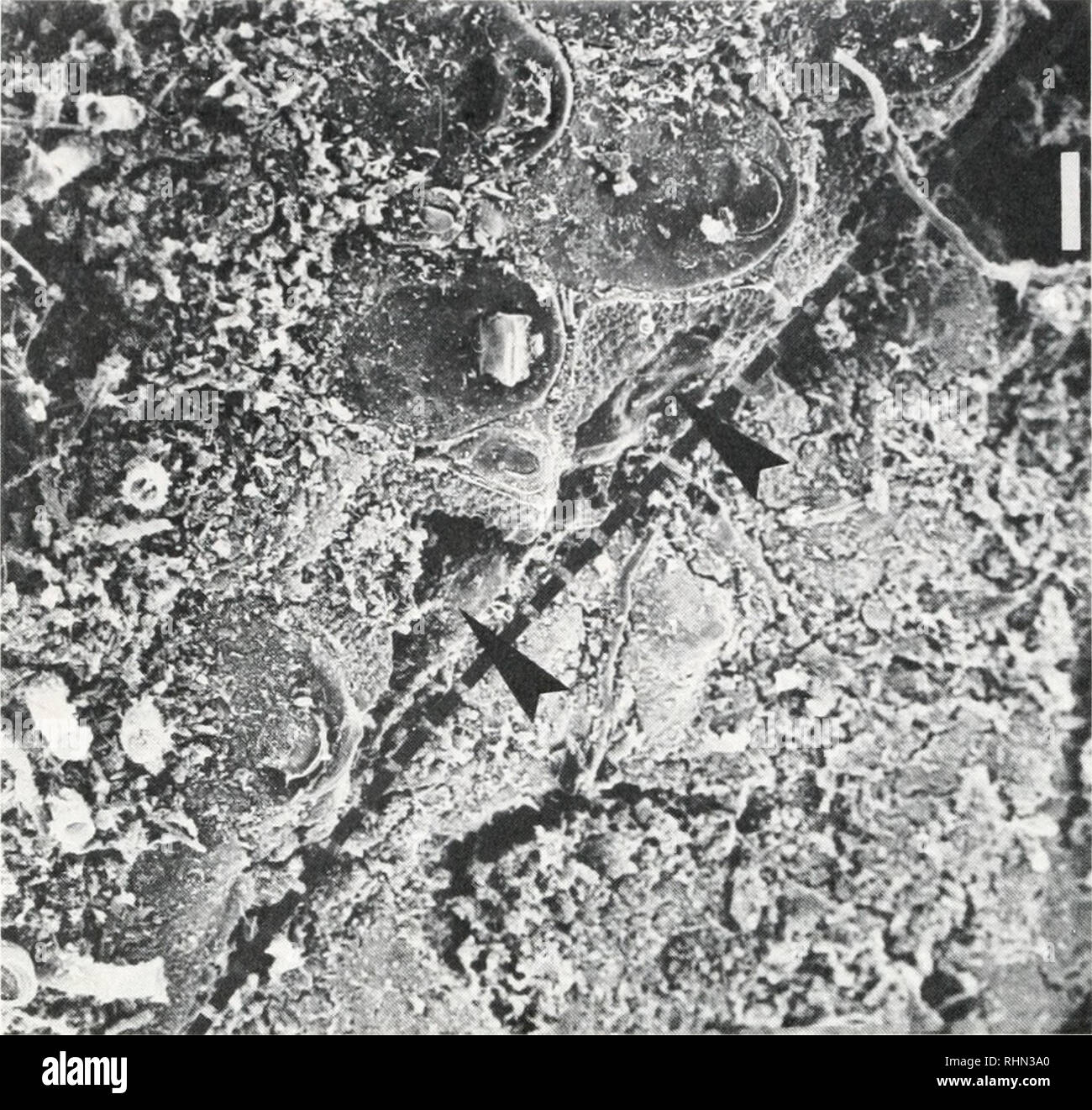
. The Biological bulletin. Biology; Zoology; Biology; Marine Biology. 100 M. LABARBERA. FIGURE 5. SEM micrograph of a bryozoan colony (Antropora tincta) adjacent to a D strigata. The brachiopod's valve margins are indicated by broken lines; note that several zooids adjacent to the brachiopod have been bisected (arrows) and that the colony has been eroded down to its basis beneath the brachiopod. Scale bar = 100 ^m. and mechanically inhibits sponge and bryozoan growth by directly damaging their tissues. The growing edge of sponges is initially thin (Ayling, 1983) and lacks spicular or fibrous r
Image details
Contributor:
Library Book Collection / Alamy Stock PhotoImage ID:
RHN3A0File size:
7.2 MB (562.2 KB Compressed download)Releases:
Model - no | Property - noDo I need a release?Dimensions:
1624 x 1539 px | 27.5 x 26.1 cm | 10.8 x 10.3 inches | 150dpiMore information:
This image is a public domain image, which means either that copyright has expired in the image or the copyright holder has waived their copyright. Alamy charges you a fee for access to the high resolution copy of the image.
This image could have imperfections as it’s either historical or reportage.
. The Biological bulletin. Biology; Zoology; Biology; Marine Biology. 100 M. LABARBERA. FIGURE 5. SEM micrograph of a bryozoan colony (Antropora tincta) adjacent to a D strigata. The brachiopod's valve margins are indicated by broken lines; note that several zooids adjacent to the brachiopod have been bisected (arrows) and that the colony has been eroded down to its basis beneath the brachiopod. Scale bar = 100 ^m. and mechanically inhibits sponge and bryozoan growth by directly damaging their tissues. The growing edge of sponges is initially thin (Ayling, 1983) and lacks spicular or fibrous reinforcement (Simpson, 1963). Although the rate of spatial coverage may increase dramatically where the sponge has been disturbed, absolute rates of coverage are maximally 6.98 mnr/cm perimeter/day (Ayling, 1983). Given the frequency with which D. strigata sweeps the vicinity with its setae and the vulnerability of sponge tissues, such low expansion rates are easily nullified. Newly budded bryozoan zooids are weakly calcified (Ryland, 1970); contrary to the usual pattern (Jackson, 1983), here the bryozoan's actively growing edge is more vulnerable than the fully calcified regions where growth is arrested. Antropora tincta exhibits frontal budding when growth is blocked (Buss, 1980, 1981; Jackson, 1983); for colonies around D. strigata, the only available agent for blockage is the brachiopod's setae. As noted above, frontal budding can be induced in A. tincta by the anterior setae of adjacent brachiopods. Since these setae are longer (thus exerting smaller forces at their tips) and lack the spines of the lateral and posterior setae, these bryozoans will be highly vulnerable to the disturbance imposed by the latter. Numerous eroded epizoans occurred under the brachiopods' ventral valves, al- though no abrasion of the valve itself was seen. The edge of the ventral valve is the most likely abrasive agent as evidenced by: (1) ground and polished regions on adjacent serpulids, vermet