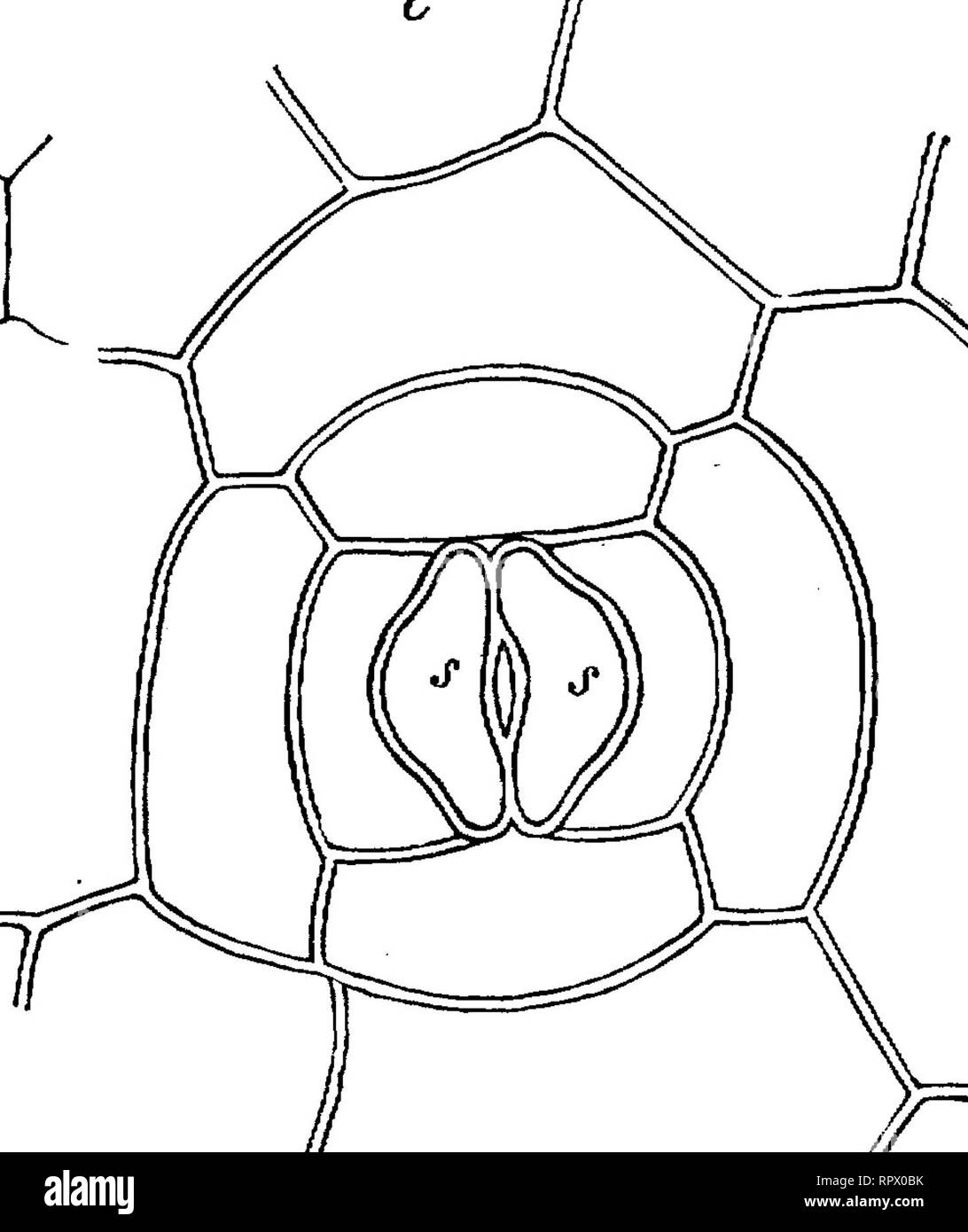
. Text-book of botany, morphological and physiological. Botany. •mc. FIG. 87.—Superficial view of a stoma of Anemia fraxini- folia with the epidermal cell completely surrounding it; e epi- dermis, ss guard-cells; cl chlorophyll-granules. FIG. 88.—Transverse section of a leaf of Pmus Pinaster (X800); s guard-cells of the stoma; p its cleft; v entrance; / air-cavity; c cuticularised layers of the epidermis; a middle lamella, * inner thickening-layers of the cells beneath the epidermis ; g parenchyma of the leaf containing chlorophyll. stoma; and thus arises the remarkable arrangement represented
Image details
Contributor:
Library Book Collection / Alamy Stock PhotoImage ID:
RPX0BKFile size:
7.2 MB (204.7 KB Compressed download)Releases:
Model - no | Property - noDo I need a release?Dimensions:
1449 x 1725 px | 24.5 x 29.2 cm | 9.7 x 11.5 inches | 150dpiMore information:
This image is a public domain image, which means either that copyright has expired in the image or the copyright holder has waived their copyright. Alamy charges you a fee for access to the high resolution copy of the image.
This image could have imperfections as it’s either historical or reportage.
. Text-book of botany, morphological and physiological. Botany. •mc. FIG. 87.—Superficial view of a stoma of Anemia fraxini- folia with the epidermal cell completely surrounding it; e epi- dermis, ss guard-cells; cl chlorophyll-granules. FIG. 88.—Transverse section of a leaf of Pmus Pinaster (X800); s guard-cells of the stoma; p its cleft; v entrance; / air-cavity; c cuticularised layers of the epidermis; a middle lamella, * inner thickening-layers of the cells beneath the epidermis ; g parenchyma of the leaf containing chlorophyll. stoma; and thus arises the remarkable arrangement represented in Fig. 87, where, as may be seen, the two guard-cells are entirely enclosed within a single epidermal cell. Similar, but more complicated, is, according to Rauter, the structure in Nipbo- bolus Lingua. By further growth of the guard-cells and of the epidermal cells which surround them, different relative positions of the former to the surface may be brought about; the guard-cells^ may, when mature, lie in one plane with those of the epidermis, or may be deeply depressed and apparently belong to a deeper layer of cells (Fig. 88); some- times they are, on the contrary, elevated above the surface of the epidermis. The stomata of Marehantieae may shortly be mentioned here in connexion with what has already been said on Fig. 65, p. 78. After the formation of the air-cavities, which are filled with outgrowths containing chlorophyll (Fig. 89, A, chl)i one cell of the 1 Strasburger, in Jahrb. fiir wiss. Bot. VII. p. 393; also Rauter, /, c.. Please note that these images are extracted from scanned page images that may have been digitally enhanced for readability - coloration and appearance of these illustrations may not perfectly resemble the original work.. Sachs, Julius, 1832-1897; Vines, Sydney Howard, 1849-1934. ed. and tr. Oxford, Clarendon press