Quick filters:
Plasma membrane Stock Photos and Images
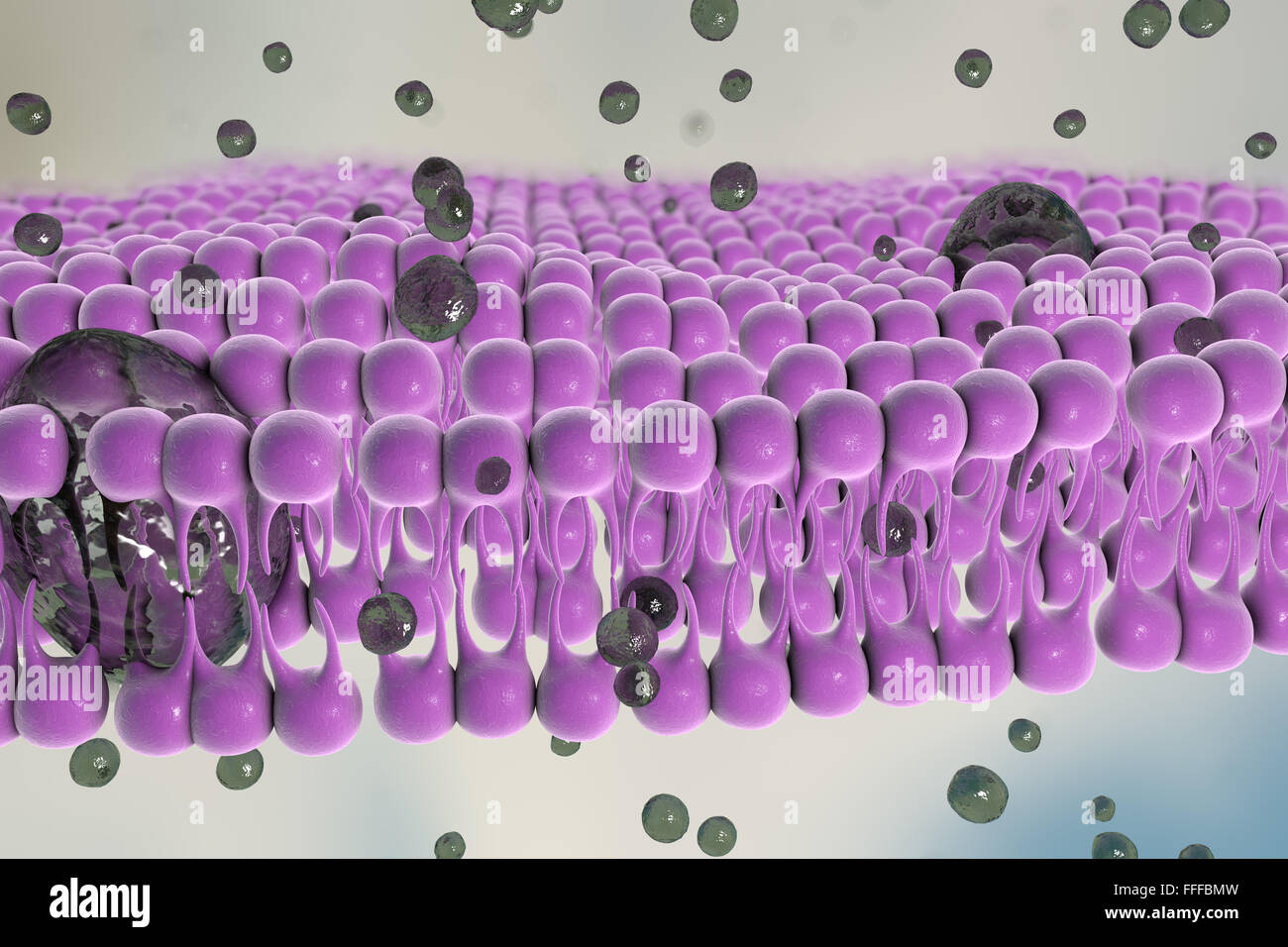 Plasma membrane. Illustration of the structure of the plasma membrane that encloses cells. The membrane is a bilayer of Stock Photohttps://www.alamy.com/image-license-details/?v=1https://www.alamy.com/stock-photo-plasma-membrane-illustration-of-the-structure-of-the-plasma-membrane-95610169.html
RFFFFBMW–Plasma membrane. Illustration of the structure of the plasma membrane that encloses cells. The membrane is a bilayer of
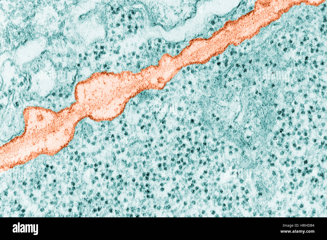 Plasma Membrane TEM Stock Photohttps://www.alamy.com/image-license-details/?v=1https://www.alamy.com/stock-photo-plasma-membrane-tem-134993352.html
RMHRHDB4–Plasma Membrane TEM
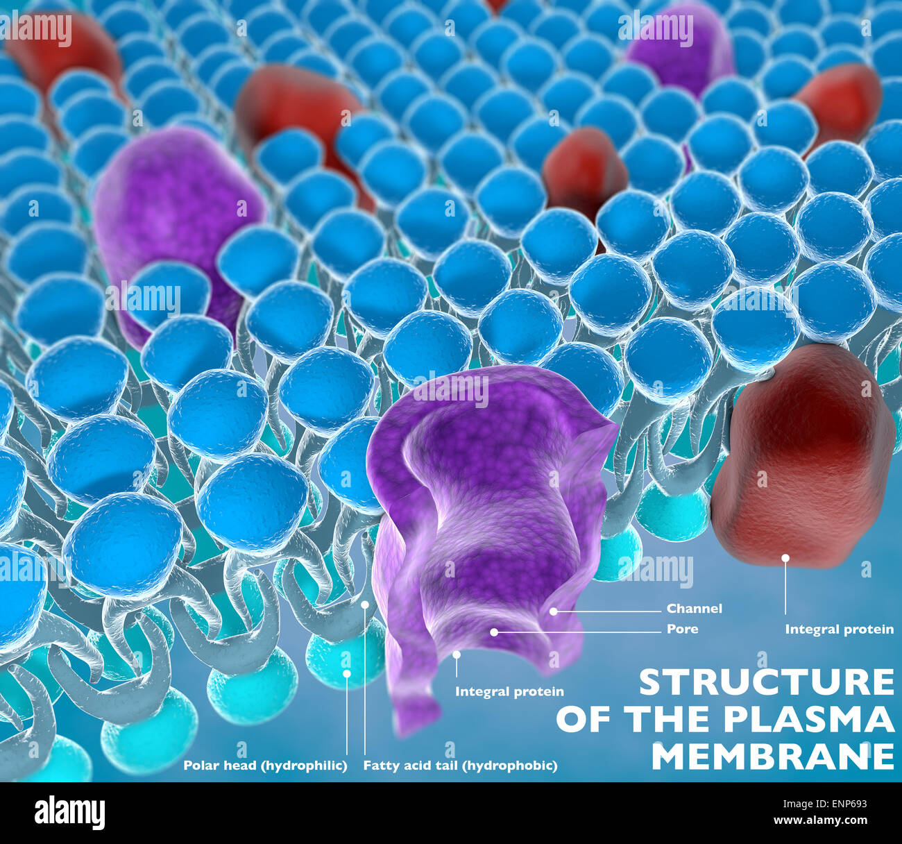 Structure of the plasma membrane of a cell Stock Photohttps://www.alamy.com/image-license-details/?v=1https://www.alamy.com/stock-photo-structure-of-the-plasma-membrane-of-a-cell-82237151.html
RMENP693–Structure of the plasma membrane of a cell
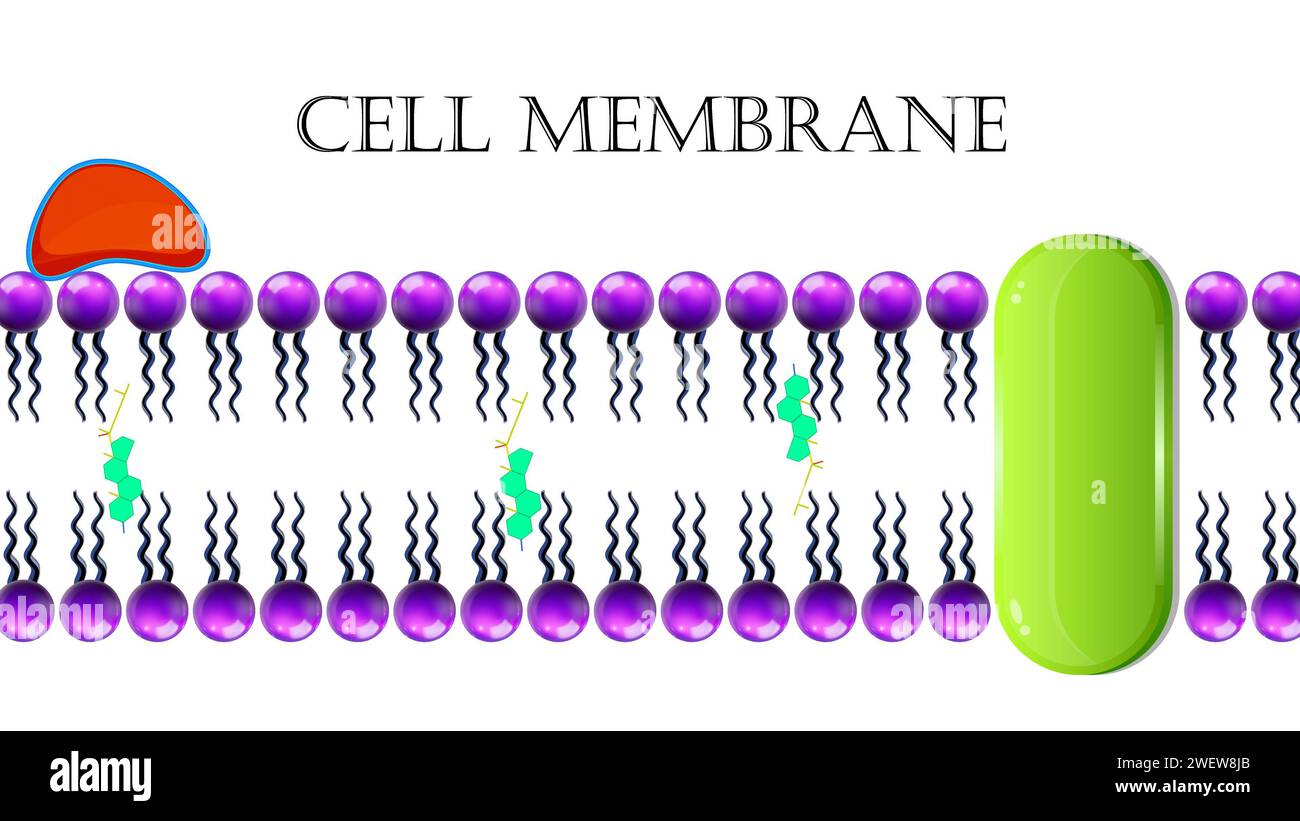 Cell Membrane Or Plasma Membrane Stock Photohttps://www.alamy.com/image-license-details/?v=1https://www.alamy.com/cell-membrane-or-plasma-membrane-image594313283.html
RF2WEW8JB–Cell Membrane Or Plasma Membrane
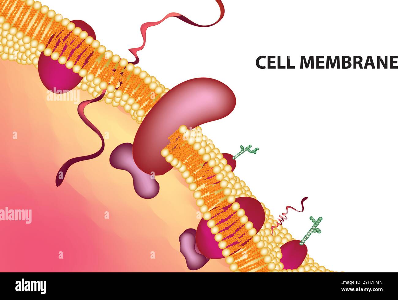 Detailed Structure of Plasma Membrane, Vector Illustration on White Background, Cell Biology Diagram, Scientific Artwork Stock Vectorhttps://www.alamy.com/image-license-details/?v=1https://www.alamy.com/detailed-structure-of-plasma-membrane-vector-illustration-on-white-background-cell-biology-diagram-scientific-artwork-image630188405.html
RF2YH7FMN–Detailed Structure of Plasma Membrane, Vector Illustration on White Background, Cell Biology Diagram, Scientific Artwork
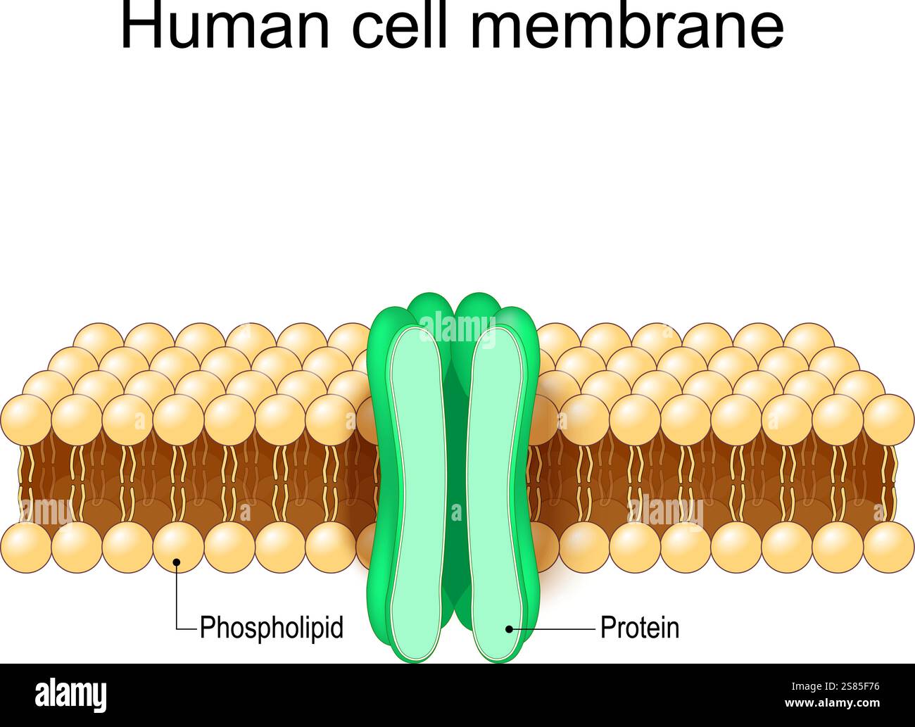 Cell membrane structure. Close-up of a plasma membrane. Anatomy of Cytoplasmic membrane. Phospholipid bilayer and protein. Plasmalemma. Vector Stock Vectorhttps://www.alamy.com/image-license-details/?v=1https://www.alamy.com/cell-membrane-structure-close-up-of-a-plasma-membrane-anatomy-of-cytoplasmic-membrane-phospholipid-bilayer-and-protein-plasmalemma-vector-image641822586.html
RF2S85F76–Cell membrane structure. Close-up of a plasma membrane. Anatomy of Cytoplasmic membrane. Phospholipid bilayer and protein. Plasmalemma. Vector
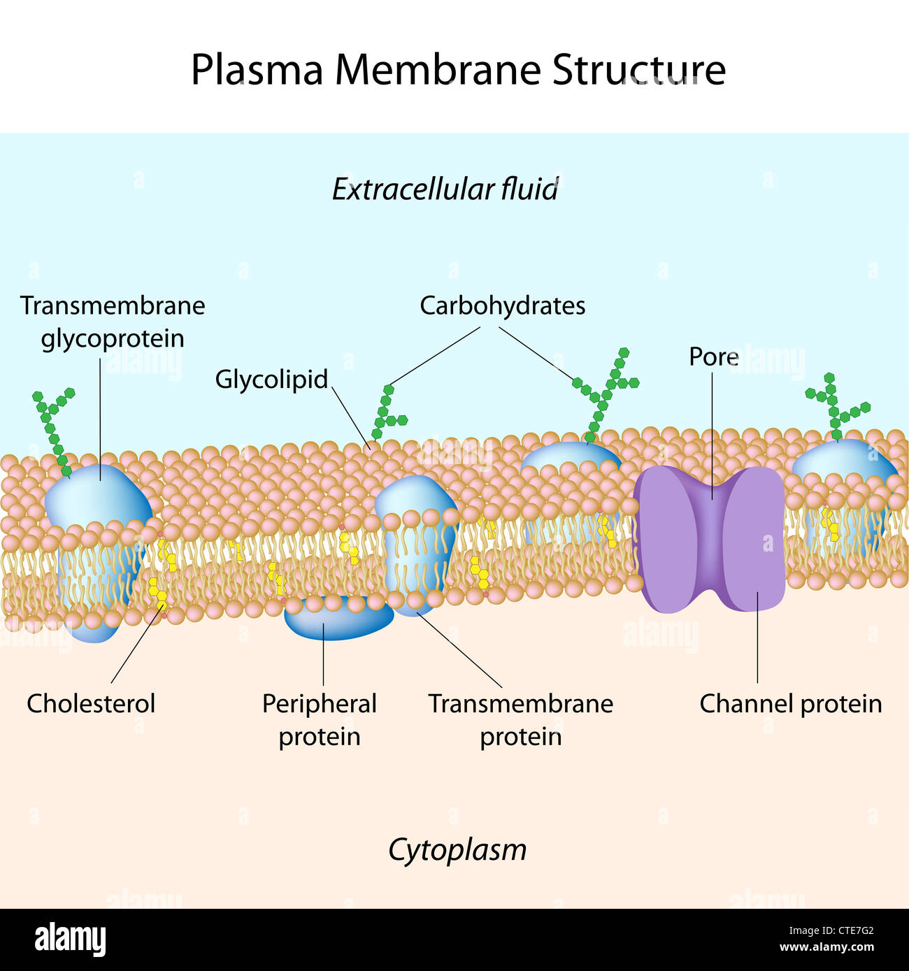 Structure of plasma membrane Stock Photohttps://www.alamy.com/image-license-details/?v=1https://www.alamy.com/stock-photo-structure-of-plasma-membrane-49485746.html
RFCTE7G2–Structure of plasma membrane
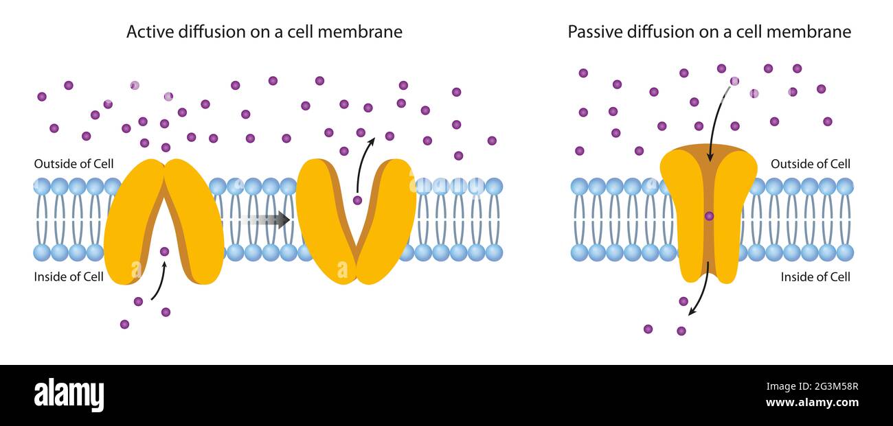 Diffusion Across the Plasma Membrane Stock Photohttps://www.alamy.com/image-license-details/?v=1https://www.alamy.com/diffusion-across-the-plasma-membrane-image432546375.html
RF2G3M58R–Diffusion Across the Plasma Membrane
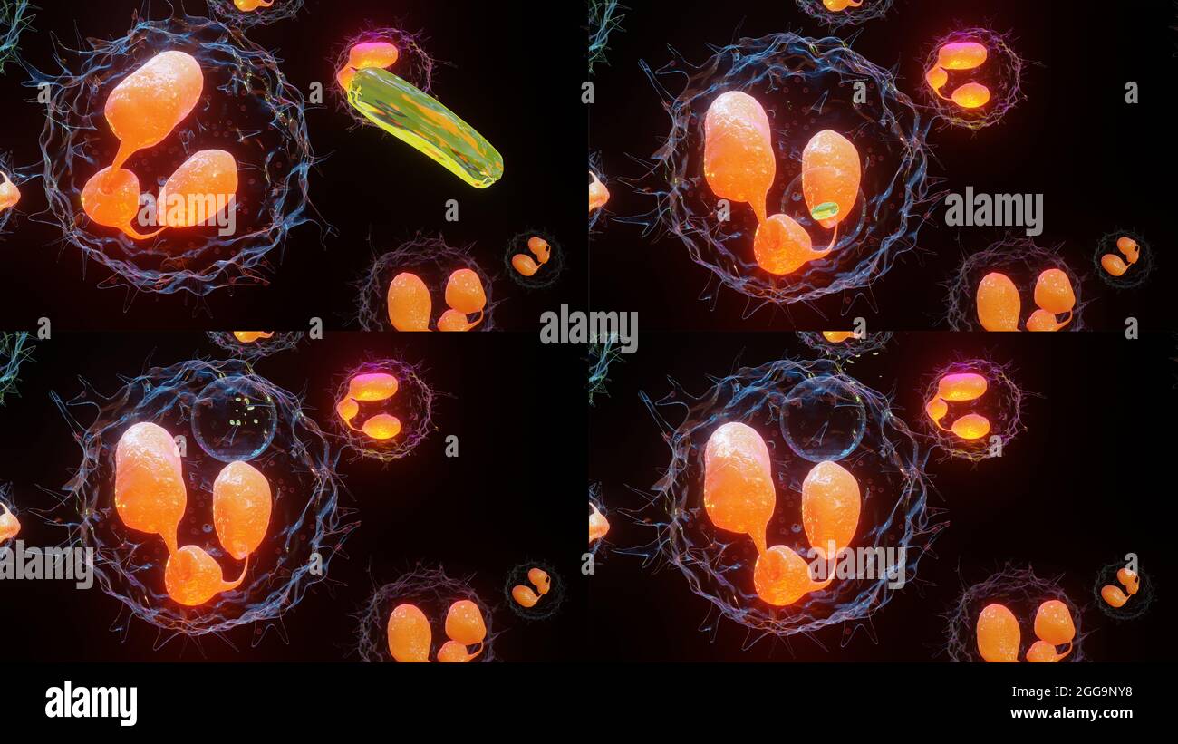 3D illustration of Phagocytosis. Neutrophe that uses its plasma membrane to engulf bacteria. From endocytosis to exocytosis. Digestion process in phag Stock Photohttps://www.alamy.com/image-license-details/?v=1https://www.alamy.com/3d-illustration-of-phagocytosis-neutrophe-that-uses-its-plasma-membrane-to-engulf-bacteria-from-endocytosis-to-exocytosis-digestion-process-in-phag-image440308492.html
RM2GG9NY8–3D illustration of Phagocytosis. Neutrophe that uses its plasma membrane to engulf bacteria. From endocytosis to exocytosis. Digestion process in phag
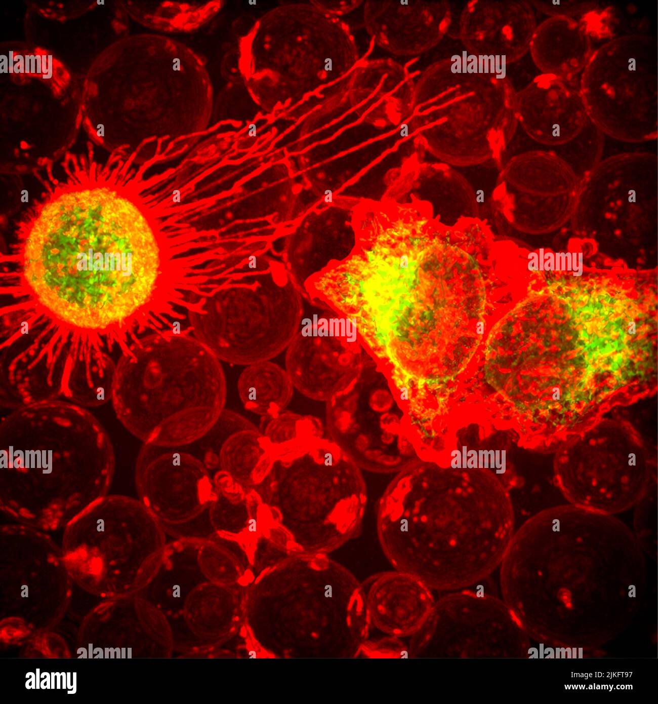 On the right, two cells (greenish yellow) are in the process of forming bubbles, or plasma membrane vesicles (PMV). During this process of being, a cell's membrane temporarily dissociates from its underlying cytoskeleton, forming a tiny pocket which, over the course of about 30 minutes, is Stock Photohttps://www.alamy.com/image-license-details/?v=1https://www.alamy.com/on-the-right-two-cells-greenish-yellow-are-in-the-process-of-forming-bubbles-or-plasma-membrane-vesicles-pmv-during-this-process-of-being-a-cells-membrane-temporarily-dissociates-from-its-underlying-cytoskeleton-forming-a-tiny-pocket-which-over-the-course-of-about-30-minutes-is-image476706755.html
RM2JKFT97–On the right, two cells (greenish yellow) are in the process of forming bubbles, or plasma membrane vesicles (PMV). During this process of being, a cell's membrane temporarily dissociates from its underlying cytoskeleton, forming a tiny pocket which, over the course of about 30 minutes, is
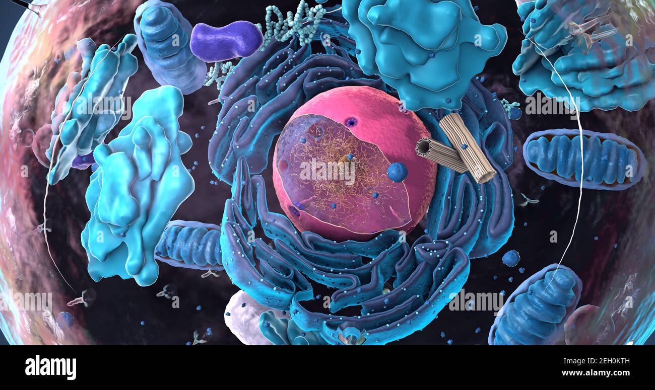 Components of Eukaryotic cell, nucleus and organelles and plasma membrane - 3d illustration Stock Photohttps://www.alamy.com/image-license-details/?v=1https://www.alamy.com/components-of-eukaryotic-cell-nucleus-and-organelles-and-plasma-membrane-3d-illustration-image406303201.html
RF2EH0KTH–Components of Eukaryotic cell, nucleus and organelles and plasma membrane - 3d illustration
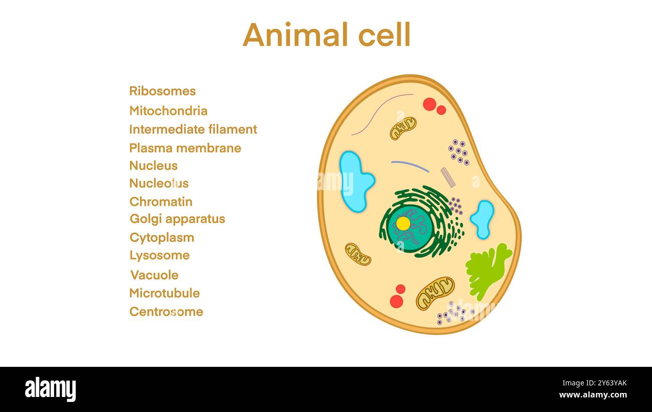 animal cell anatomy, biological animal cell with organelles cross section, Animal cell with placed text annotations to all organelles, Animal cell Stock Photohttps://www.alamy.com/image-license-details/?v=1https://www.alamy.com/animal-cell-anatomy-biological-animal-cell-with-organelles-cross-section-animal-cell-with-placed-text-annotations-to-all-organelles-animal-cell-image623348507.html
RF2Y63YAK–animal cell anatomy, biological animal cell with organelles cross section, Animal cell with placed text annotations to all organelles, Animal cell
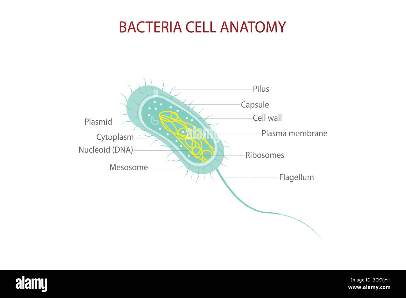 Bacteria Cell Anatomy Diagram with Labeled Parts Including Nucleoid, Ribosomes, Flagellum, and Plasma Membrane. Stock Vectorhttps://www.alamy.com/image-license-details/?v=1https://www.alamy.com/bacteria-cell-anatomy-diagram-with-labeled-parts-including-nucleoid-ribosomes-flagellum-and-plasma-membrane-image700700773.html
RF3CKYJYH–Bacteria Cell Anatomy Diagram with Labeled Parts Including Nucleoid, Ribosomes, Flagellum, and Plasma Membrane.
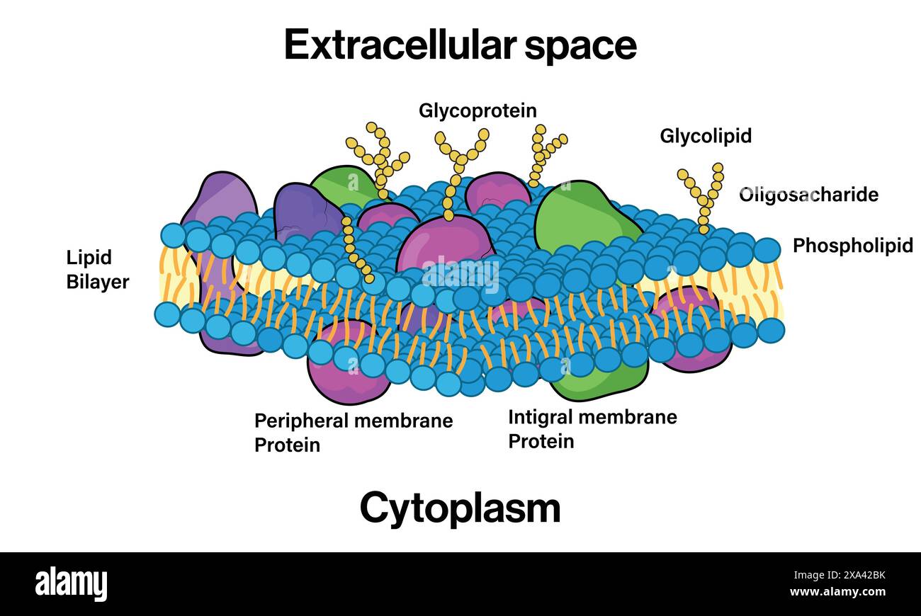 Detailed Vector Illustration of Plasma Membrane Structure for Cell Biology and Biochemistry Education on White Background. Stock Vectorhttps://www.alamy.com/image-license-details/?v=1https://www.alamy.com/detailed-vector-illustration-of-plasma-membrane-structure-for-cell-biology-and-biochemistry-education-on-white-background-image608599143.html
RF2XA42BK–Detailed Vector Illustration of Plasma Membrane Structure for Cell Biology and Biochemistry Education on White Background.
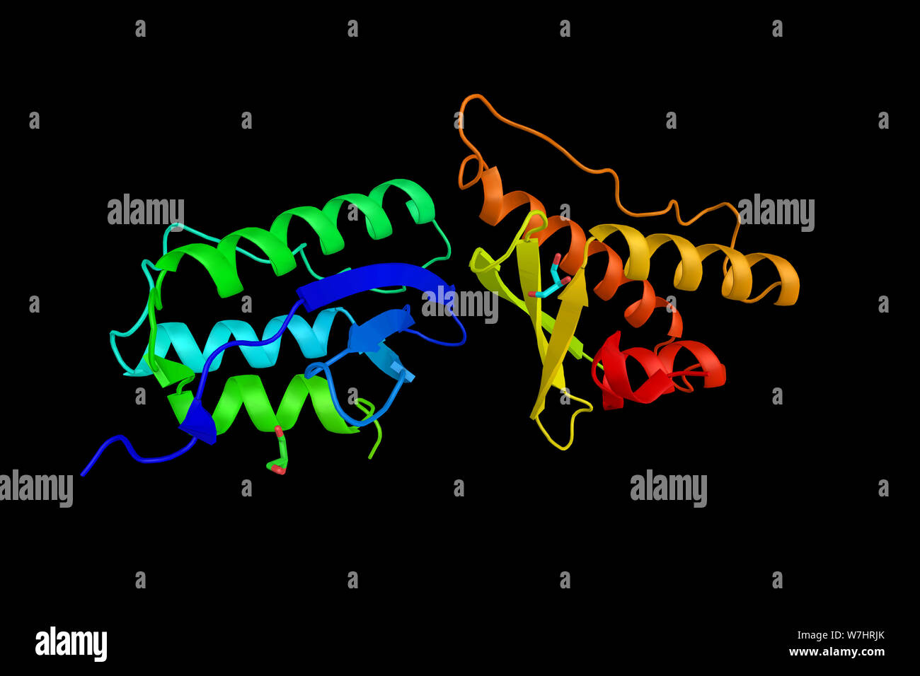 Nischarin, a nonadrenergic imidazoline-1 receptor protein that localizes to the cytosol and anchors to the inner layer of the plasma membrane. 3d rend Stock Photohttps://www.alamy.com/image-license-details/?v=1https://www.alamy.com/nischarin-a-nonadrenergic-imidazoline-1-receptor-protein-that-localizes-to-the-cytosol-and-anchors-to-the-inner-layer-of-the-plasma-membrane-3d-rend-image262849851.html
RFW7HRJK–Nischarin, a nonadrenergic imidazoline-1 receptor protein that localizes to the cytosol and anchors to the inner layer of the plasma membrane. 3d rend
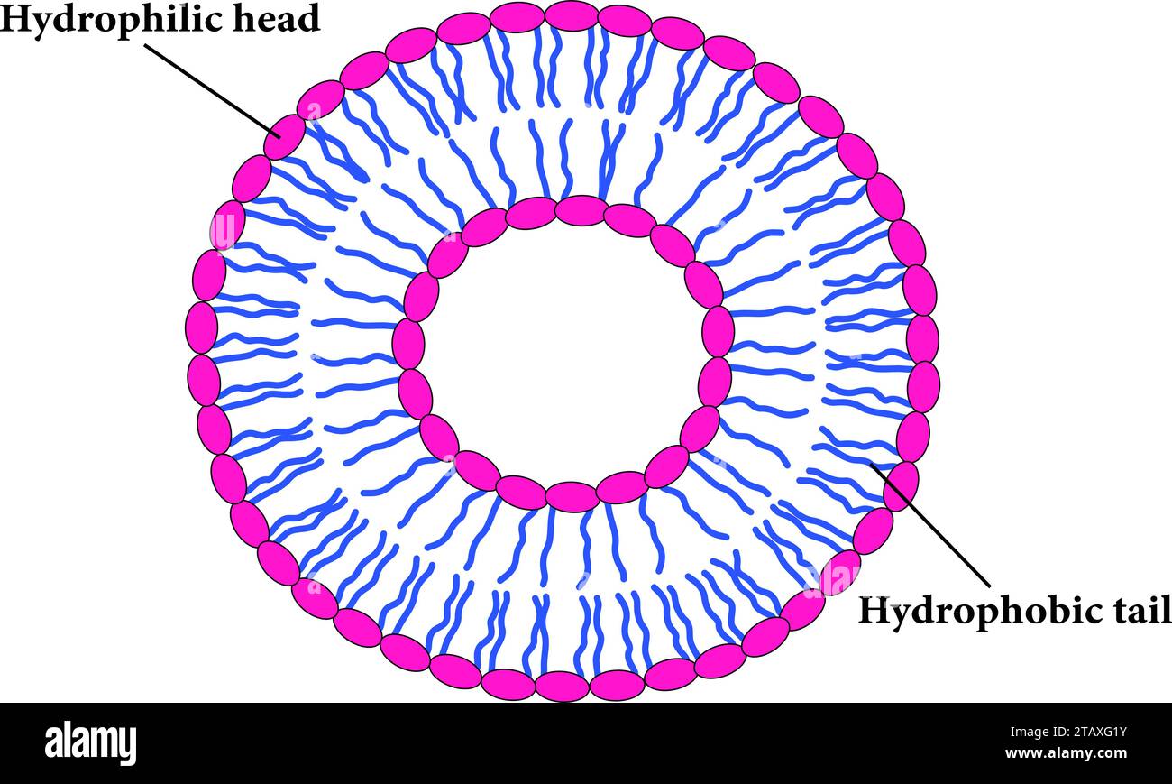 The Scheme of a liposome formed by phospholipids.Vector illustration. Stock Vectorhttps://www.alamy.com/image-license-details/?v=1https://www.alamy.com/the-scheme-of-a-liposome-formed-by-phospholipidsvector-illustration-image574672055.html
RF2TAXG1Y–The Scheme of a liposome formed by phospholipids.Vector illustration.
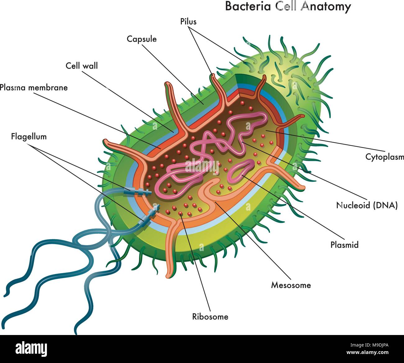 vector medical illustration of the bacteria cell anatomy Stock Vectorhttps://www.alamy.com/image-license-details/?v=1https://www.alamy.com/vector-medical-illustration-of-the-bacteria-cell-anatomy-image177935698.html
RFM9DJPA–vector medical illustration of the bacteria cell anatomy
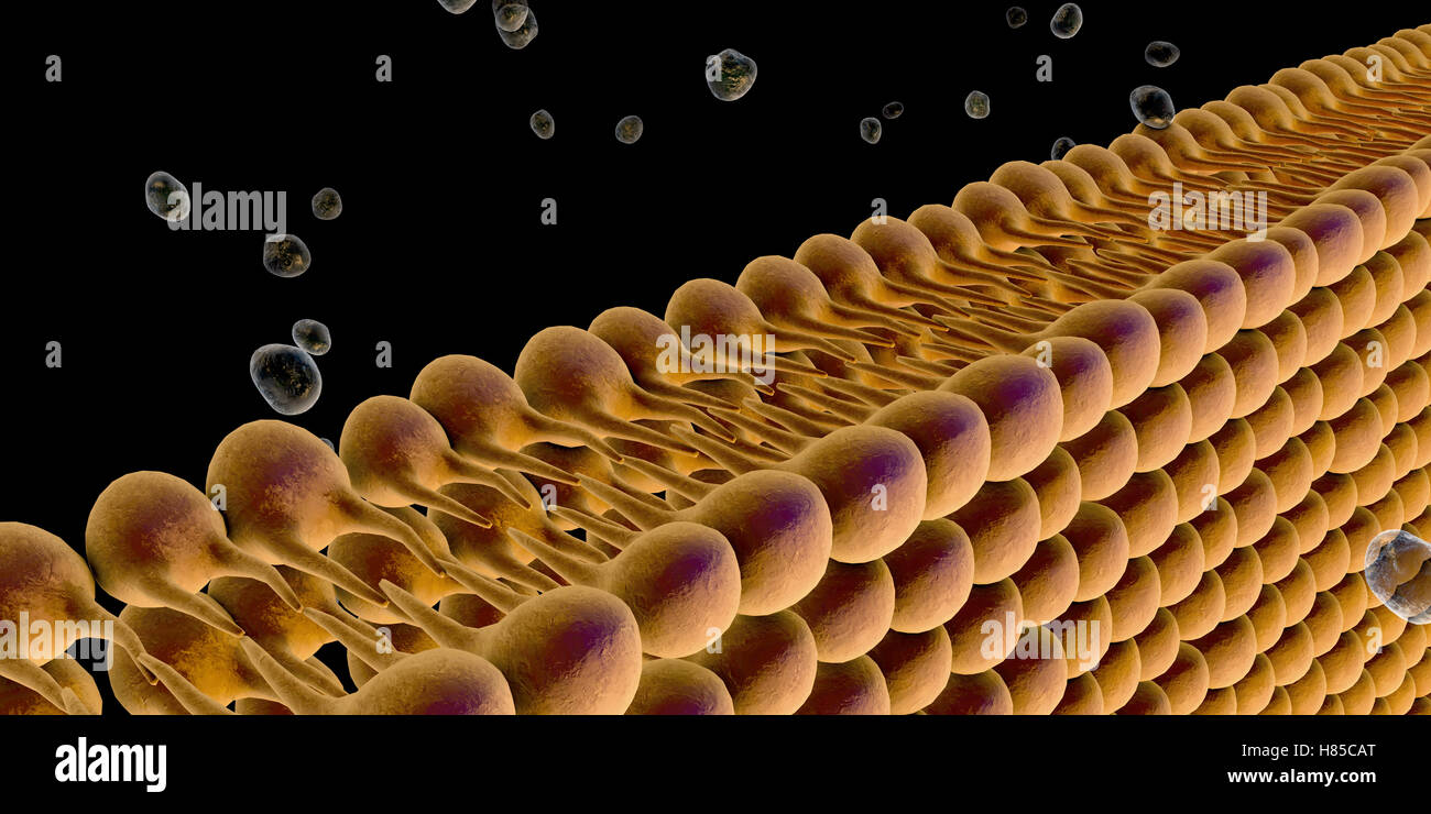 Plasma Membrane Of Cell With other molecules, 3d render Stock Photohttps://www.alamy.com/image-license-details/?v=1https://www.alamy.com/stock-photo-plasma-membrane-of-cell-with-other-molecules-3d-render-125509296.html
RFH85CAT–Plasma Membrane Of Cell With other molecules, 3d render
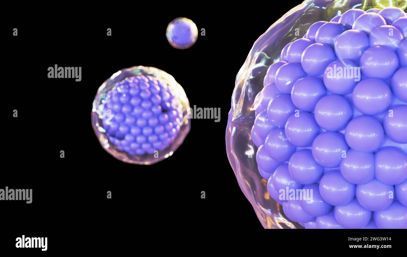 3d rendering of cells are surrounded by a plasma membrane, which is a thin, flexible barrier that separates the cell from its environment. Stock Photohttps://www.alamy.com/image-license-details/?v=1https://www.alamy.com/3d-rendering-of-cells-are-surrounded-by-a-plasma-membrane-which-is-a-thin-flexible-barrier-that-separates-the-cell-from-its-environment-image595072496.html
RF2WG3W14–3d rendering of cells are surrounded by a plasma membrane, which is a thin, flexible barrier that separates the cell from its environment.
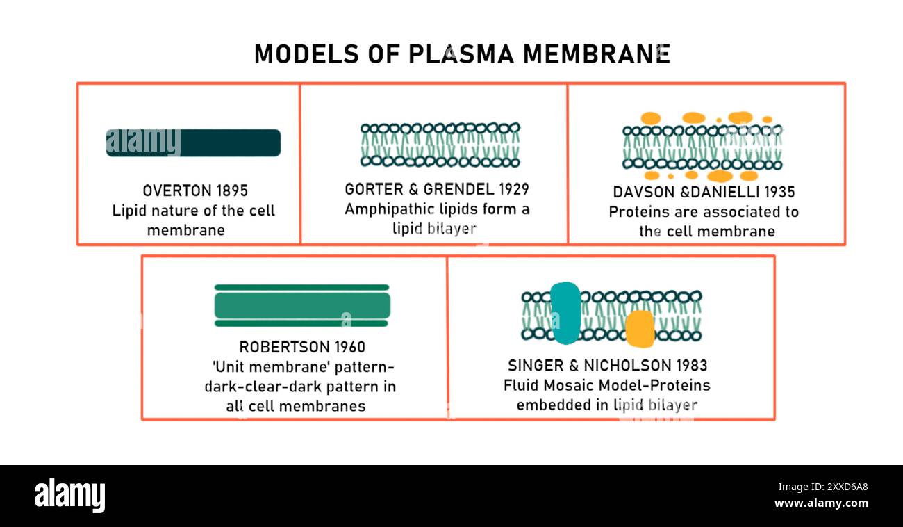 Plasma membrane models devised over history, illustration. Stock Photohttps://www.alamy.com/image-license-details/?v=1https://www.alamy.com/plasma-membrane-models-devised-over-history-illustration-image618634304.html
RF2XXD6A8–Plasma membrane models devised over history, illustration.
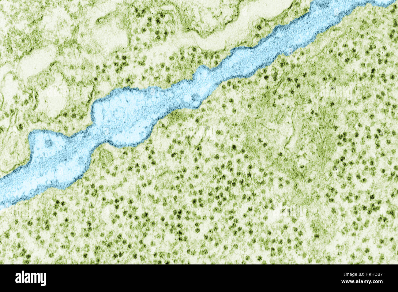 Plasma Membrane TEM Stock Photohttps://www.alamy.com/image-license-details/?v=1https://www.alamy.com/stock-photo-plasma-membrane-tem-134993355.html
RMHRHDB7–Plasma Membrane TEM
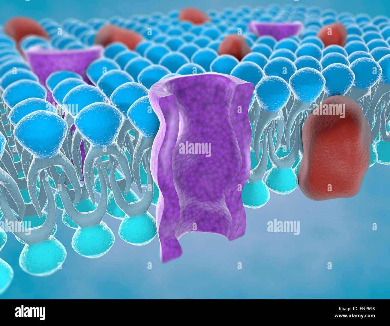 Structure of the plasma membrane of a cell Stock Photohttps://www.alamy.com/image-license-details/?v=1https://www.alamy.com/stock-photo-structure-of-the-plasma-membrane-of-a-cell-82237156.html
RMENP698–Structure of the plasma membrane of a cell
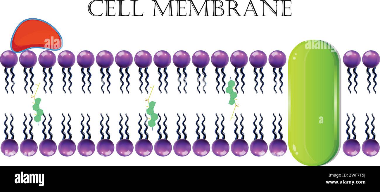 Cell Membrane Or Plasma Membrane Stock Vectorhttps://www.alamy.com/image-license-details/?v=1https://www.alamy.com/cell-membrane-or-plasma-membrane-image594544990.html
RF2WF7T5J–Cell Membrane Or Plasma Membrane
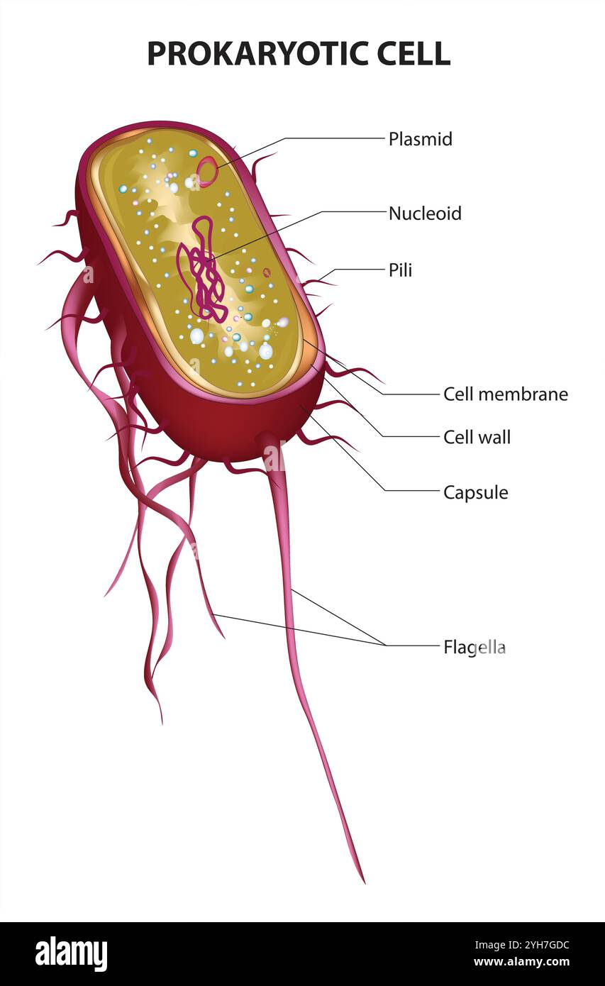 Prokaryotic Cell Structure Chart, vector medical illustration, online education material. English translation text Stock Vectorhttps://www.alamy.com/image-license-details/?v=1https://www.alamy.com/prokaryotic-cell-structure-chart-vector-medical-illustration-online-education-material-english-translation-text-image630188984.html
RF2YH7GDC–Prokaryotic Cell Structure Chart, vector medical illustration, online education material. English translation text
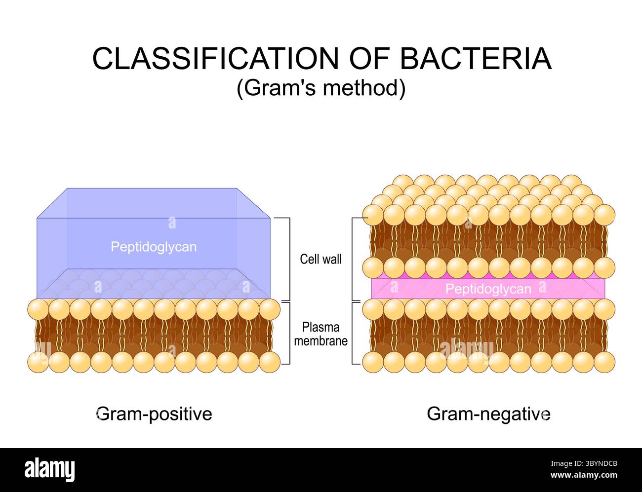 Classification of bacteria. Grams method. Gram-positive and Gram-negative bacterium. Cross section of Cell wall, Plasma membrane and Peptidoglycan. De Stock Vectorhttps://www.alamy.com/image-license-details/?v=1https://www.alamy.com/classification-of-bacteria-grams-method-gram-positive-and-gram-negative-bacterium-cross-section-of-cell-wall-plasma-membrane-and-peptidoglycan-de-image688271595.html
RF3BYNDCB–Classification of bacteria. Grams method. Gram-positive and Gram-negative bacterium. Cross section of Cell wall, Plasma membrane and Peptidoglycan. De
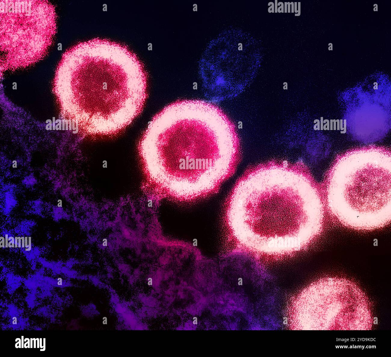 Transmission electron micrograph of HIV-1 virus particles pink replicating from the plasma membrane of an infected H9 T cell purple. HIV-1 Virus 016867 217 Stock Photohttps://www.alamy.com/image-license-details/?v=1https://www.alamy.com/transmission-electron-micrograph-of-hiv-1-virus-particles-pink-replicating-from-the-plasma-membrane-of-an-infected-h9-t-cell-purple-hiv-1-virus-016867-217-image627776616.html
RM2YD9KDC–Transmission electron micrograph of HIV-1 virus particles pink replicating from the plasma membrane of an infected H9 T cell purple. HIV-1 Virus 016867 217
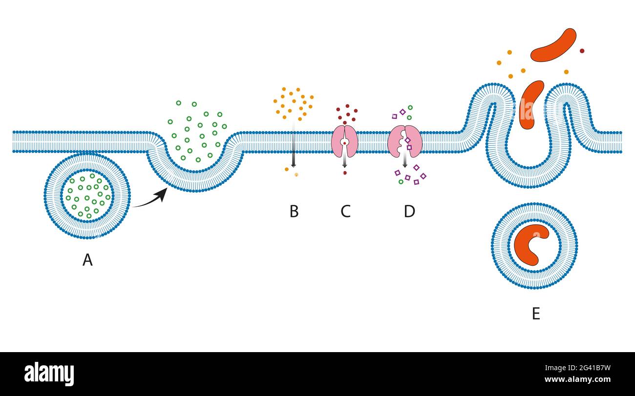 Movement through the Plasma Membrane methods Stock Photohttps://www.alamy.com/image-license-details/?v=1https://www.alamy.com/movement-through-the-plasma-membrane-methods-image432748621.html
RF2G41B7W–Movement through the Plasma Membrane methods
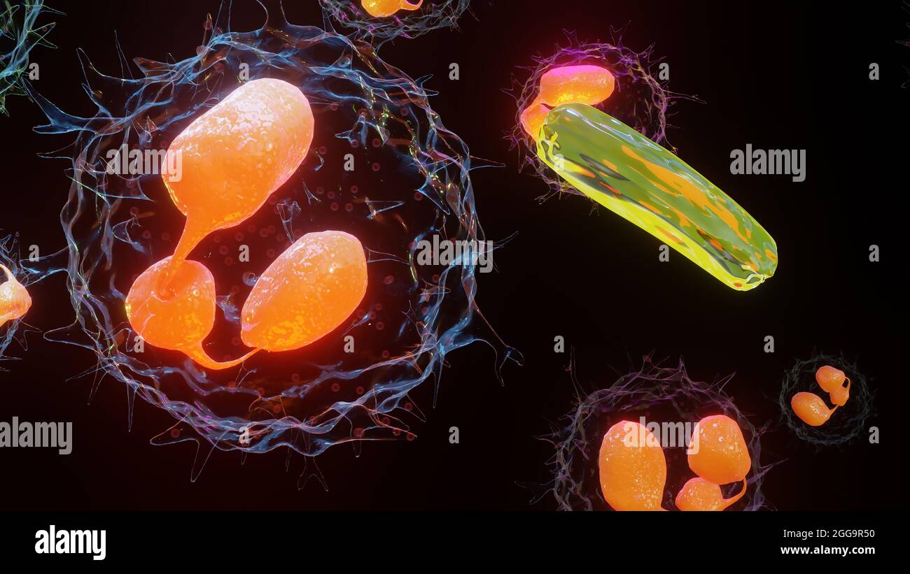 3D illustration of Phagocytosis. Neutrophe that uses its plasma membrane to engulf bacteria. From endocytosis to exocytosis. Digestion process in phag Stock Photohttps://www.alamy.com/image-license-details/?v=1https://www.alamy.com/3d-illustration-of-phagocytosis-neutrophe-that-uses-its-plasma-membrane-to-engulf-bacteria-from-endocytosis-to-exocytosis-digestion-process-in-phag-image440309436.html
RM2GG9R50–3D illustration of Phagocytosis. Neutrophe that uses its plasma membrane to engulf bacteria. From endocytosis to exocytosis. Digestion process in phag
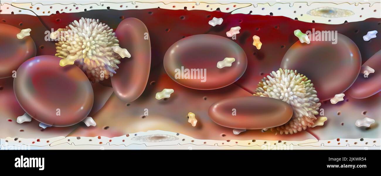 Blood vessel containing red blood cells, white blood cells, platelets, and plasma. Stock Photohttps://www.alamy.com/image-license-details/?v=1https://www.alamy.com/blood-vessel-containing-red-blood-cells-white-blood-cells-platelets-and-plasma-image476925376.html
RF2JKWR54–Blood vessel containing red blood cells, white blood cells, platelets, and plasma.
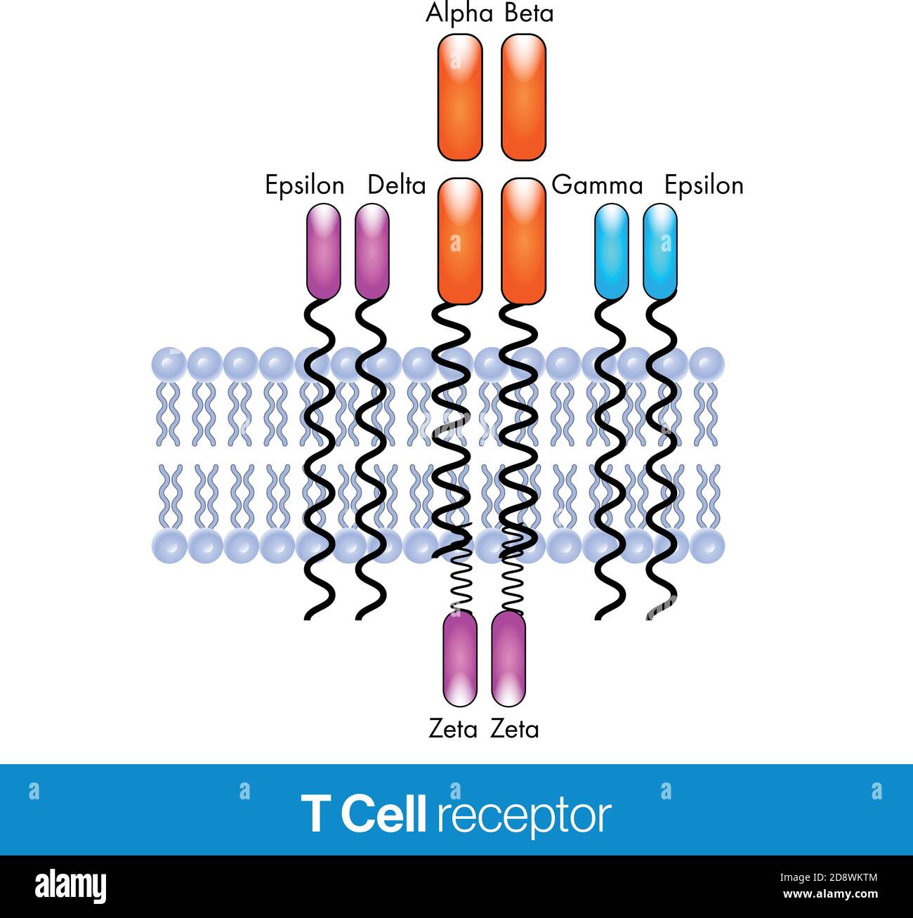 T cell receptor vector design illustration with the plasma membrane in the background. Stock Vectorhttps://www.alamy.com/image-license-details/?v=1https://www.alamy.com/t-cell-receptor-vector-design-illustration-with-the-plasma-membrane-in-the-background-image384109732.html
RF2D8WKTM–T cell receptor vector design illustration with the plasma membrane in the background.
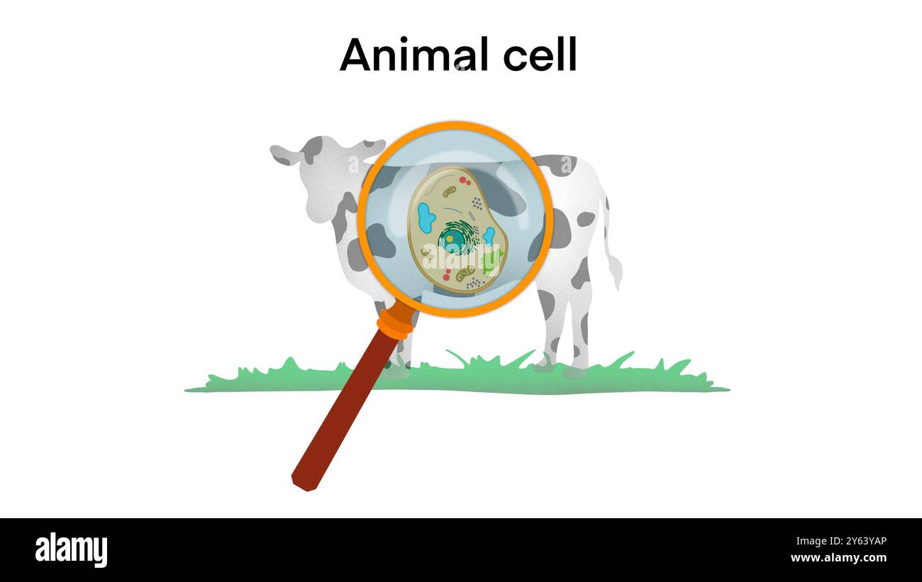 animal cell anatomy, biological animal cell with organelles cross section, Animal cell structure. Educational material, Anatomy of animal cell Stock Photohttps://www.alamy.com/image-license-details/?v=1https://www.alamy.com/animal-cell-anatomy-biological-animal-cell-with-organelles-cross-section-animal-cell-structure-educational-material-anatomy-of-animal-cell-image623348510.html
RF2Y63YAP–animal cell anatomy, biological animal cell with organelles cross section, Animal cell structure. Educational material, Anatomy of animal cell
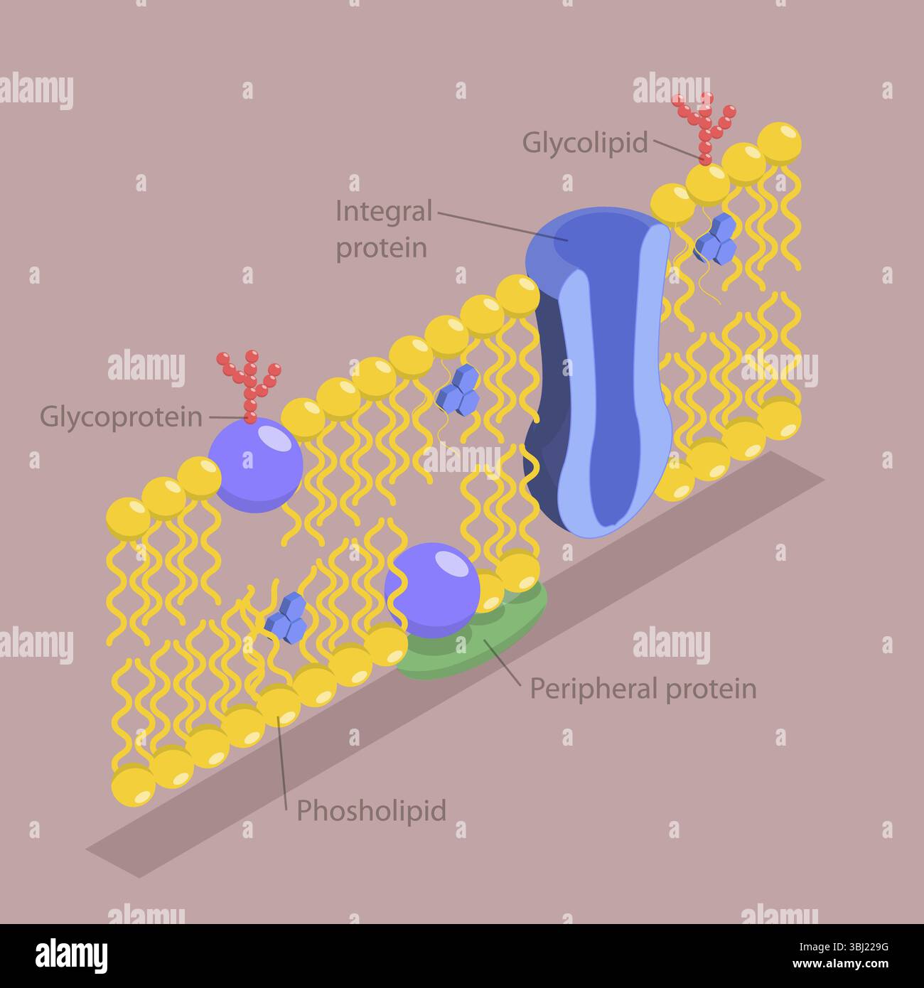 3D Isometric Flat Illustration of Structure of Plasma Membrane, Anatomical Structure According To The Fluid Mosaic Model Stock Photohttps://www.alamy.com/image-license-details/?v=1https://www.alamy.com/3d-isometric-flat-illustration-of-structure-of-plasma-membrane-anatomical-structure-according-to-the-fluid-mosaic-model-image682313900.html
RF3BJ229G–3D Isometric Flat Illustration of Structure of Plasma Membrane, Anatomical Structure According To The Fluid Mosaic Model
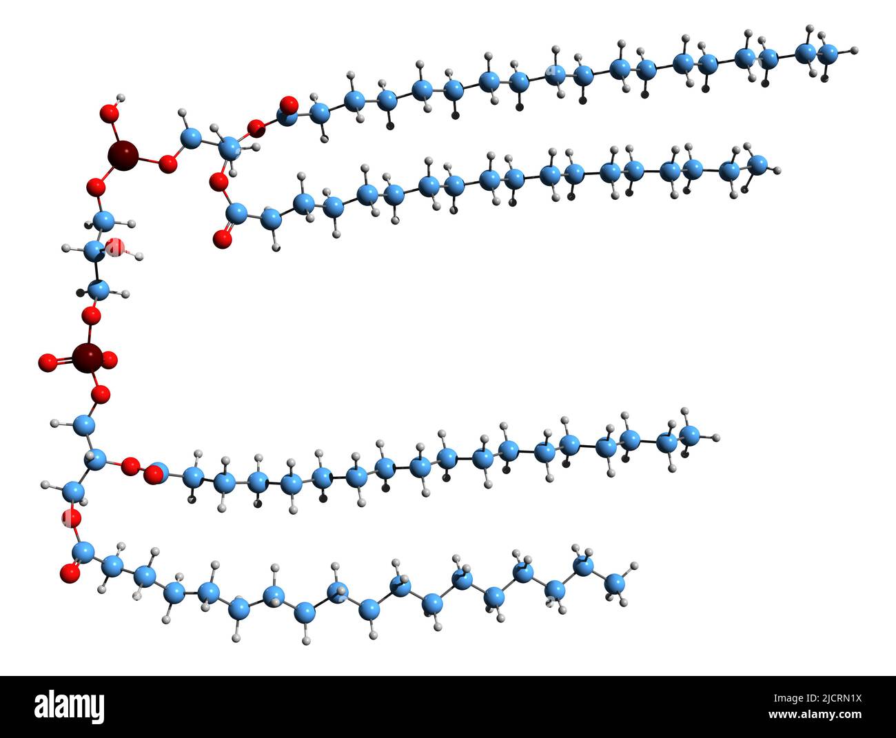 3D image of bisphosphatidylglycerol skeletal formula - molecular chemical structure of glycerophospholipid isolated on white background Stock Photohttps://www.alamy.com/image-license-details/?v=1https://www.alamy.com/3d-image-of-bisphosphatidylglycerol-skeletal-formula-molecular-chemical-structure-of-glycerophospholipid-isolated-on-white-background-image472577222.html
RF2JCRN1X–3D image of bisphosphatidylglycerol skeletal formula - molecular chemical structure of glycerophospholipid isolated on white background
 Conceptual image of human cell. Stock Photohttps://www.alamy.com/image-license-details/?v=1https://www.alamy.com/stock-photo-conceptual-image-of-human-cell-57644214.html
RFD9NWNX–Conceptual image of human cell.
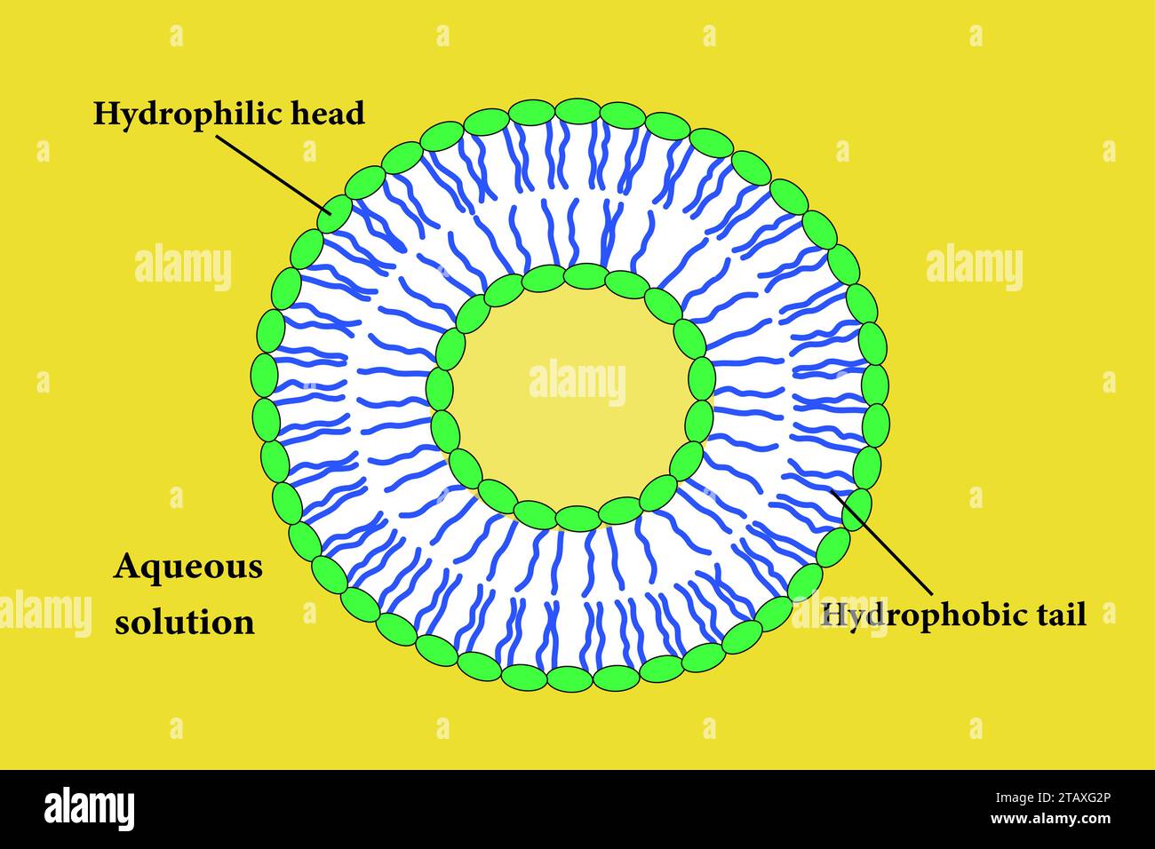 The Scheme of a liposome formed by phospholipids.Vector illustration. Stock Vectorhttps://www.alamy.com/image-license-details/?v=1https://www.alamy.com/the-scheme-of-a-liposome-formed-by-phospholipidsvector-illustration-image574672078.html
RF2TAXG2P–The Scheme of a liposome formed by phospholipids.Vector illustration.
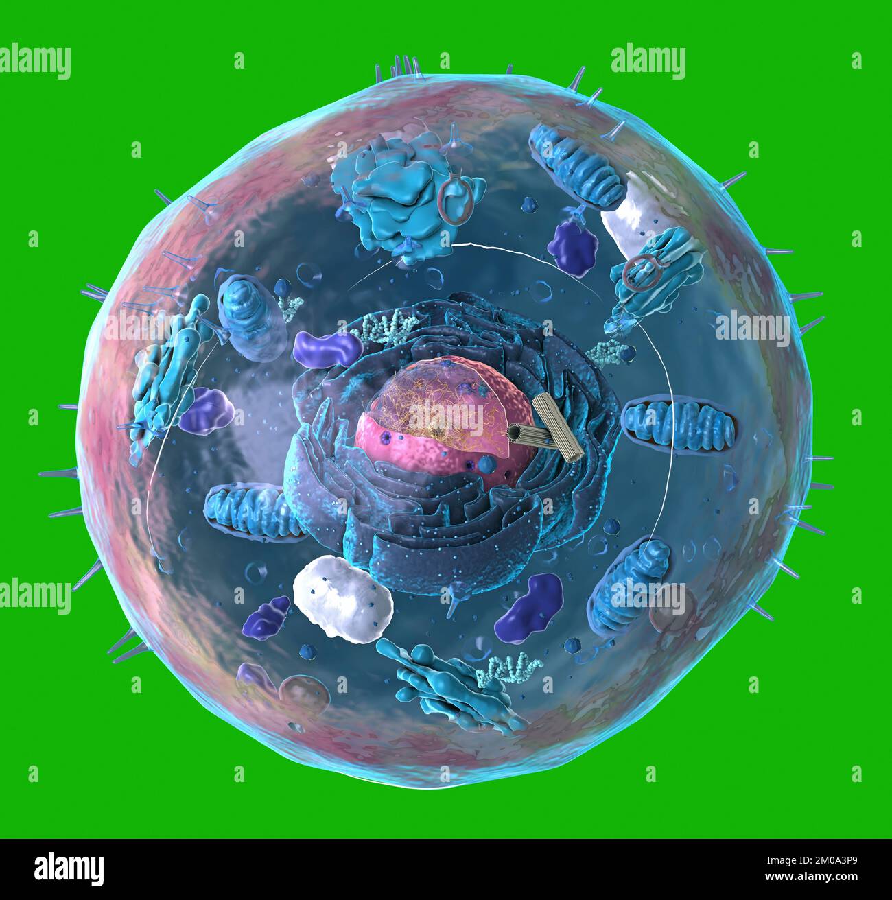 Components of Eukaryotic cell, nucleus and organelles and plasma membrane - 3d illustration Stock Photohttps://www.alamy.com/image-license-details/?v=1https://www.alamy.com/components-of-eukaryotic-cell-nucleus-and-organelles-and-plasma-membrane-3d-illustration-image499323169.html
RF2M0A3P9–Components of Eukaryotic cell, nucleus and organelles and plasma membrane - 3d illustration
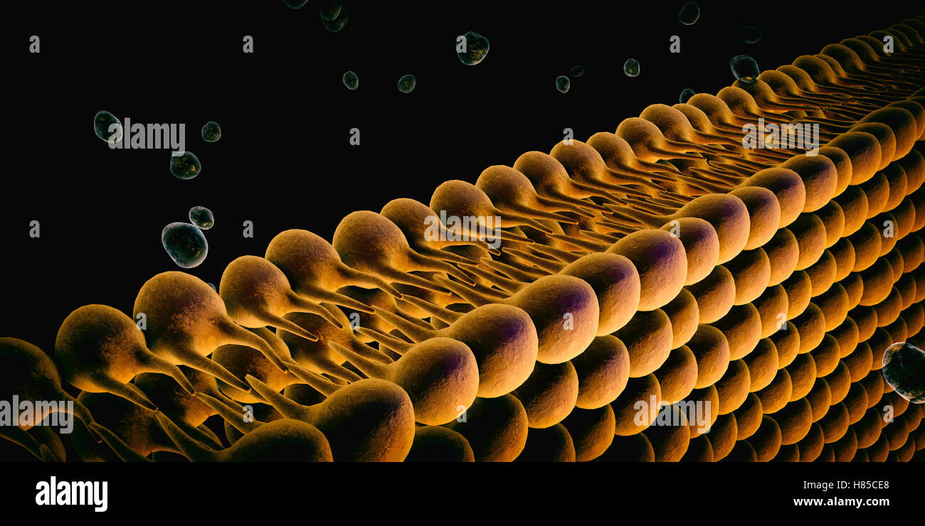 Plasma Membrane Of Cell With other molecules, 3d render Stock Photohttps://www.alamy.com/image-license-details/?v=1https://www.alamy.com/stock-photo-plasma-membrane-of-cell-with-other-molecules-3d-render-125509392.html
RFH85CE8–Plasma Membrane Of Cell With other molecules, 3d render
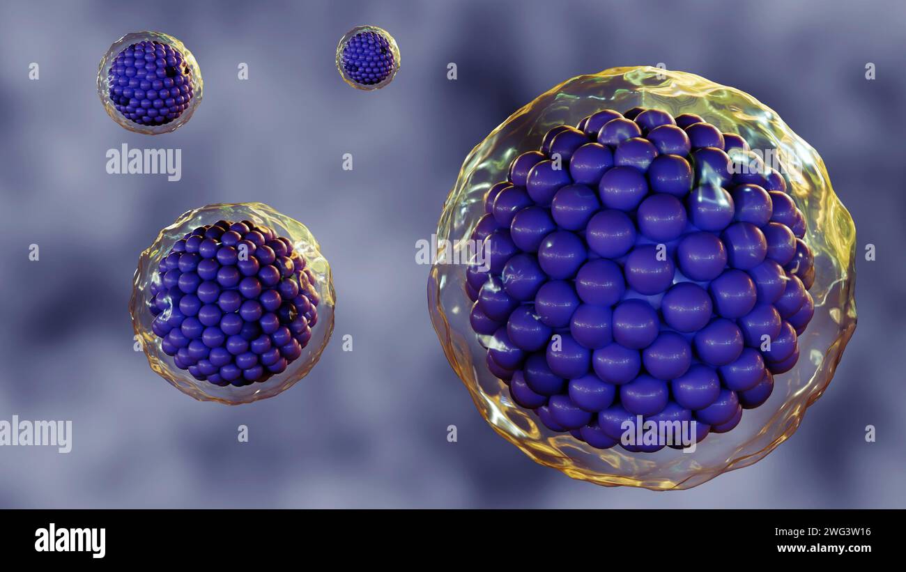 3d rendering of cells are surrounded by a plasma membrane, which is a thin, flexible barrier that separates the cell from its environment. Stock Photohttps://www.alamy.com/image-license-details/?v=1https://www.alamy.com/3d-rendering-of-cells-are-surrounded-by-a-plasma-membrane-which-is-a-thin-flexible-barrier-that-separates-the-cell-from-its-environment-image595072498.html
RF2WG3W16–3d rendering of cells are surrounded by a plasma membrane, which is a thin, flexible barrier that separates the cell from its environment.
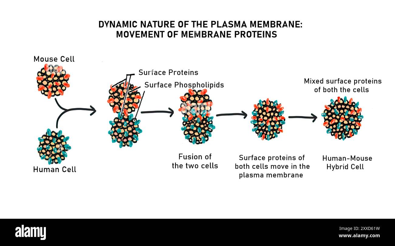 Lateral movement of protein in the plasma membrane, illustration. The phospholipids and proteins present in the plasma membrane are free to move around laterally. Stock Photohttps://www.alamy.com/image-license-details/?v=1https://www.alamy.com/lateral-movement-of-protein-in-the-plasma-membrane-illustration-the-phospholipids-and-proteins-present-in-the-plasma-membrane-are-free-to-move-around-laterally-image618634069.html
RF2XXD61W–Lateral movement of protein in the plasma membrane, illustration. The phospholipids and proteins present in the plasma membrane are free to move around laterally.
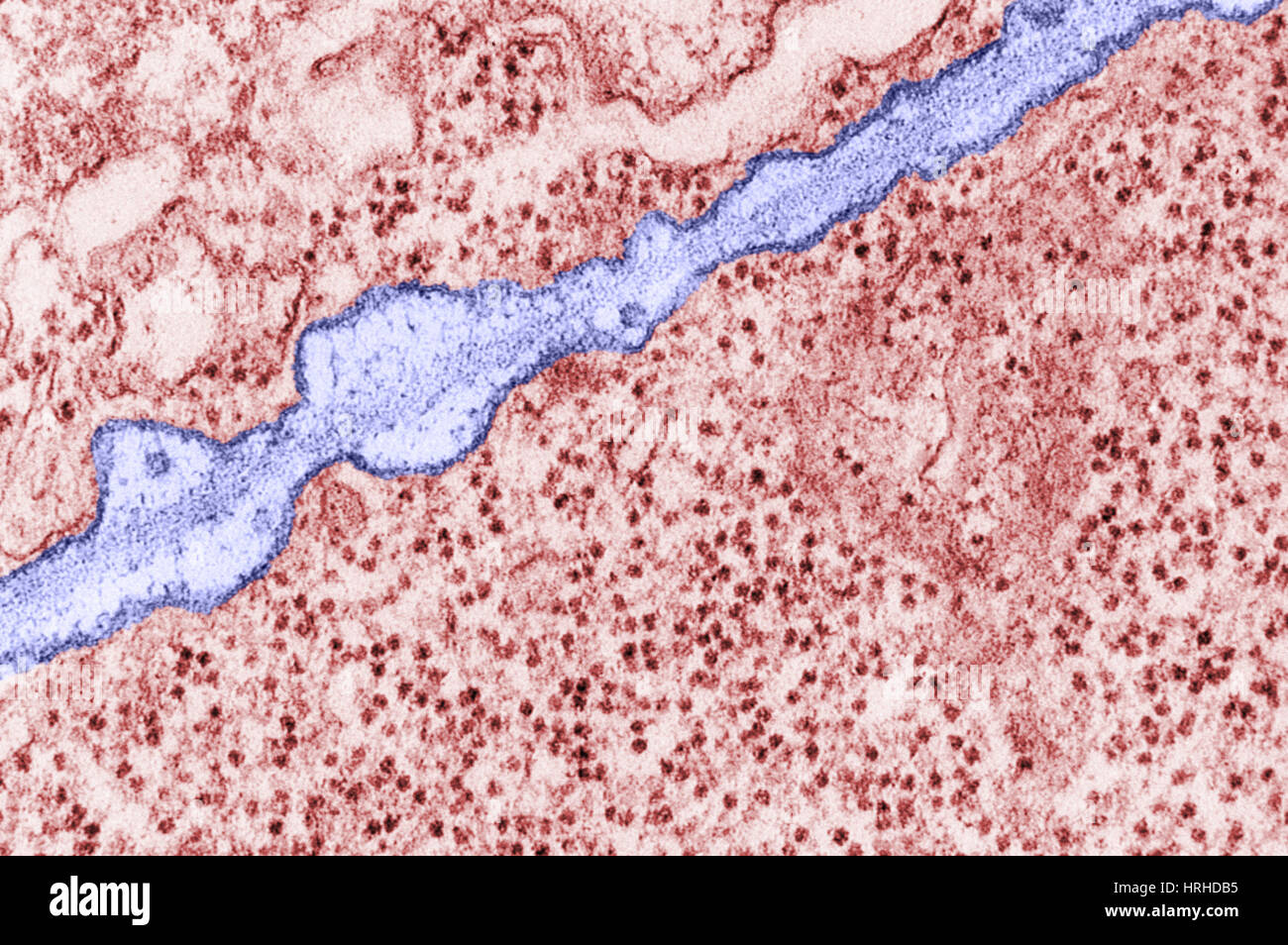 Plasma Membrane TEM Stock Photohttps://www.alamy.com/image-license-details/?v=1https://www.alamy.com/stock-photo-plasma-membrane-tem-134993353.html
RMHRHDB5–Plasma Membrane TEM
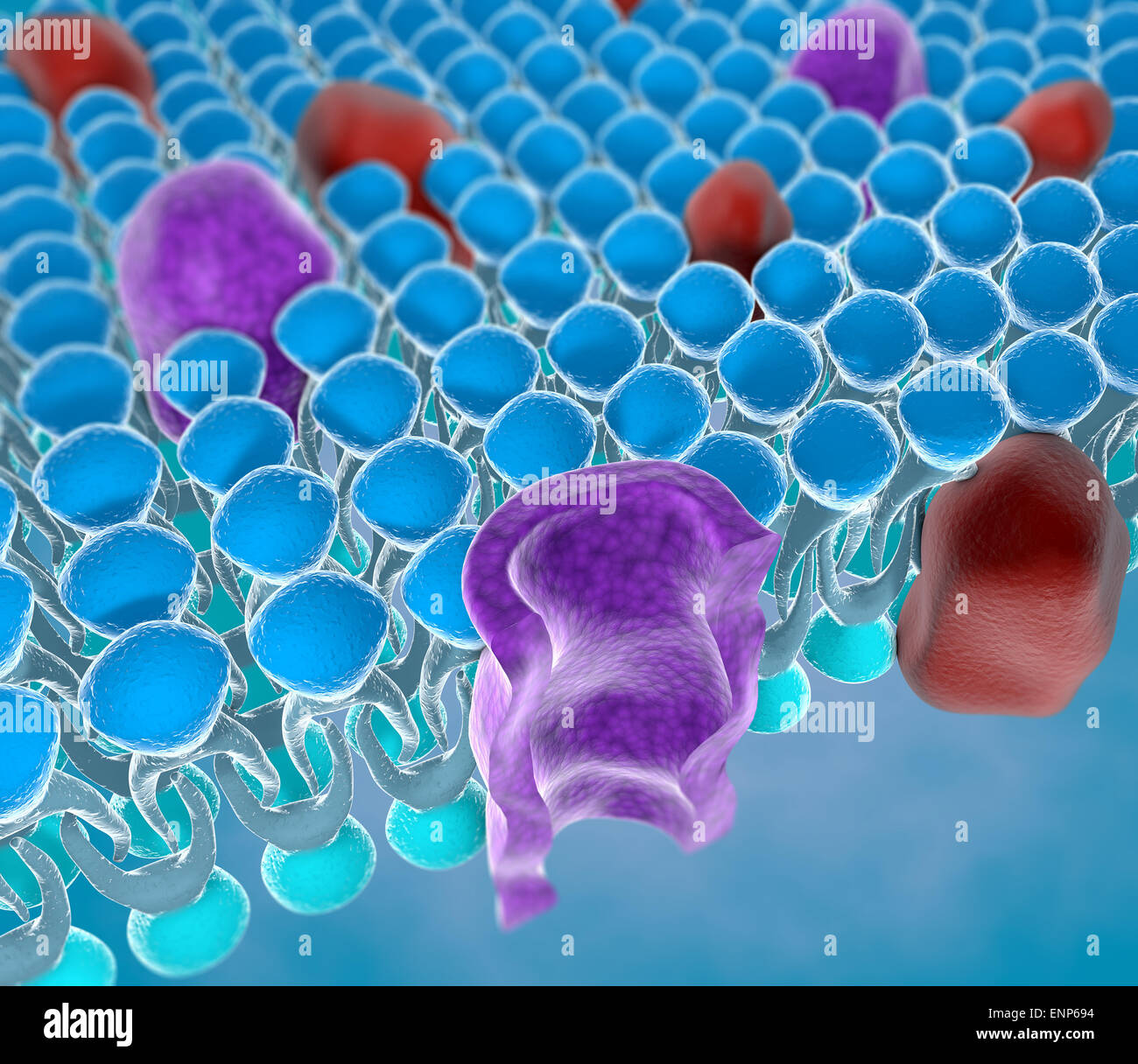 Structure of the plasma membrane of a cell Stock Photohttps://www.alamy.com/image-license-details/?v=1https://www.alamy.com/stock-photo-structure-of-the-plasma-membrane-of-a-cell-82237152.html
RMENP694–Structure of the plasma membrane of a cell
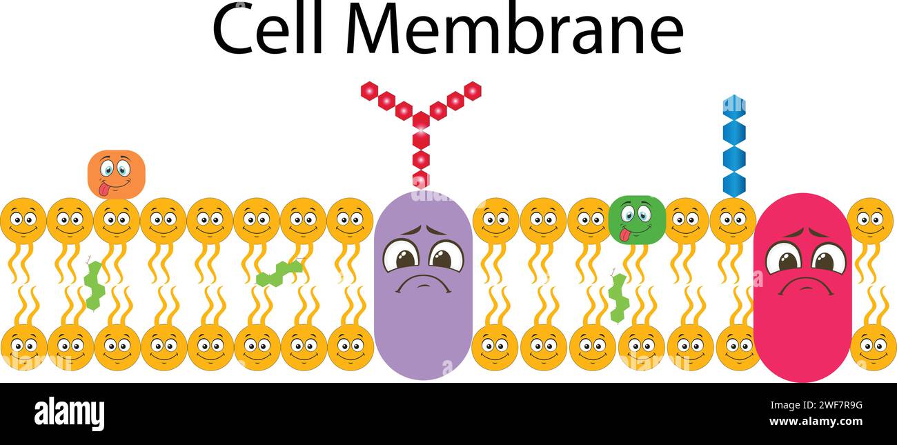 Plasma Membrane Or Cell Membrane Or Plasmalemma Stock Vectorhttps://www.alamy.com/image-license-details/?v=1https://www.alamy.com/plasma-membrane-or-cell-membrane-or-plasmalemma-image594544316.html
RF2WF7R9G–Plasma Membrane Or Cell Membrane Or Plasmalemma
 Mitosis stages Stock Photohttps://www.alamy.com/image-license-details/?v=1https://www.alamy.com/mitosis-stages-image476853409.html
RF2JKPFAW–Mitosis stages
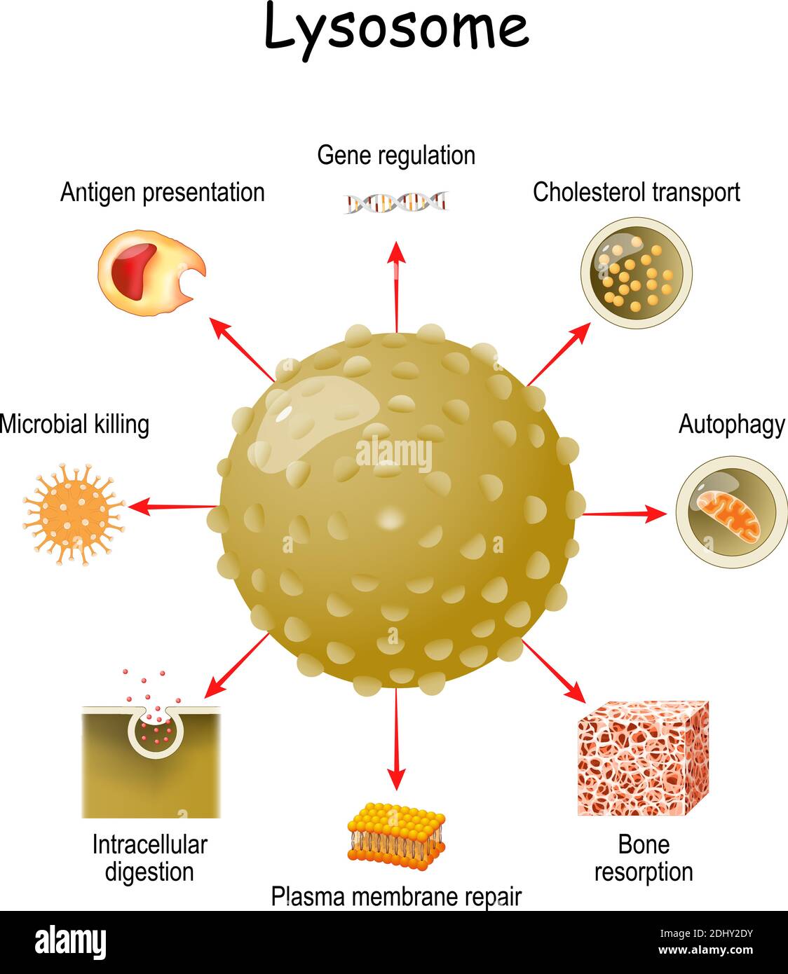 lysosome Function. multitask lysosome from intracellular digestion, and autophagy to antigen presentation, bone matrix resorption, and plasma membrane Stock Vectorhttps://www.alamy.com/image-license-details/?v=1https://www.alamy.com/lysosome-function-multitask-lysosome-from-intracellular-digestion-and-autophagy-to-antigen-presentation-bone-matrix-resorption-and-plasma-membrane-image389671911.html
RF2DHY2DY–lysosome Function. multitask lysosome from intracellular digestion, and autophagy to antigen presentation, bone matrix resorption, and plasma membrane
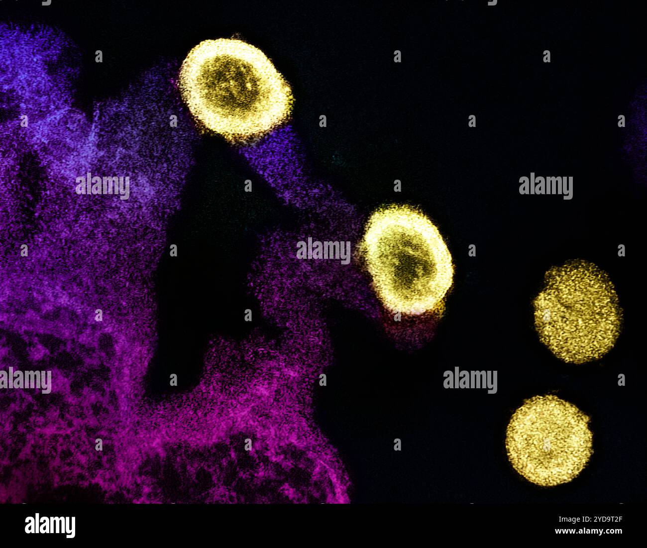 Transmission electron micrograph of HIV-1 virus particles yellow replicating from the plasma membrane of an H9 T cell purple and pink. 2024 : Semaglutide Reduces Severity of Common Liver Disease in People with HIV. HIV Virus Particles 016867 275 Stock Photohttps://www.alamy.com/image-license-details/?v=1https://www.alamy.com/transmission-electron-micrograph-of-hiv-1-virus-particles-yellow-replicating-from-the-plasma-membrane-of-an-h9-t-cell-purple-and-pink-2024-semaglutide-reduces-severity-of-common-liver-disease-in-people-with-hiv-hiv-virus-particles-016867-275-image627780231.html
RM2YD9T2F–Transmission electron micrograph of HIV-1 virus particles yellow replicating from the plasma membrane of an H9 T cell purple and pink. 2024 : Semaglutide Reduces Severity of Common Liver Disease in People with HIV. HIV Virus Particles 016867 275
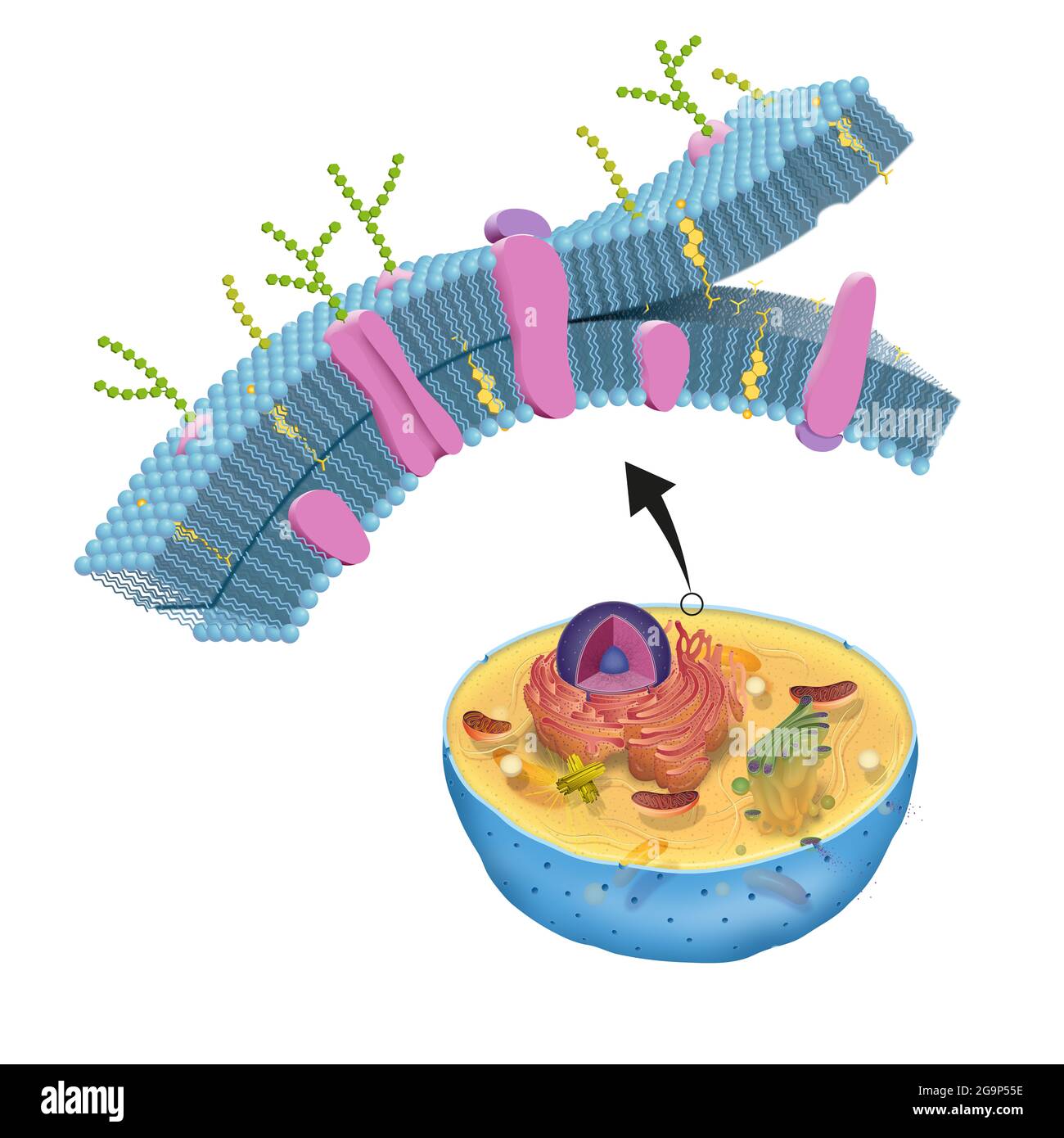 The cell membrane, also called the plasma membrane, is found in all cells and separates the interior of the cell from the outside environment Stock Photohttps://www.alamy.com/image-license-details/?v=1https://www.alamy.com/the-cell-membrane-also-called-the-plasma-membrane-is-found-in-all-cells-and-separates-the-interior-of-the-cell-from-the-outside-environment-image436278122.html
RF2G9P55E–The cell membrane, also called the plasma membrane, is found in all cells and separates the interior of the cell from the outside environment
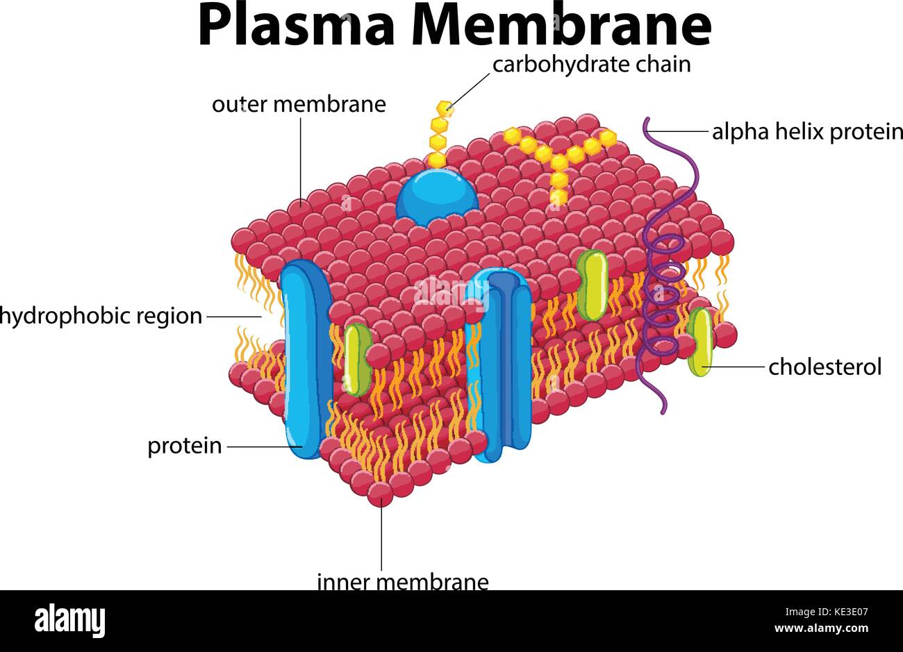 Diagram with plasma membrane illustration Stock Vectorhttps://www.alamy.com/image-license-details/?v=1https://www.alamy.com/stock-image-diagram-with-plasma-membrane-illustration-163575335.html
RFKE3E07–Diagram with plasma membrane illustration
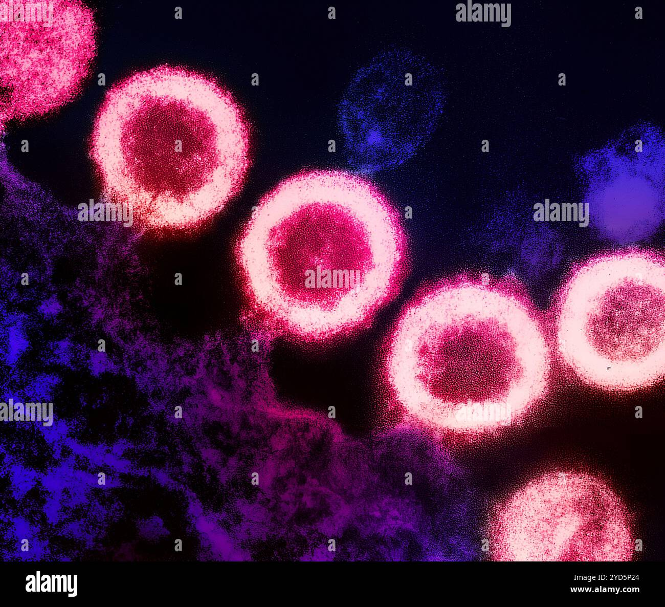 Transmission electron micrograph of HIV-1 virus particles (pink) replicating from the plasma membrane of an infected H9 T cell (purple). Stock Photohttps://www.alamy.com/image-license-details/?v=1https://www.alamy.com/transmission-electron-micrograph-of-hiv-1-virus-particles-pink-replicating-from-the-plasma-membrane-of-an-infected-h9-t-cell-purple-image627690844.html
RM2YD5P24–Transmission electron micrograph of HIV-1 virus particles (pink) replicating from the plasma membrane of an infected H9 T cell (purple).
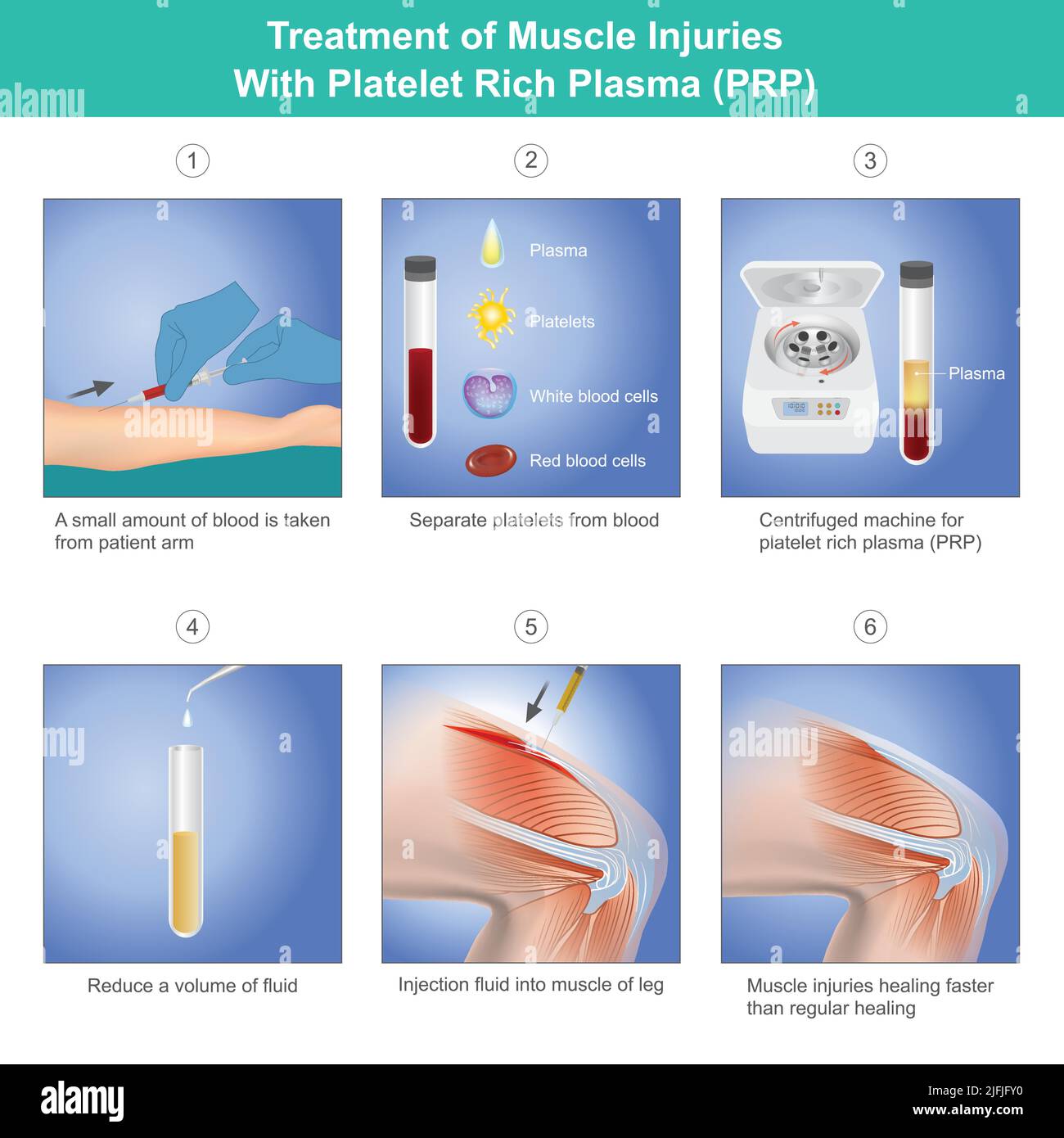 Treatment of Muscle Injuries With Platelet Rich Plasma. Diagram treatment of muscle injuries a knee from blood platelet a patient. Stock Vectorhttps://www.alamy.com/image-license-details/?v=1https://www.alamy.com/treatment-of-muscle-injuries-with-platelet-rich-plasma-diagram-treatment-of-muscle-injuries-a-knee-from-blood-platelet-a-patient-image474307428.html
RF2JFJFY0–Treatment of Muscle Injuries With Platelet Rich Plasma. Diagram treatment of muscle injuries a knee from blood platelet a patient.
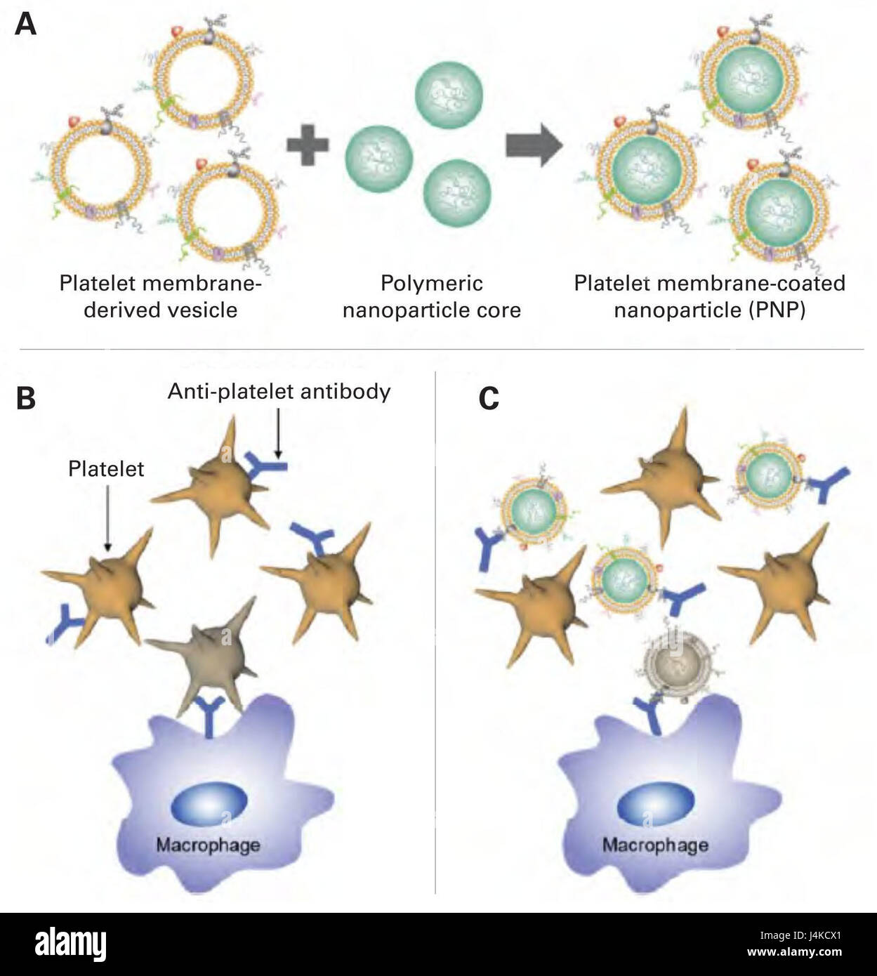 Schematic of platelet membrane-coated nanoparticles (PNPs) for the treatment of immune thrombocytopenia purpura (ITP). (A) To fabricate PNPs, the plasma membrane from fresh platelets is derived and then coated onto poly (lactic-co-glycolic acid (PLGA) polymeric nanoparticle cores, transferring the surface antigenic material from the original cells onto the outside of the nanoparticles. (B) Without treatment, ITP is characterized by the binding of pathological autoantibodies to healthy platelets, resulting in their clearance by the reticuloendothelial system. (C) When PNPs are administered, th Stock Photohttps://www.alamy.com/image-license-details/?v=1https://www.alamy.com/stock-photo-schematic-of-platelet-membrane-coated-nanoparticles-pnps-for-the-treatment-140568793.html
RMJ4KCX1–Schematic of platelet membrane-coated nanoparticles (PNPs) for the treatment of immune thrombocytopenia purpura (ITP). (A) To fabricate PNPs, the plasma membrane from fresh platelets is derived and then coated onto poly (lactic-co-glycolic acid (PLGA) polymeric nanoparticle cores, transferring the surface antigenic material from the original cells onto the outside of the nanoparticles. (B) Without treatment, ITP is characterized by the binding of pathological autoantibodies to healthy platelets, resulting in their clearance by the reticuloendothelial system. (C) When PNPs are administered, th
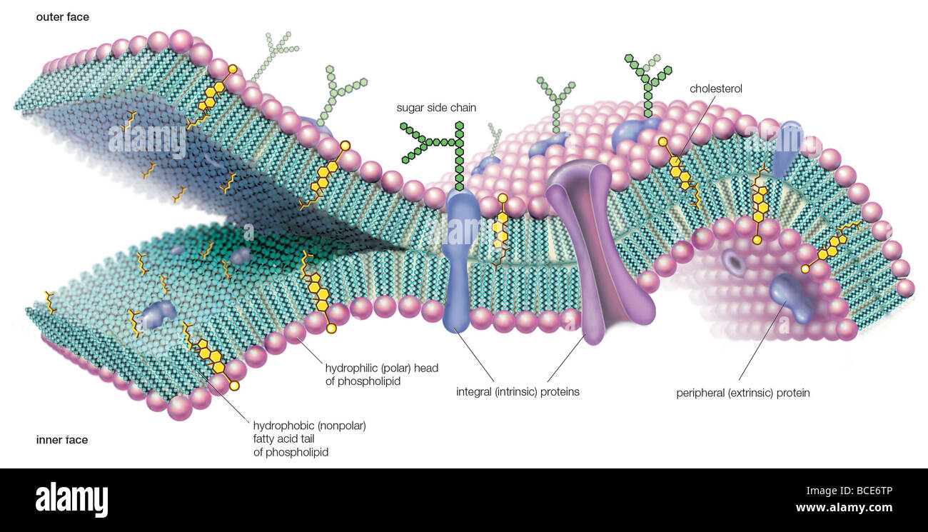 A molecular view of the cell membrane highlighting phospholipids, cholesterol, and intrinsic and extrinsic proteins. Stock Photohttps://www.alamy.com/image-license-details/?v=1https://www.alamy.com/stock-photo-a-molecular-view-of-the-cell-membrane-highlighting-phospholipids-cholesterol-24898966.html
RMBCE6TP–A molecular view of the cell membrane highlighting phospholipids, cholesterol, and intrinsic and extrinsic proteins.
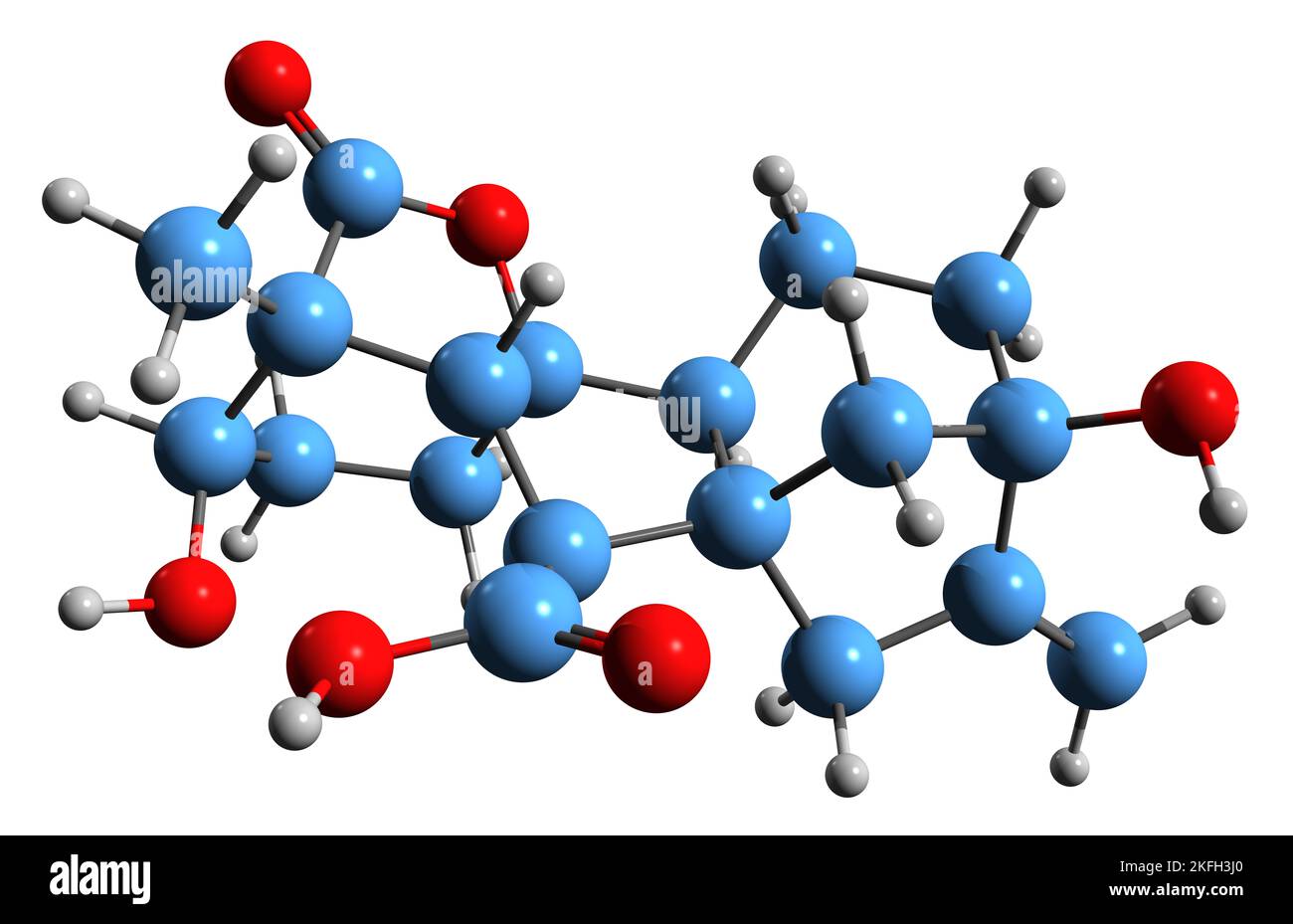 3D image of Gibberellin A1 skeletal formula - molecular chemical structure of plant hormone isolated on white background Stock Photohttps://www.alamy.com/image-license-details/?v=1https://www.alamy.com/3d-image-of-gibberellin-a1-skeletal-formula-molecular-chemical-structure-of-plant-hormone-isolated-on-white-background-image491486184.html
RF2KFH3J0–3D image of Gibberellin A1 skeletal formula - molecular chemical structure of plant hormone isolated on white background
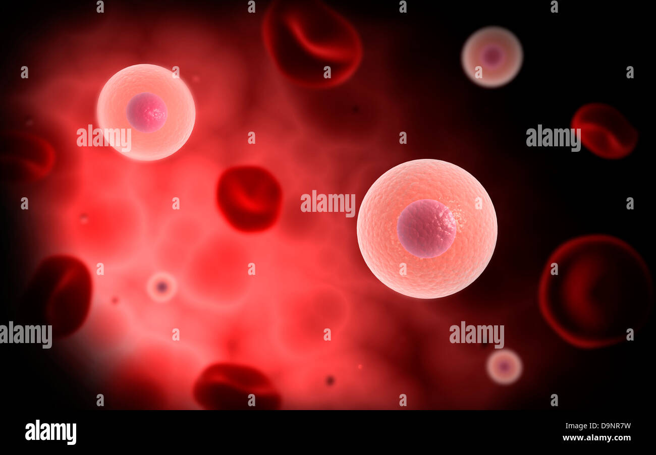 Microscopic view of plasma cell inside blood vessel. Stock Photohttps://www.alamy.com/image-license-details/?v=1https://www.alamy.com/stock-photo-microscopic-view-of-plasma-cell-inside-blood-vessel-57642253.html
RFD9NR7W–Microscopic view of plasma cell inside blood vessel.
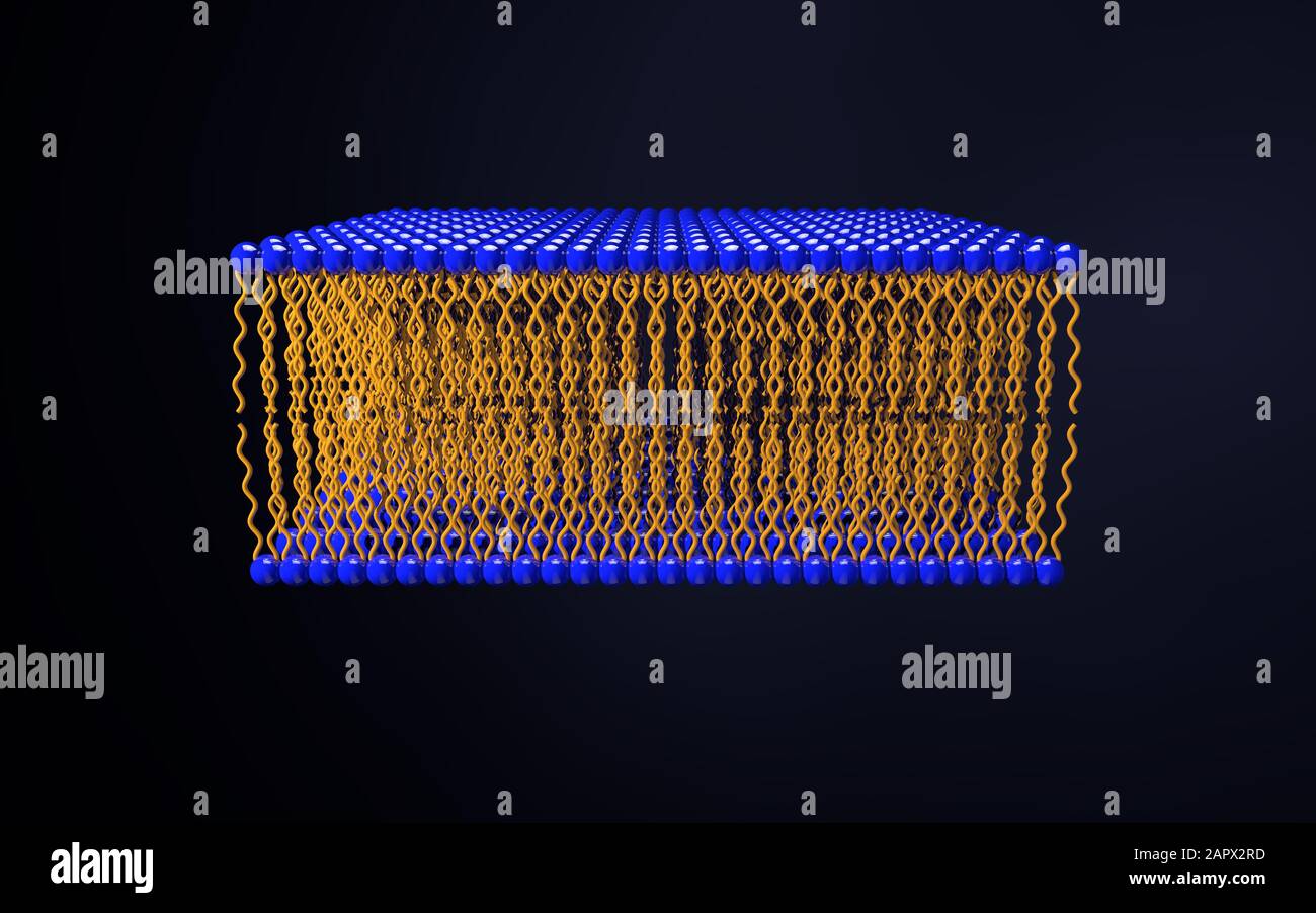 Omega-3 Phospholipid skin cell membrane lipid layer structure. 3D rendering medical illustration Stock Photohttps://www.alamy.com/image-license-details/?v=1https://www.alamy.com/omega-3-phospholipid-skin-cell-membrane-lipid-layer-structure-3d-rendering-medical-illustration-image341092401.html
RF2APX2RD–Omega-3 Phospholipid skin cell membrane lipid layer structure. 3D rendering medical illustration
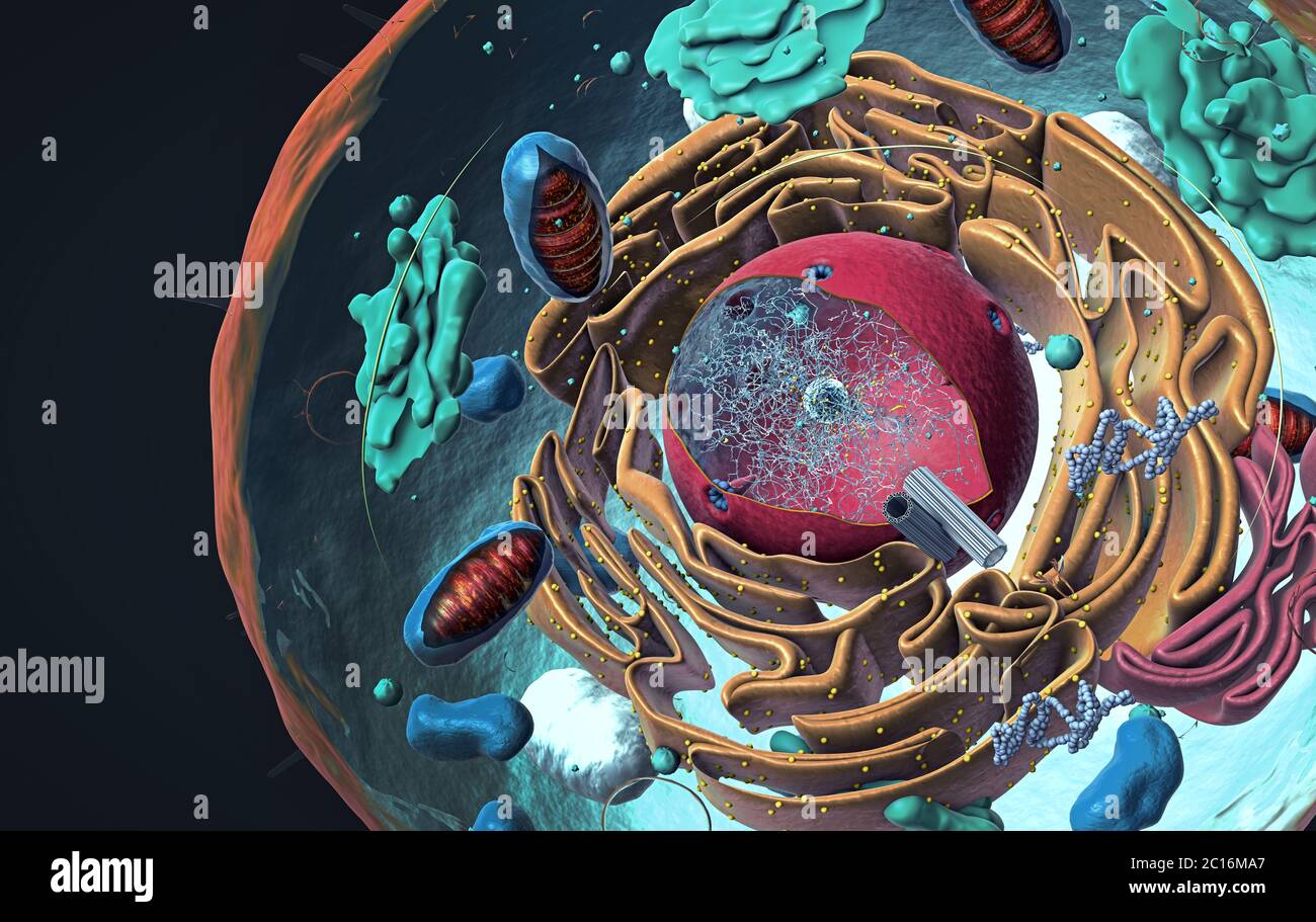 Components of Eukaryotic cell, nucleus and organelles and plasma membrane - 3d illustration Stock Photohttps://www.alamy.com/image-license-details/?v=1https://www.alamy.com/components-of-eukaryotic-cell-nucleus-and-organelles-and-plasma-membrane-3d-illustration-image362180063.html
RF2C16MA7–Components of Eukaryotic cell, nucleus and organelles and plasma membrane - 3d illustration
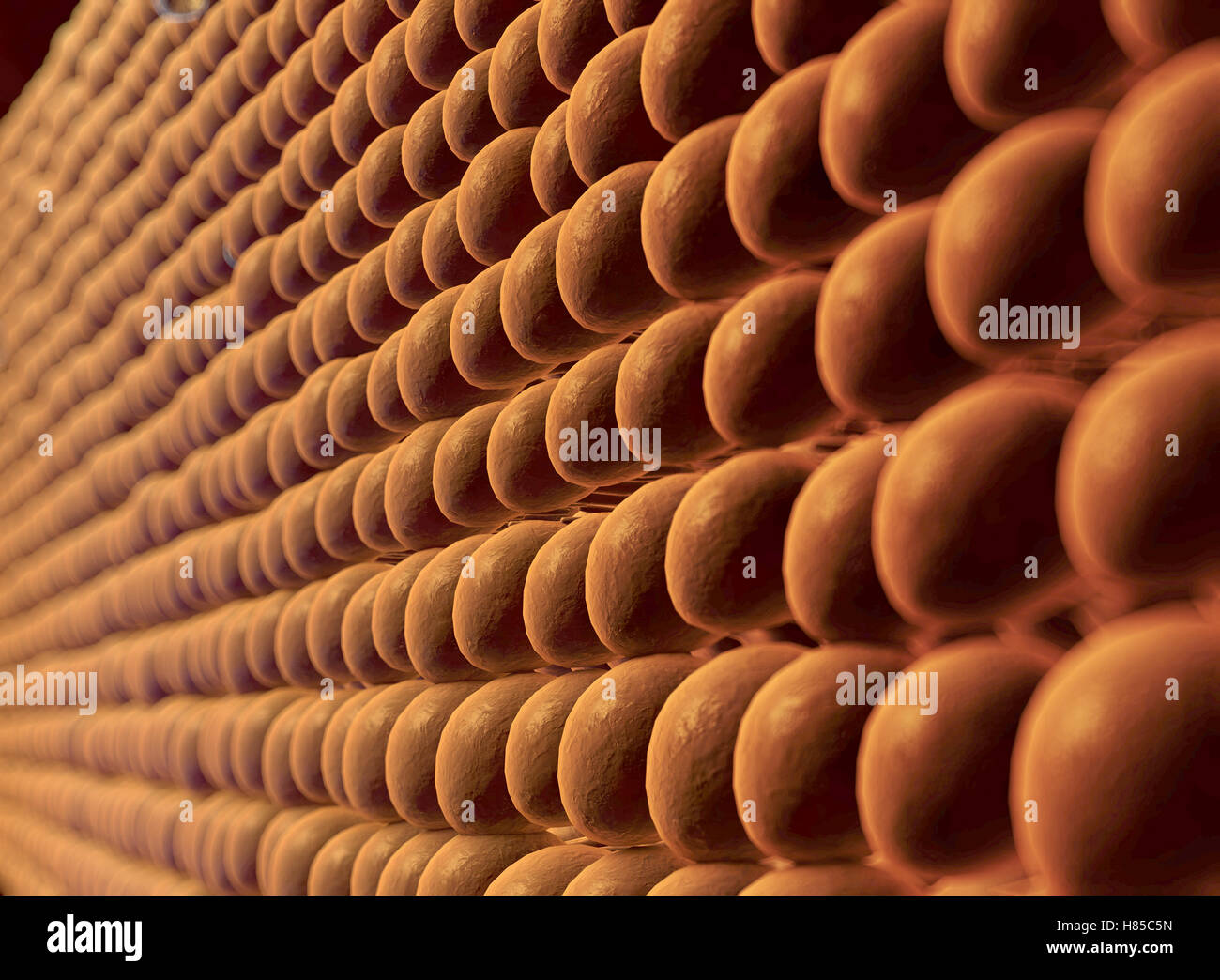 Plasma Membrane Of Cell With other molecules, 3d render Stock Photohttps://www.alamy.com/image-license-details/?v=1https://www.alamy.com/stock-photo-plasma-membrane-of-cell-with-other-molecules-3d-render-125509153.html
RFH85C5N–Plasma Membrane Of Cell With other molecules, 3d render
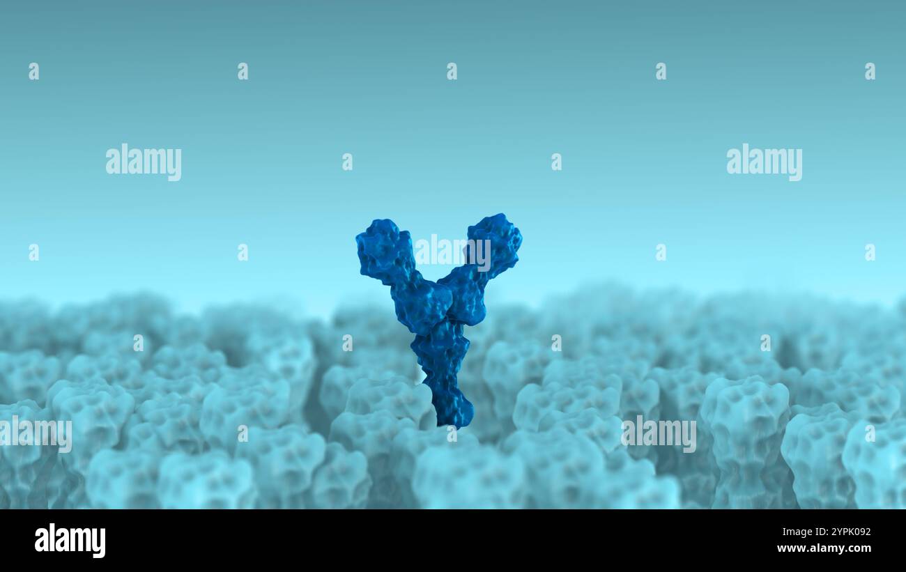 Receptor Binding by Antibodies Regulating Cellular Activity Stock Photohttps://www.alamy.com/image-license-details/?v=1https://www.alamy.com/receptor-binding-by-antibodies-regulating-cellular-activity-image633513022.html
RF2YPK092–Receptor Binding by Antibodies Regulating Cellular Activity
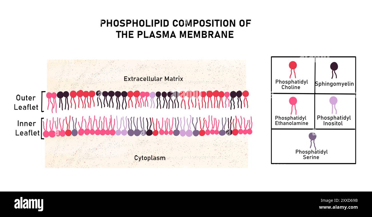 Composition of plasma membrane, illustration. The plasma membrane is made of a bilayer of phospholipids. The amount of specific phospholipids present in the inner and outer sides of the plasma membrane is different. Stock Photohttps://www.alamy.com/image-license-details/?v=1https://www.alamy.com/composition-of-plasma-membrane-illustration-the-plasma-membrane-is-made-of-a-bilayer-of-phospholipids-the-amount-of-specific-phospholipids-present-in-the-inner-and-outer-sides-of-the-plasma-membrane-is-different-image618634279.html
RF2XXD69B–Composition of plasma membrane, illustration. The plasma membrane is made of a bilayer of phospholipids. The amount of specific phospholipids present in the inner and outer sides of the plasma membrane is different.
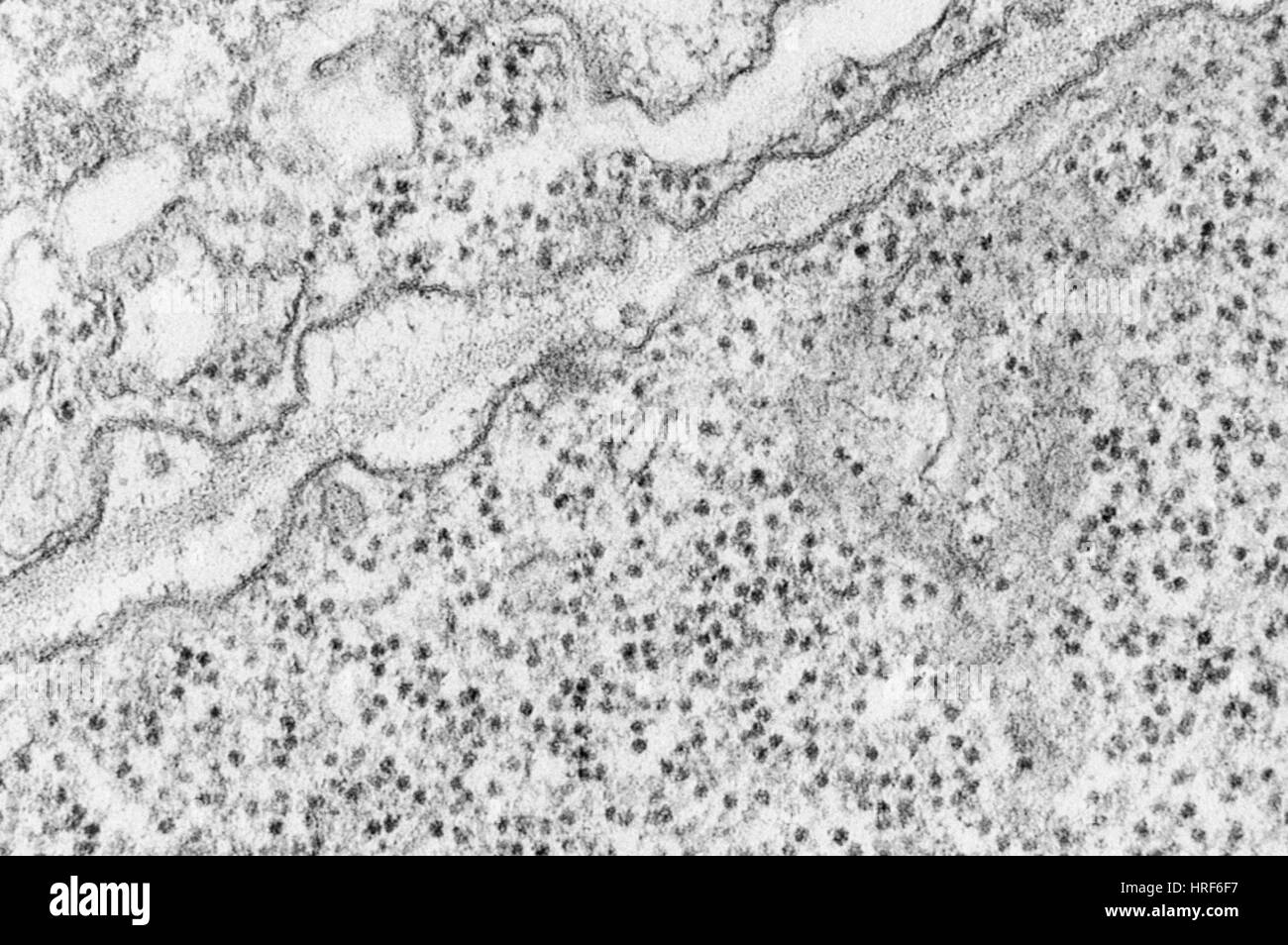 Plasma Membrane TEM Stock Photohttps://www.alamy.com/image-license-details/?v=1https://www.alamy.com/stock-photo-plasma-membrane-tem-134944075.html
RMHRF6F7–Plasma Membrane TEM
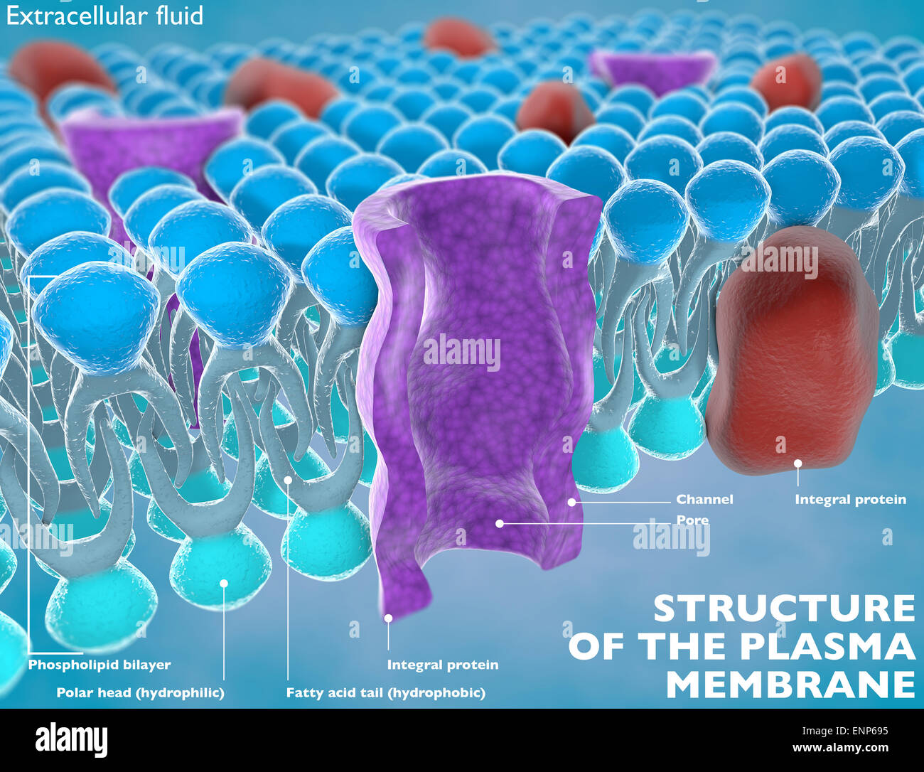 Structure of the plasma membrane of a cell Stock Photohttps://www.alamy.com/image-license-details/?v=1https://www.alamy.com/stock-photo-structure-of-the-plasma-membrane-of-a-cell-82237153.html
RMENP695–Structure of the plasma membrane of a cell
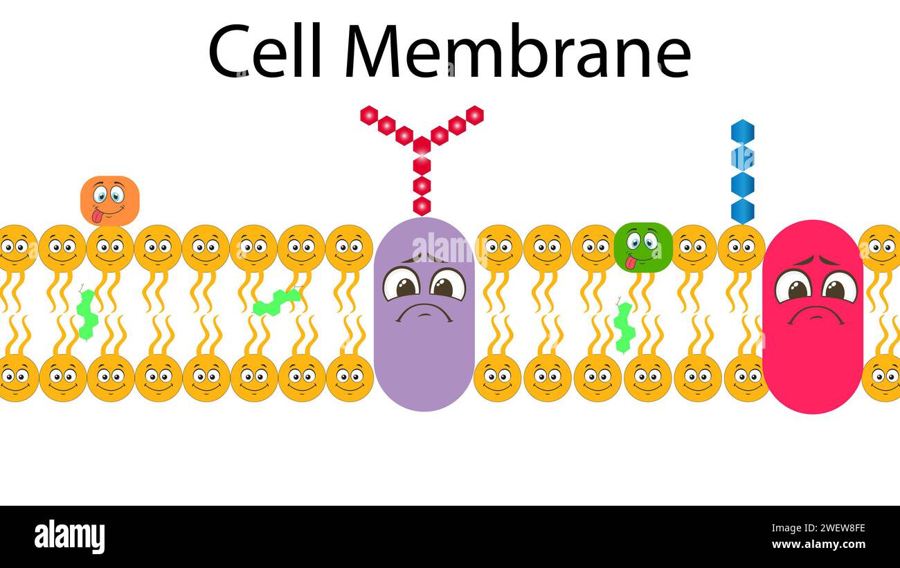 Plasma Membrane Or Cell Membrane Or Plasmalemma Stock Photohttps://www.alamy.com/image-license-details/?v=1https://www.alamy.com/plasma-membrane-or-cell-membrane-or-plasmalemma-image594313202.html
RF2WEW8FE–Plasma Membrane Or Cell Membrane Or Plasmalemma
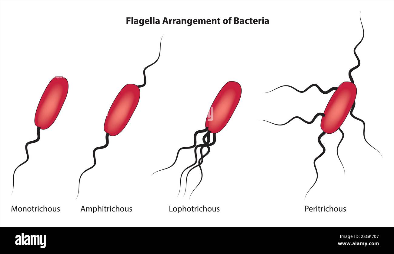 Flagella arrangement of bacteria Stock Vectorhttps://www.alamy.com/image-license-details/?v=1https://www.alamy.com/flagella-arrangement-of-bacteria-image647040695.html
RF2SGK707–Flagella arrangement of bacteria
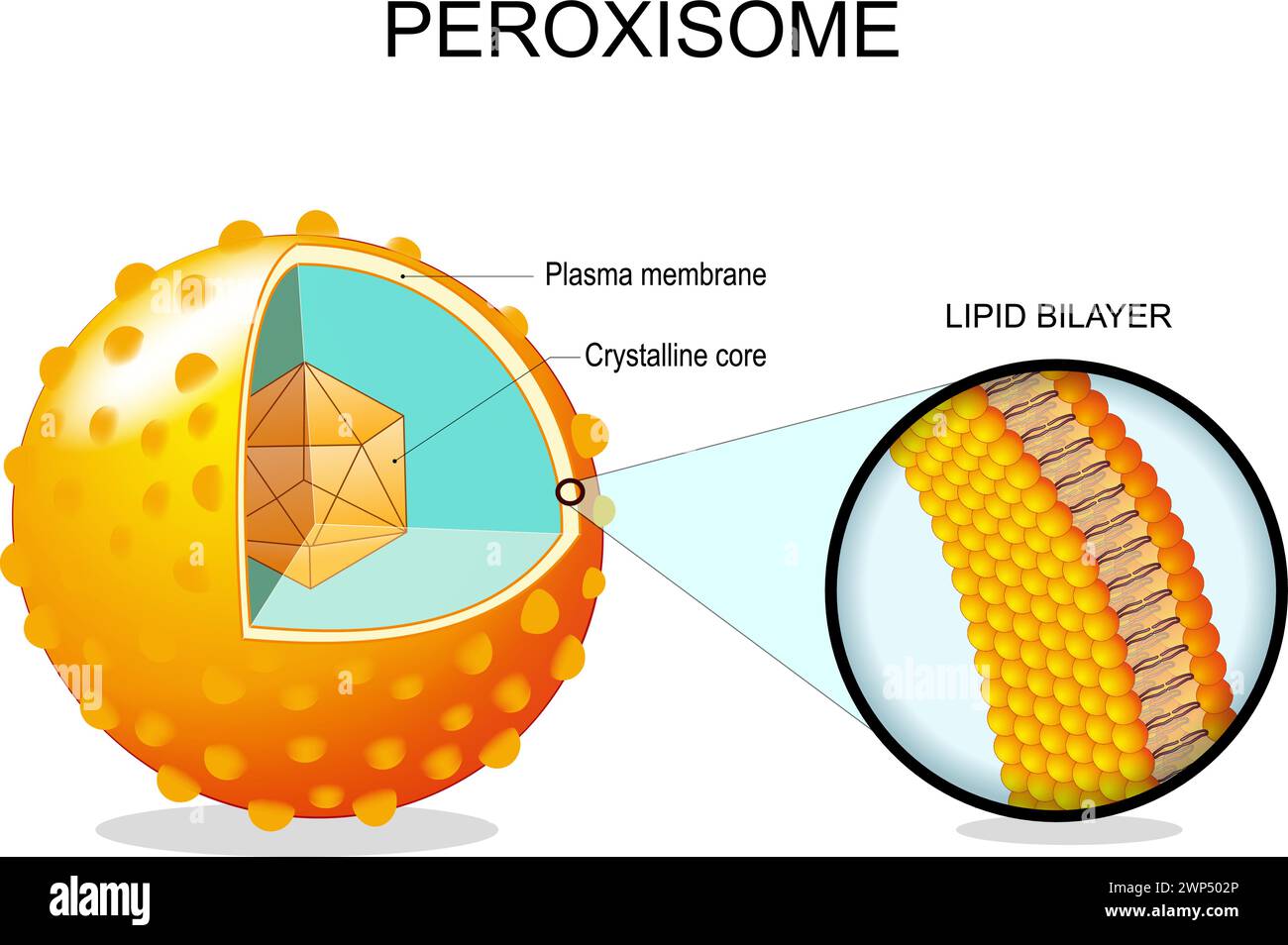 Peroxisome anatomy. Cross section of a cell organelle. Close-up of a Lipid bilayer Plasma membrane, Crystalline core, transport proteins. Vector illus Stock Vectorhttps://www.alamy.com/image-license-details/?v=1https://www.alamy.com/peroxisome-anatomy-cross-section-of-a-cell-organelle-close-up-of-a-lipid-bilayer-plasma-membrane-crystalline-core-transport-proteins-vector-illus-image598784782.html
RF2WP502P–Peroxisome anatomy. Cross section of a cell organelle. Close-up of a Lipid bilayer Plasma membrane, Crystalline core, transport proteins. Vector illus
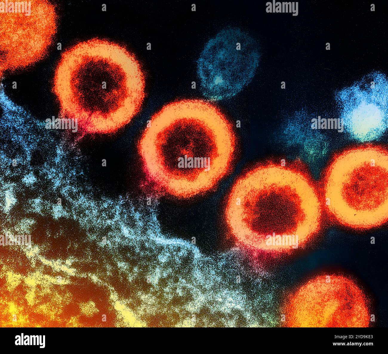 Transmission electron micrograph of HIV-1 virus particles orange replicating from the plasma membrane of an infected H9 T cell. HIV-1 Virus 016867 216 Stock Photohttps://www.alamy.com/image-license-details/?v=1https://www.alamy.com/transmission-electron-micrograph-of-hiv-1-virus-particles-orange-replicating-from-the-plasma-membrane-of-an-infected-h9-t-cell-hiv-1-virus-016867-216-image627776635.html
RM2YD9KE3–Transmission electron micrograph of HIV-1 virus particles orange replicating from the plasma membrane of an infected H9 T cell. HIV-1 Virus 016867 216
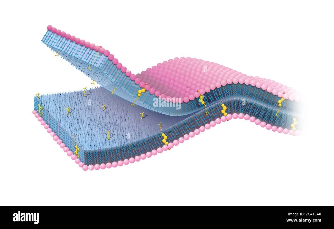 Cell membrane or cytoplasmic membrane with cholesterol Stock Photohttps://www.alamy.com/image-license-details/?v=1https://www.alamy.com/cell-membrane-or-cytoplasmic-membrane-with-cholesterol-image432749472.html
RF2G41CA8–Cell membrane or cytoplasmic membrane with cholesterol
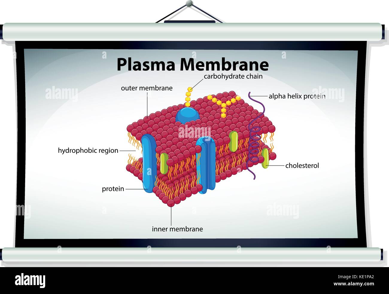 Chart showing plasma membrane illustration Stock Vectorhttps://www.alamy.com/image-license-details/?v=1https://www.alamy.com/stock-image-chart-showing-plasma-membrane-illustration-163537978.html
RFKE1PA2–Chart showing plasma membrane illustration
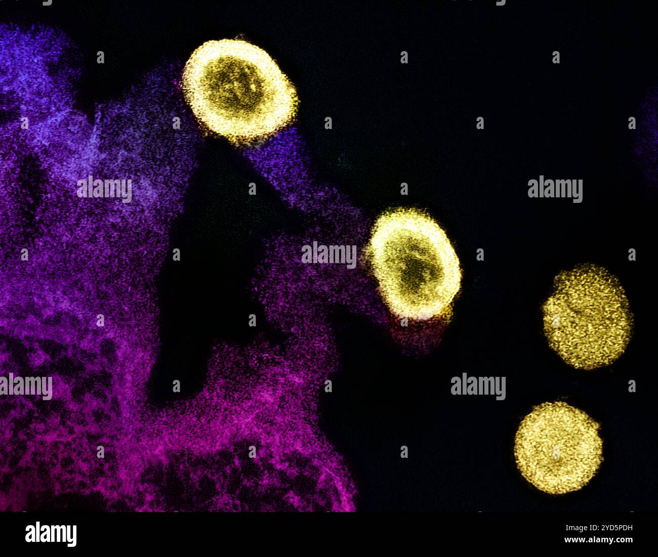 Transmission electron micrograph of HIV-1 virus particles (yellow) replicating from the plasma membrane of an H9 T cell (purple and pink). Stock Photohttps://www.alamy.com/image-license-details/?v=1https://www.alamy.com/transmission-electron-micrograph-of-hiv-1-virus-particles-yellow-replicating-from-the-plasma-membrane-of-an-h9-t-cell-purple-and-pink-image627691165.html
RM2YD5PDH–Transmission electron micrograph of HIV-1 virus particles (yellow) replicating from the plasma membrane of an H9 T cell (purple and pink).
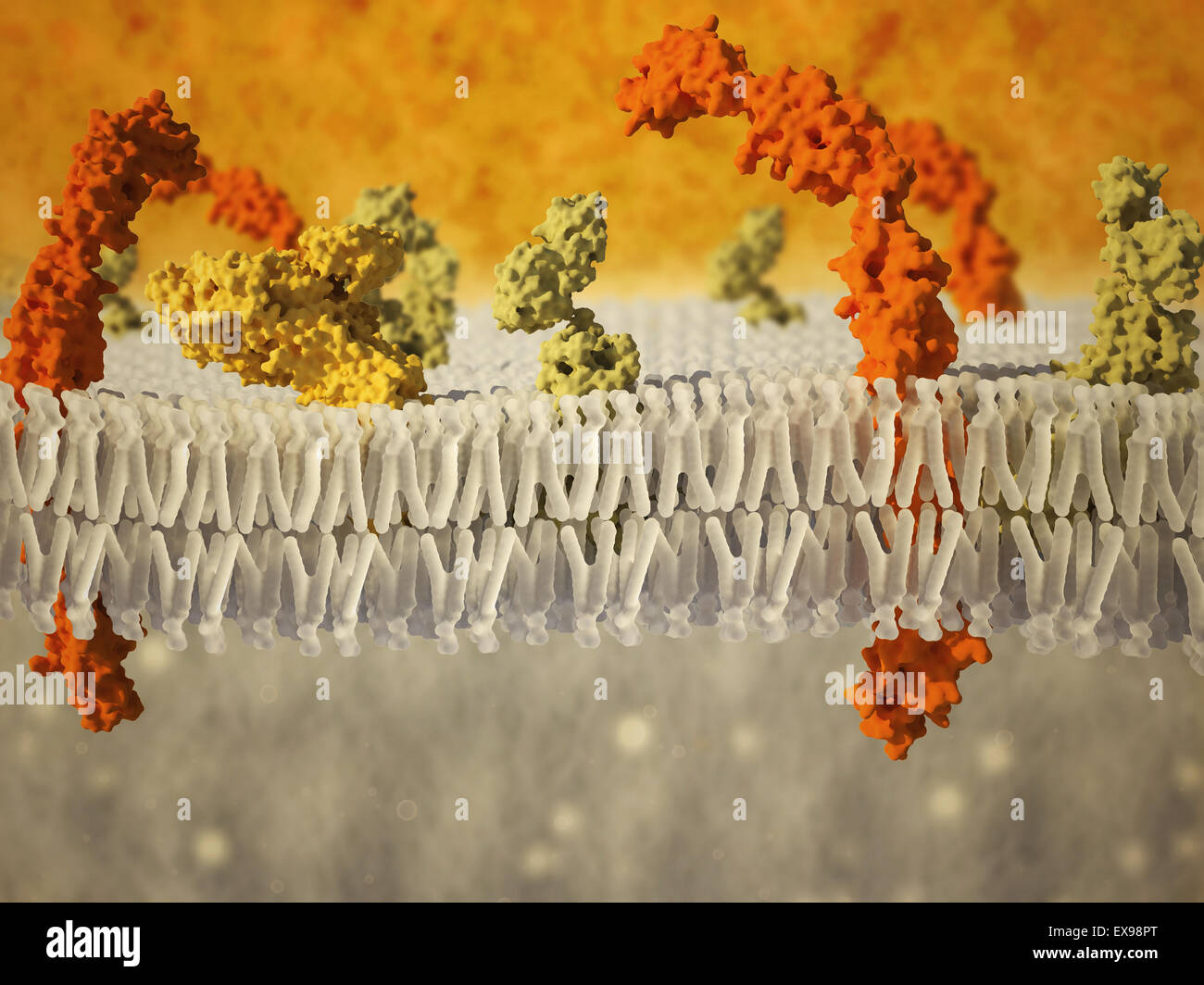 Phospholipid bilayer of the cell membrane. Stock Photohttps://www.alamy.com/image-license-details/?v=1https://www.alamy.com/stock-photo-phospholipid-bilayer-of-the-cell-membrane-85027008.html
RFEX98PT–Phospholipid bilayer of the cell membrane.
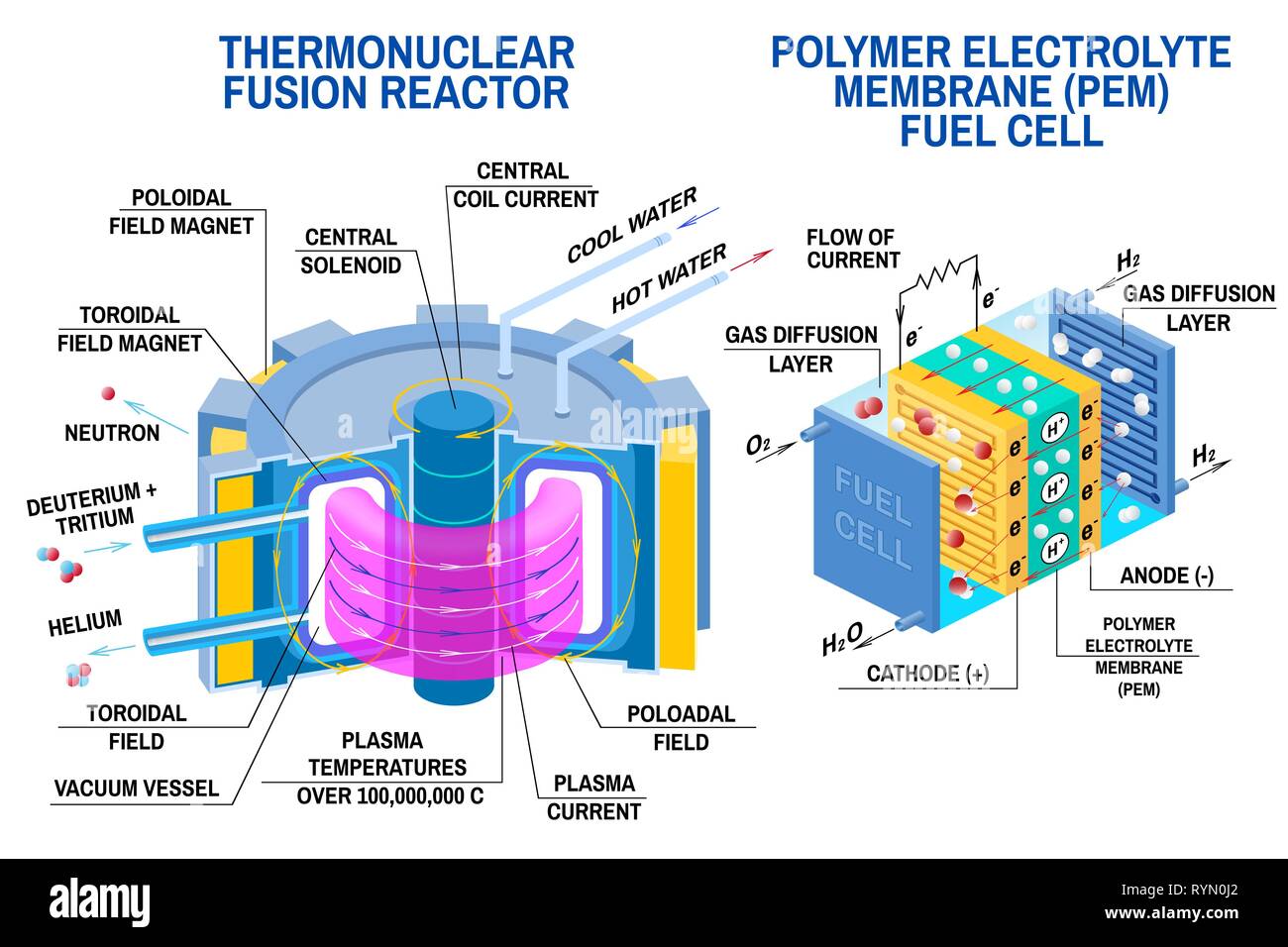 Fuel cell and Thermonuclear fusion reactor diagram. Vector. Devices that receives energy from thermonuclear fusion of hydrogen into helium and converts chemical potential energy into electrical energy Stock Vectorhttps://www.alamy.com/image-license-details/?v=1https://www.alamy.com/fuel-cell-and-thermonuclear-fusion-reactor-diagram-vector-devices-that-receives-energy-from-thermonuclear-fusion-of-hydrogen-into-helium-and-converts-chemical-potential-energy-into-electrical-energy-image240791994.html
RFRYN0J2–Fuel cell and Thermonuclear fusion reactor diagram. Vector. Devices that receives energy from thermonuclear fusion of hydrogen into helium and converts chemical potential energy into electrical energy
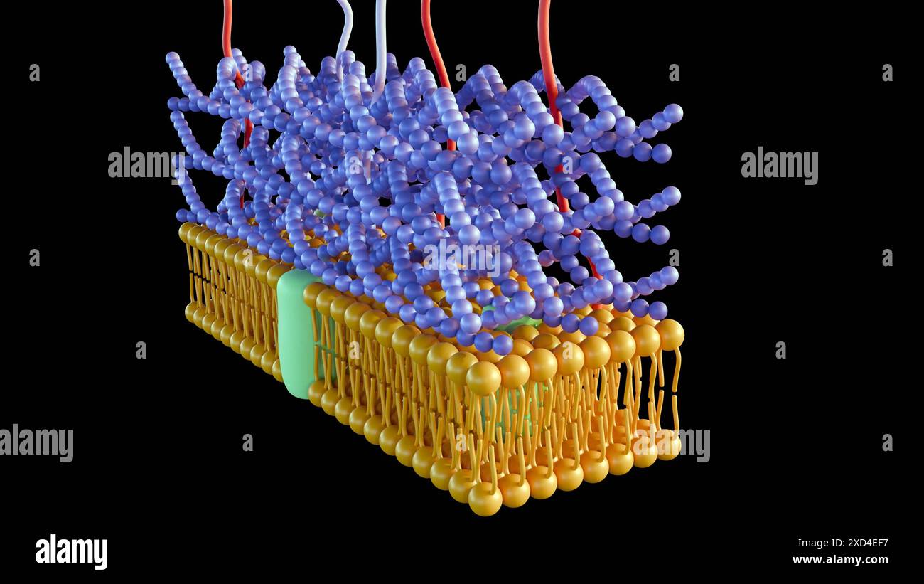 3d rendering of Gram positive bacteria have a thick peptidoglycan layer and no outer lipid membrane Stock Photohttps://www.alamy.com/image-license-details/?v=1https://www.alamy.com/3d-rendering-of-gram-positive-bacteria-have-a-thick-peptidoglycan-layer-and-no-outer-lipid-membrane-image610452619.html
RF2XD4EF7–3d rendering of Gram positive bacteria have a thick peptidoglycan layer and no outer lipid membrane
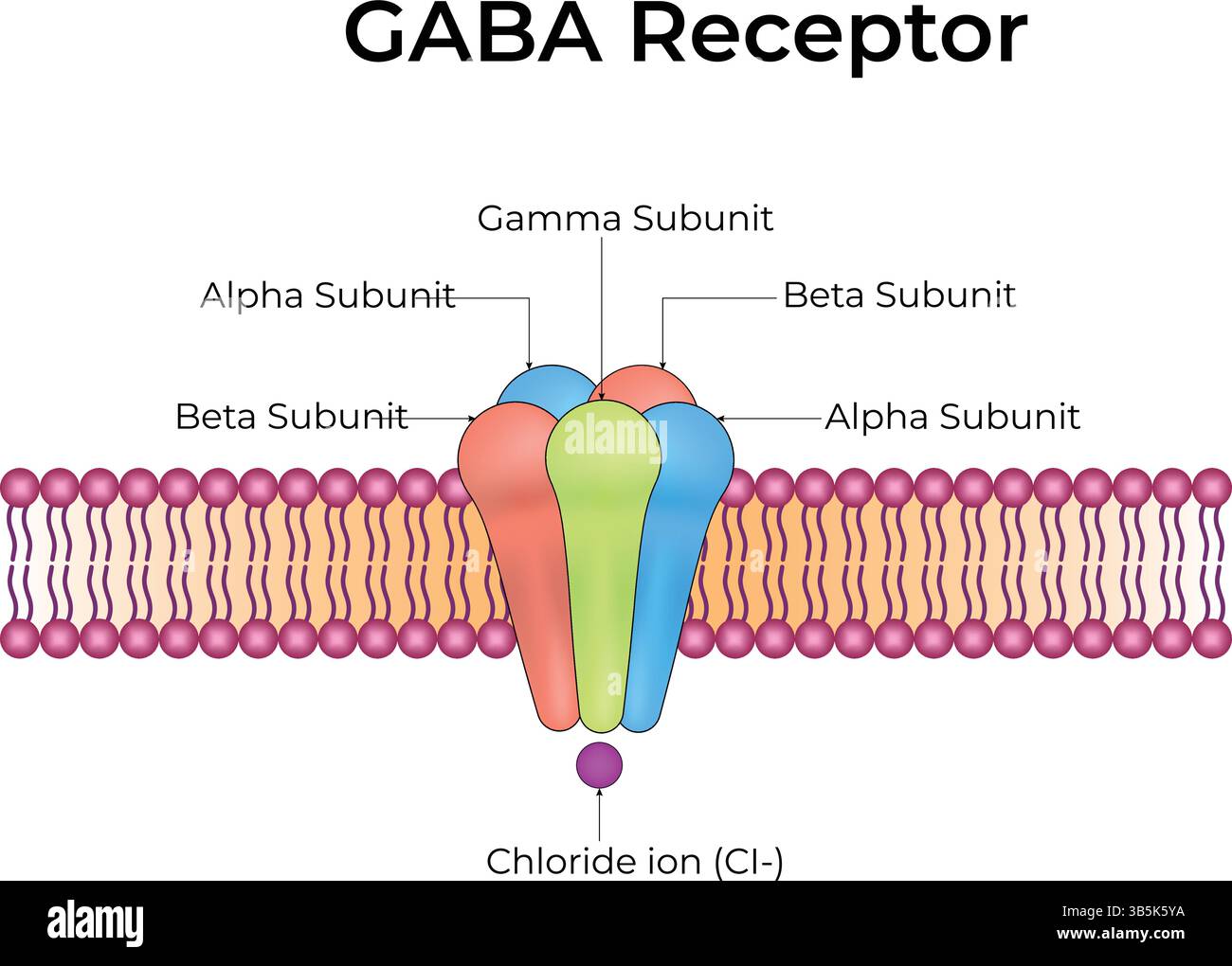 Ultrastructure of GABA receptors in the cell membrane: Neurotransmitter gamma-aminobutyric acid, GABA A and GABA B. Infographic on pharmacology. Stock Vectorhttps://www.alamy.com/image-license-details/?v=1https://www.alamy.com/ultrastructure-of-gaba-receptors-in-the-cell-membrane-neurotransmitter-gamma-aminobutyric-acid-gaba-a-and-gaba-b-infographic-on-pharmacology-image674699406.html
RF3B5K5YA–Ultrastructure of GABA receptors in the cell membrane: Neurotransmitter gamma-aminobutyric acid, GABA A and GABA B. Infographic on pharmacology.
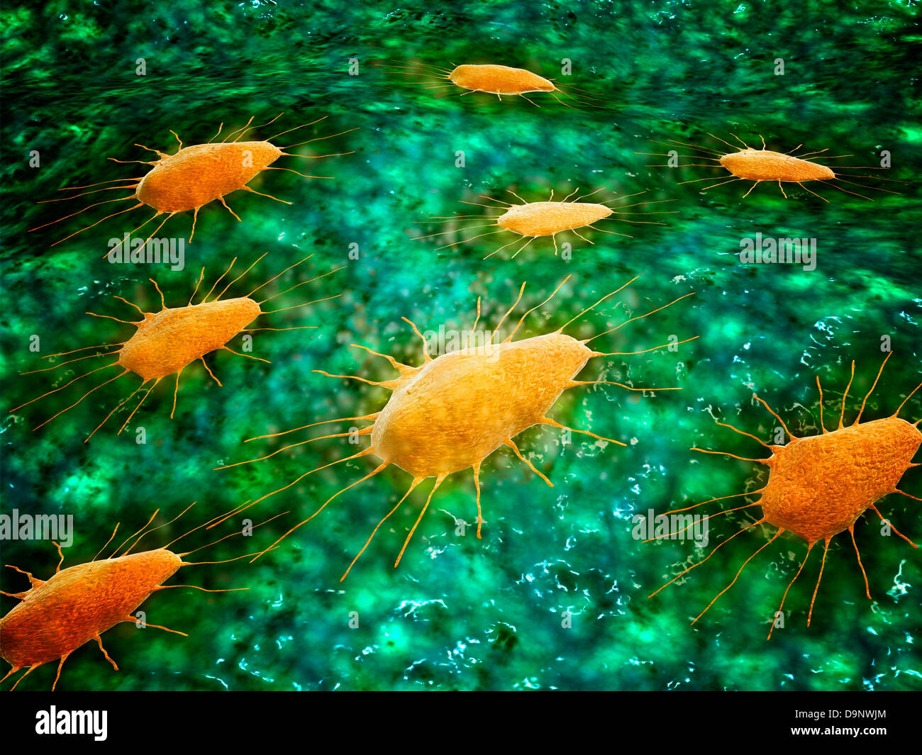 Microscopic view of a group of macrophages. Stock Photohttps://www.alamy.com/image-license-details/?v=1https://www.alamy.com/stock-photo-microscopic-view-of-a-group-of-macrophages-57644124.html
RFD9NWJM–Microscopic view of a group of macrophages.
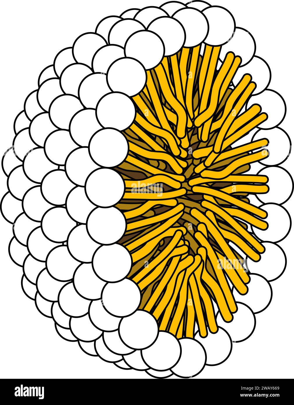 Structure of Phospholipid Molecule in Micelle .Vector illustration. Stock Vectorhttps://www.alamy.com/image-license-details/?v=1https://www.alamy.com/structure-of-phospholipid-molecule-in-micelle-vector-illustration-image591896657.html
RF2WAY669–Structure of Phospholipid Molecule in Micelle .Vector illustration.
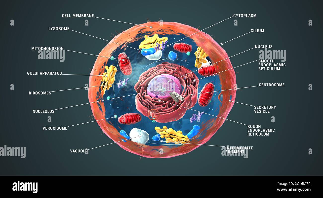 Labeled Eukaryotic cell, nucleus and organelles and plasma membrane - 3d illustration Stock Photohttps://www.alamy.com/image-license-details/?v=1https://www.alamy.com/labeled-eukaryotic-cell-nucleus-and-organelles-and-plasma-membrane-3d-illustration-image362179995.html
RF2C16M7R–Labeled Eukaryotic cell, nucleus and organelles and plasma membrane - 3d illustration
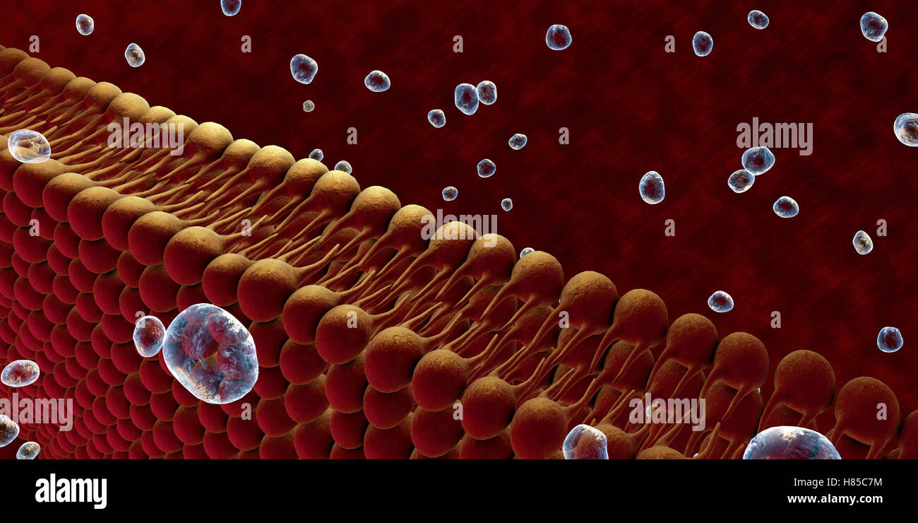 Plasma Membrane Of Cell With other molecules, 3d render Stock Photohttps://www.alamy.com/image-license-details/?v=1https://www.alamy.com/stock-photo-plasma-membrane-of-cell-with-other-molecules-3d-render-125509208.html
RFH85C7M–Plasma Membrane Of Cell With other molecules, 3d render
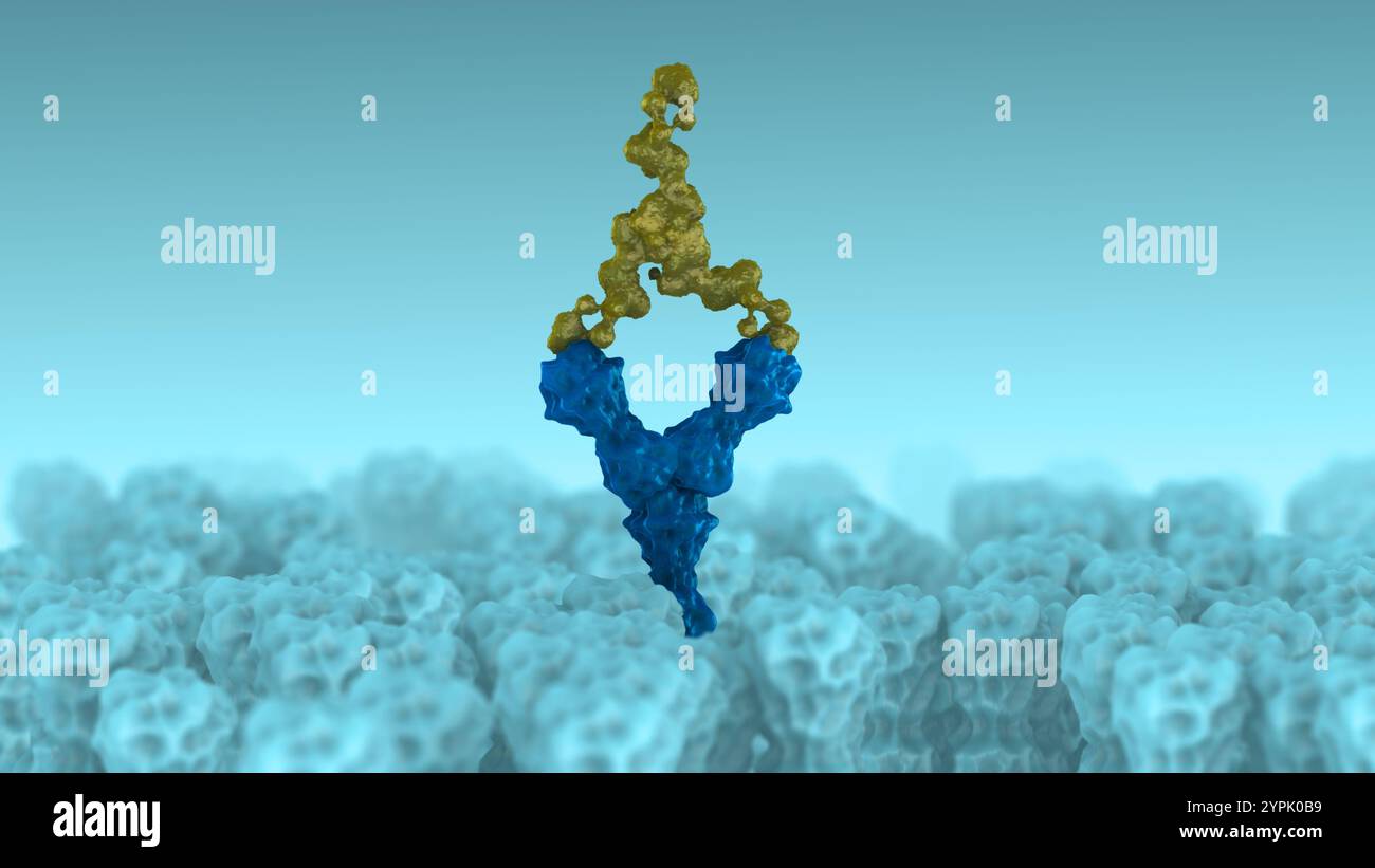 Receptor Binding by Antibodies Regulating Cellular Activity Stock Photohttps://www.alamy.com/image-license-details/?v=1https://www.alamy.com/receptor-binding-by-antibodies-regulating-cellular-activity-image633513085.html
RF2YPK0B9–Receptor Binding by Antibodies Regulating Cellular Activity
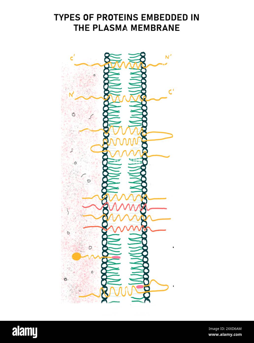 Types of protein in the plasma membrane, illustration. There are six categories of protein in the plasma membrane, on the basis of their arrangement. five are transmembrane proteins and one is a peripheral protein. Stock Photohttps://www.alamy.com/image-license-details/?v=1https://www.alamy.com/types-of-protein-in-the-plasma-membrane-illustration-there-are-six-categories-of-protein-in-the-plasma-membrane-on-the-basis-of-their-arrangement-five-are-transmembrane-proteins-and-one-is-a-peripheral-protein-image618634316.html
RF2XXD6AM–Types of protein in the plasma membrane, illustration. There are six categories of protein in the plasma membrane, on the basis of their arrangement. five are transmembrane proteins and one is a peripheral protein.
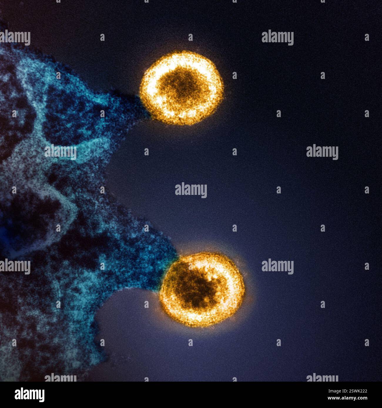 Colorized transmission electron micrograph of two HIV-1 virus particles (yellow) budding from the plasma membrane of an infected H9 T cell (blue). Image captured at the NIAID Integrated Research Facility (IRF) in Fort Detrick, Maryland. Stock Photohttps://www.alamy.com/image-license-details/?v=1https://www.alamy.com/colorized-transmission-electron-micrograph-of-two-hiv-1-virus-particles-yellow-budding-from-the-plasma-membrane-of-an-infected-h9-t-cell-blue-image-captured-at-the-niaid-integrated-research-facility-irf-in-fort-detrick-maryland-image652568730.html
RM2SWK222–Colorized transmission electron micrograph of two HIV-1 virus particles (yellow) budding from the plasma membrane of an infected H9 T cell (blue). Image captured at the NIAID Integrated Research Facility (IRF) in Fort Detrick, Maryland.
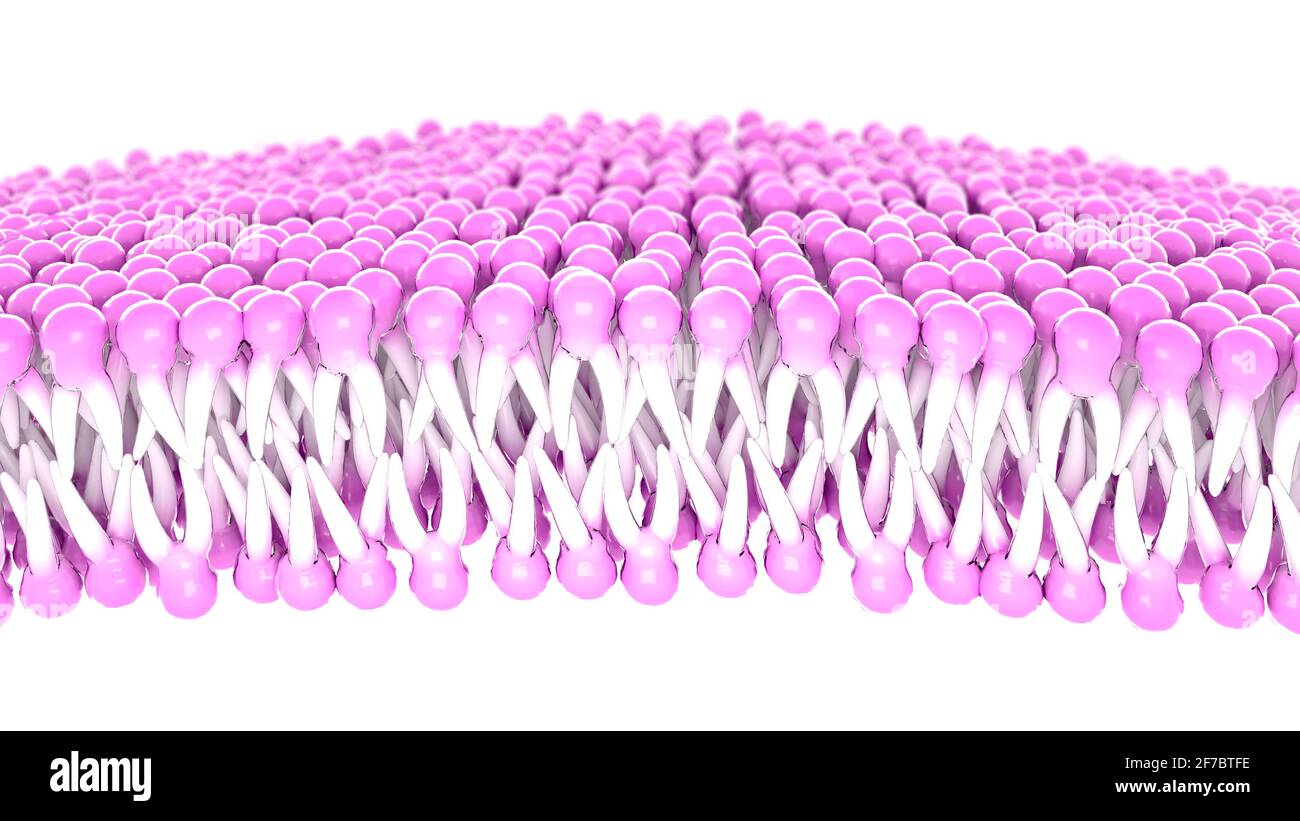 Structure of the plasma membrane of a cell. Lipids and fats viewed under a microscope. 3d render Stock Photohttps://www.alamy.com/image-license-details/?v=1https://www.alamy.com/structure-of-the-plasma-membrane-of-a-cell-lipids-and-fats-viewed-under-a-microscope-3d-render-image417612146.html
RF2F7BTFE–Structure of the plasma membrane of a cell. Lipids and fats viewed under a microscope. 3d render
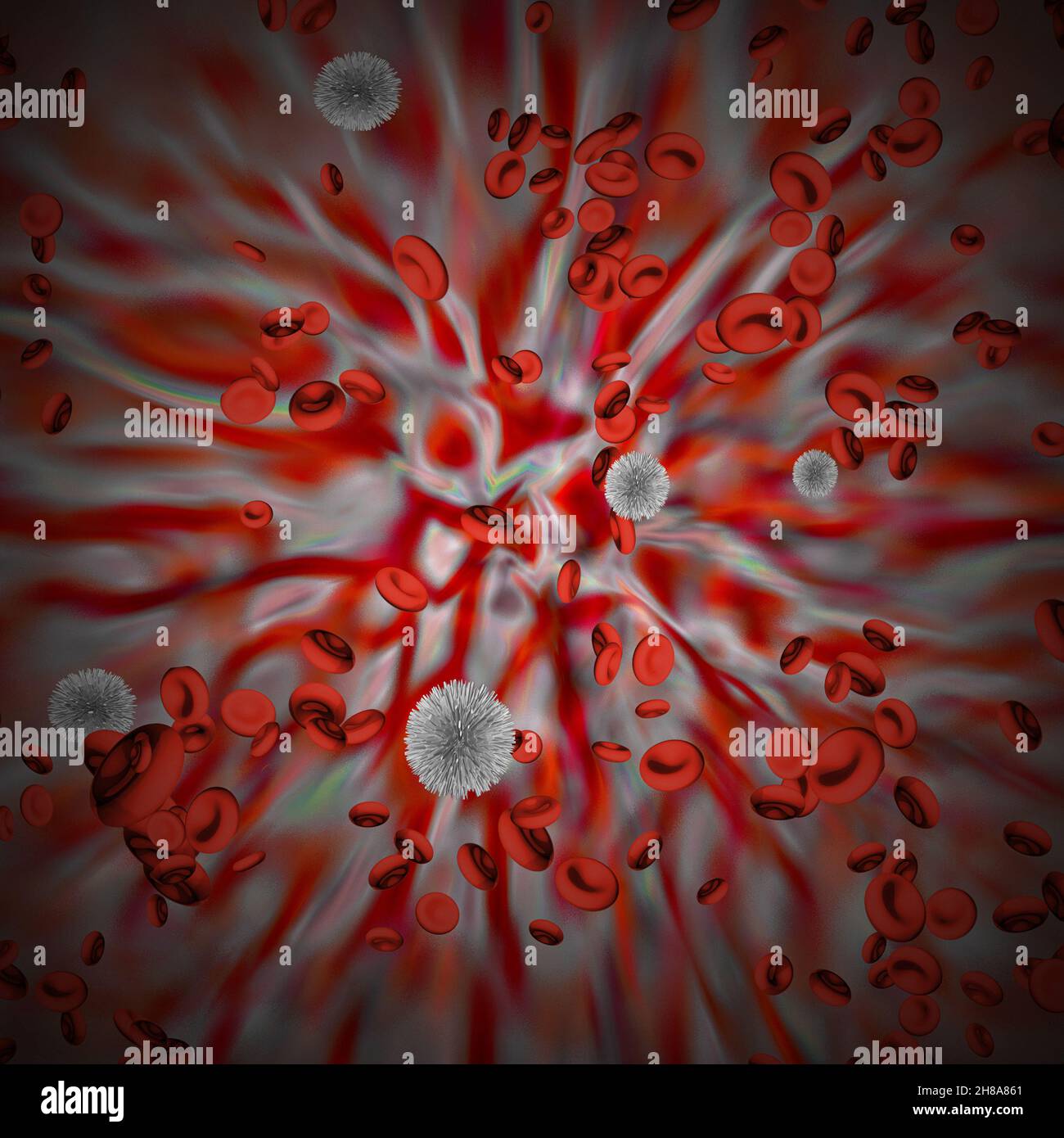 Red Blood cells with virus cells. 3D rendering Stock Photohttps://www.alamy.com/image-license-details/?v=1https://www.alamy.com/red-blood-cells-with-virus-cells-3d-rendering-image452612777.html
RF2H8A861–Red Blood cells with virus cells. 3D rendering
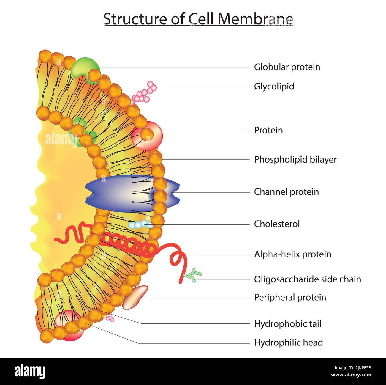 Structure of cell membrane Stock Photohttps://www.alamy.com/image-license-details/?v=1https://www.alamy.com/structure-of-cell-membrane-image476853255.html
RF2JKPF5B–Structure of cell membrane
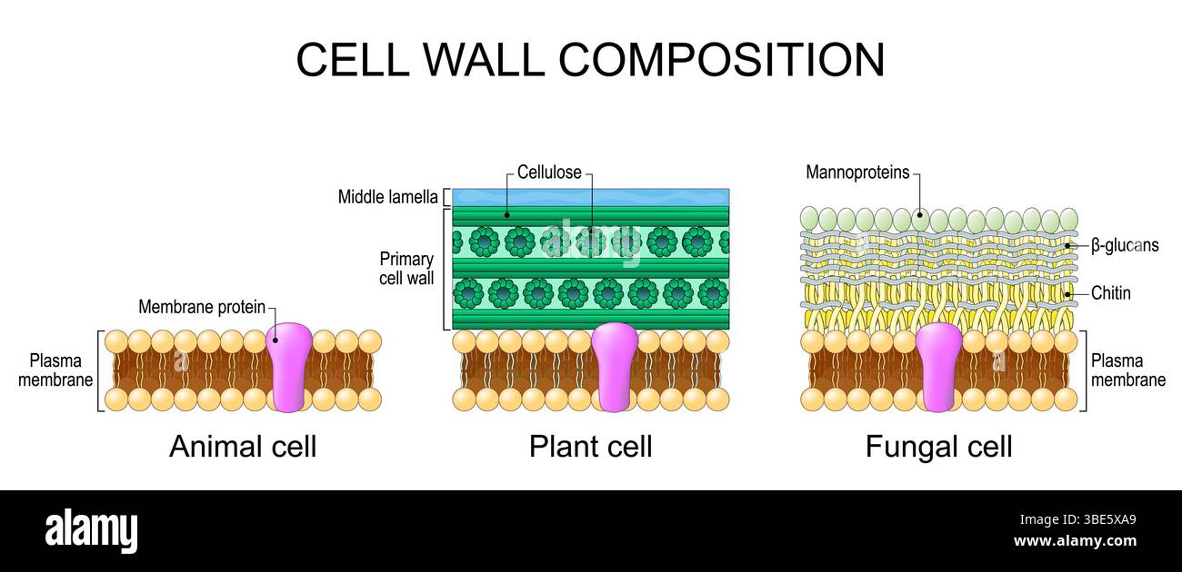 Cell wall composition. Comparison structure and anatomy of lipid bilayer plasma membrane in animal. Middle lamella, primary cell wall with Cellulose i Stock Vectorhttps://www.alamy.com/image-license-details/?v=1https://www.alamy.com/cell-wall-composition-comparison-structure-and-anatomy-of-lipid-bilayer-plasma-membrane-in-animal-middle-lamella-primary-cell-wall-with-cellulose-i-image679939969.html
RF3BE5XA9–Cell wall composition. Comparison structure and anatomy of lipid bilayer plasma membrane in animal. Middle lamella, primary cell wall with Cellulose i
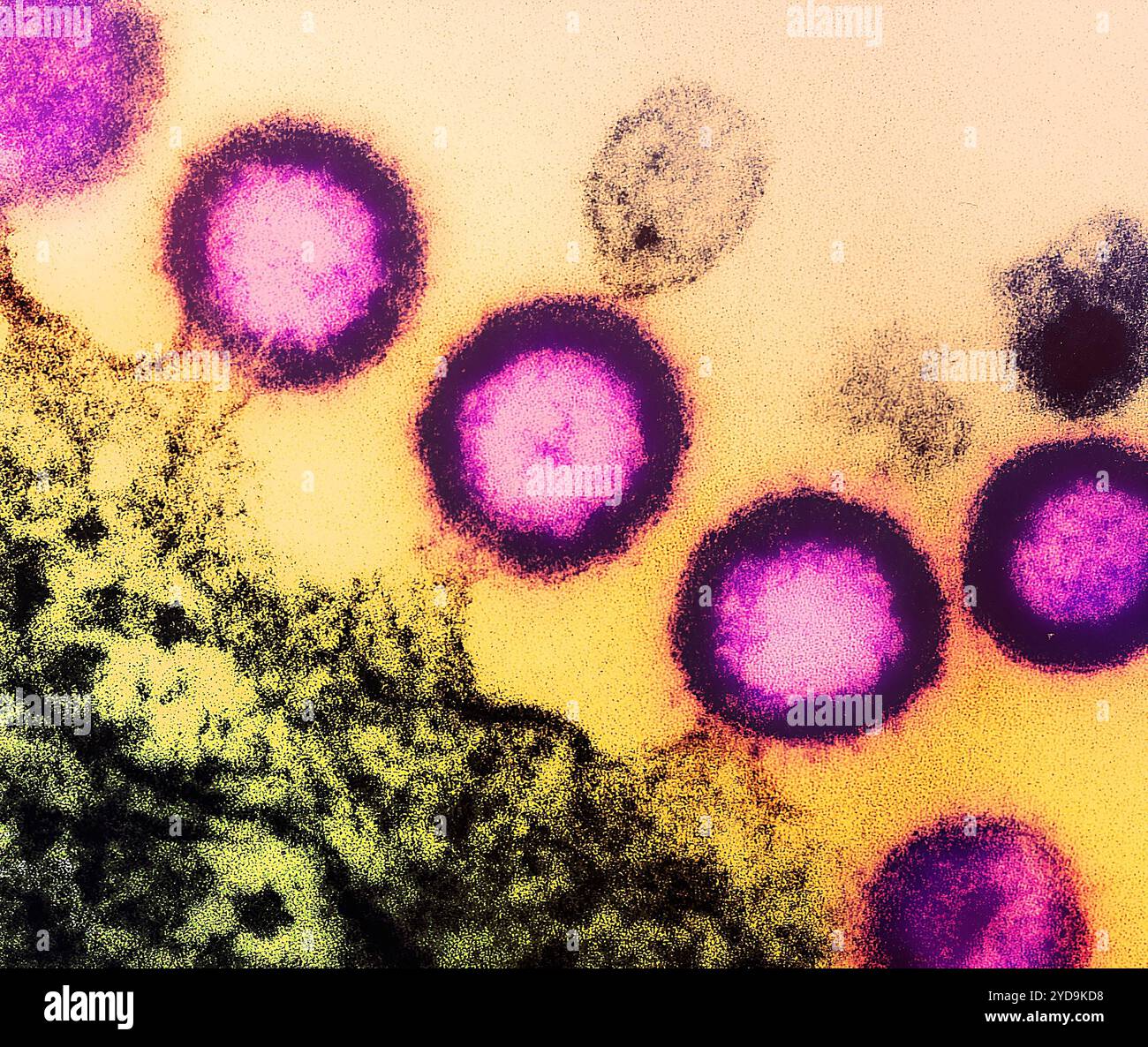 Transmission electron micrograph of HIV-1 virus particles pink replicating from the plasma membrane of an infected H9 T cell. HIV-1 Virus 016867 218 Stock Photohttps://www.alamy.com/image-license-details/?v=1https://www.alamy.com/transmission-electron-micrograph-of-hiv-1-virus-particles-pink-replicating-from-the-plasma-membrane-of-an-infected-h9-t-cell-hiv-1-virus-016867-218-image627776612.html
RM2YD9KD8–Transmission electron micrograph of HIV-1 virus particles pink replicating from the plasma membrane of an infected H9 T cell. HIV-1 Virus 016867 218
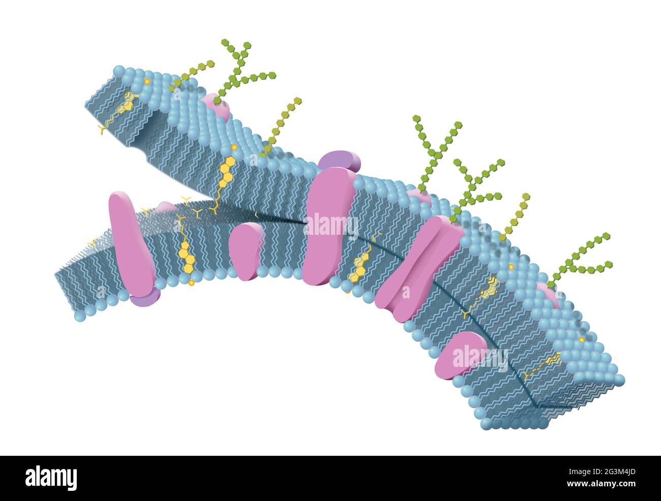 Cell membrane with phospholipids, cholesterol, intrinsic and extrinsic proteins. 3D illustration Stock Photohttps://www.alamy.com/image-license-details/?v=1https://www.alamy.com/cell-membrane-with-phospholipids-cholesterol-intrinsic-and-extrinsic-proteins-3d-illustration-image432545861.html
RF2G3M4JD–Cell membrane with phospholipids, cholesterol, intrinsic and extrinsic proteins. 3D illustration
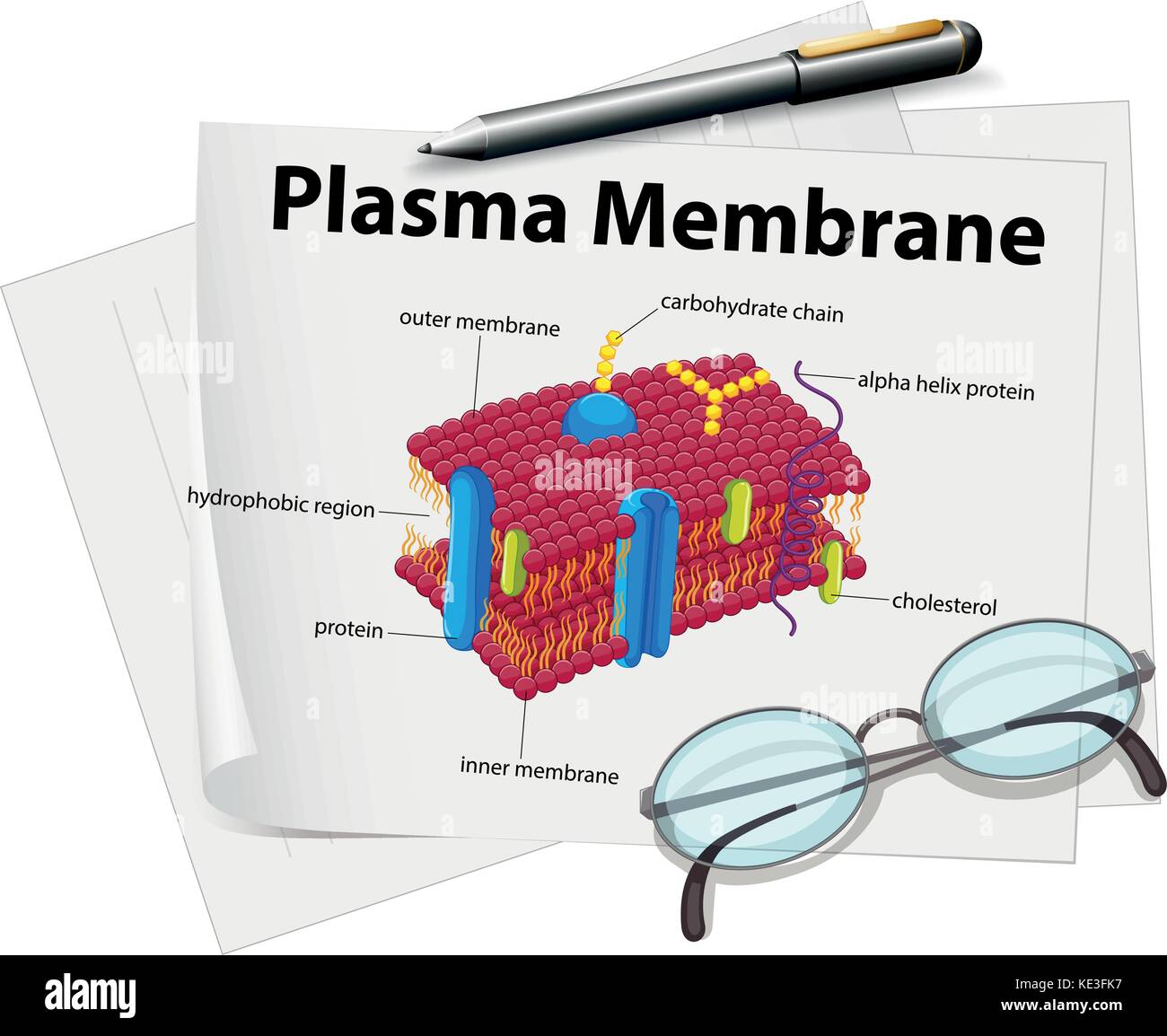 Paper showing plasma membrane drawing illustration Stock Vectorhttps://www.alamy.com/image-license-details/?v=1https://www.alamy.com/stock-image-paper-showing-plasma-membrane-drawing-illustration-163576651.html
RFKE3FK7–Paper showing plasma membrane drawing illustration
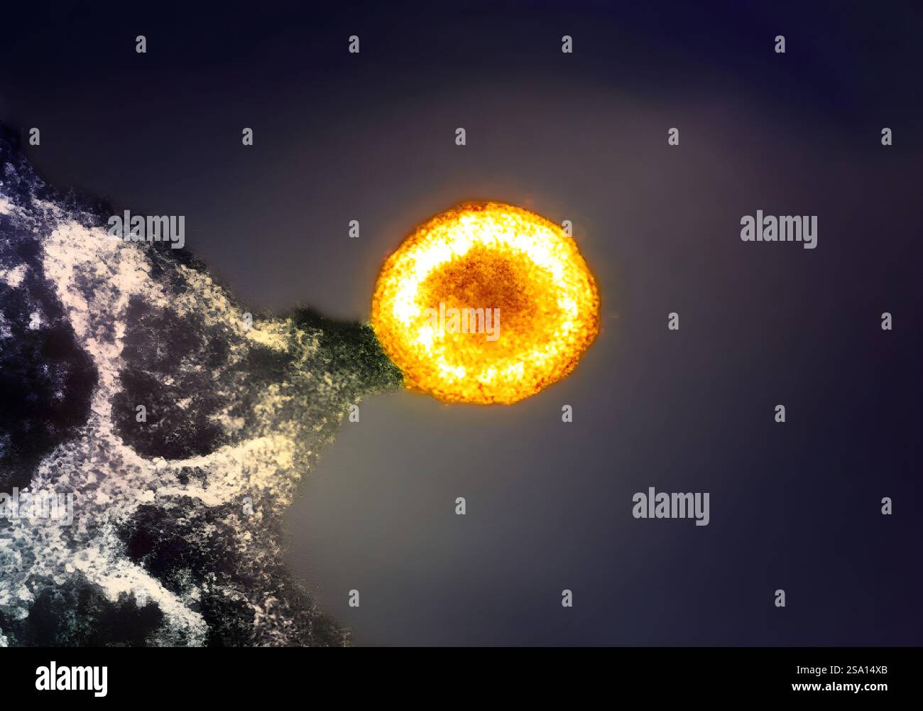 Colorized transmission electron micrograph of an HIV-1 virus particle (yellow/gold) budding from the plasma membrane of an infected H9 T cell. Stock Photohttps://www.alamy.com/image-license-details/?v=1https://www.alamy.com/colorized-transmission-electron-micrograph-of-an-hiv-1-virus-particle-yellowgold-budding-from-the-plasma-membrane-of-an-infected-h9-t-cell-image642956003.html
RM2SA14XB–Colorized transmission electron micrograph of an HIV-1 virus particle (yellow/gold) budding from the plasma membrane of an infected H9 T cell.
 Neuron stimulated by a chemical, local potential generated at plasma membrane Stock Photohttps://www.alamy.com/image-license-details/?v=1https://www.alamy.com/stock-photo-neuron-stimulated-by-a-chemical-local-potential-generated-at-plasma-49738182.html
RFCTWNFJ–Neuron stimulated by a chemical, local potential generated at plasma membrane
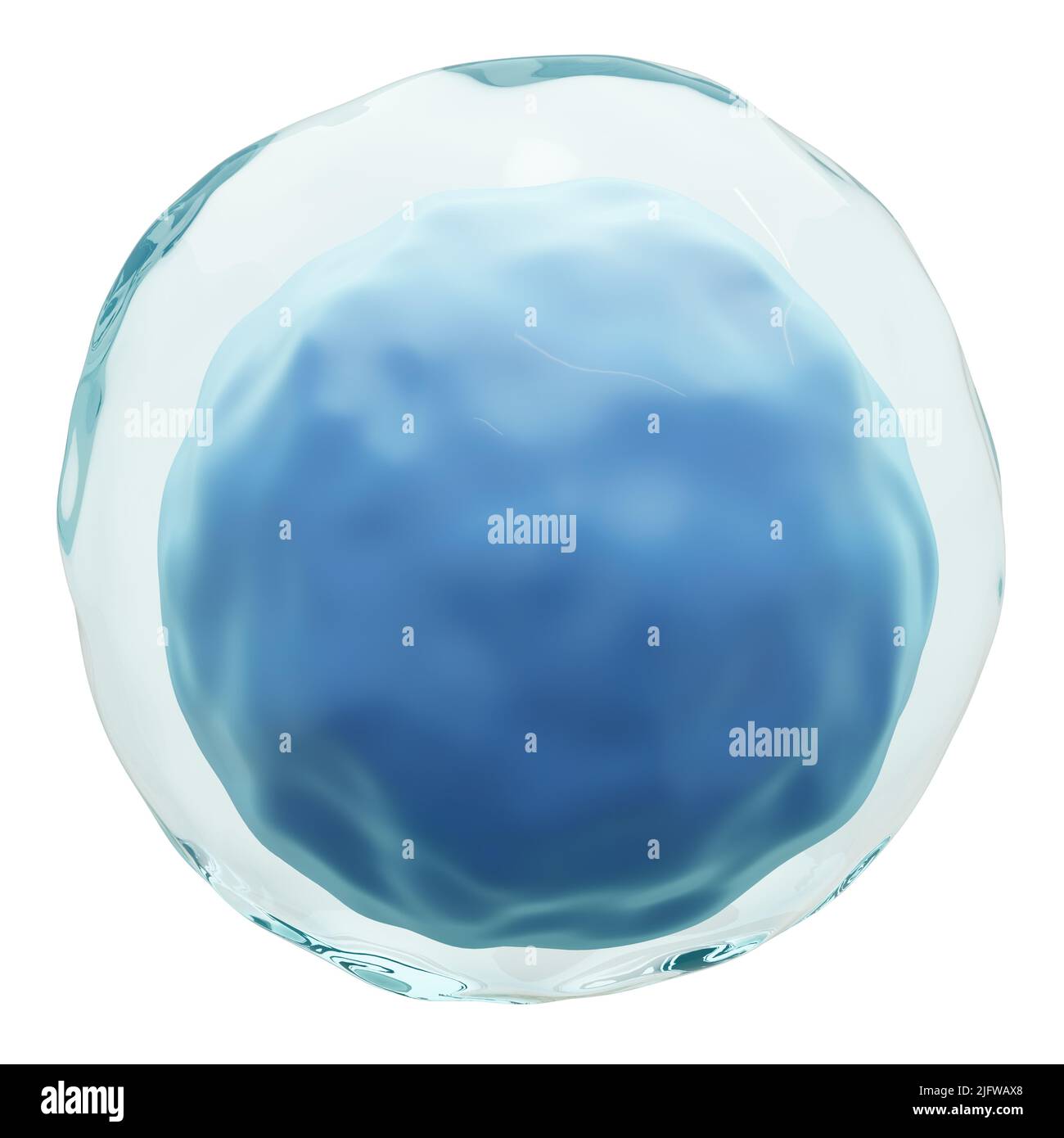 Lymphocyte . White blood cells with transparency membrane and large nucleus . Isolated white background . 3D render . Stock Photohttps://www.alamy.com/image-license-details/?v=1https://www.alamy.com/lymphocyte-white-blood-cells-with-transparency-membrane-and-large-nucleus-isolated-white-background-3d-render-image474457152.html
RF2JFWAX8–Lymphocyte . White blood cells with transparency membrane and large nucleus . Isolated white background . 3D render .
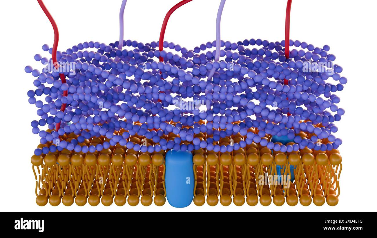 3d rendering of Gram positive bacteria have a thick peptidoglycan layer and no outer lipid membrane Stock Photohttps://www.alamy.com/image-license-details/?v=1https://www.alamy.com/3d-rendering-of-gram-positive-bacteria-have-a-thick-peptidoglycan-layer-and-no-outer-lipid-membrane-image610452628.html
RF2XD4EFG–3d rendering of Gram positive bacteria have a thick peptidoglycan layer and no outer lipid membrane
 Ultrastructure of GABA receptors in the cell membrane: Neurotransmitter gamma-aminobutyric acid, GABA A and GABA B. Infographic on pharmacology. Stock Vectorhttps://www.alamy.com/image-license-details/?v=1https://www.alamy.com/ultrastructure-of-gaba-receptors-in-the-cell-membrane-neurotransmitter-gamma-aminobutyric-acid-gaba-a-and-gaba-b-infographic-on-pharmacology-image674699904.html
RF3B5K6H4–Ultrastructure of GABA receptors in the cell membrane: Neurotransmitter gamma-aminobutyric acid, GABA A and GABA B. Infographic on pharmacology.
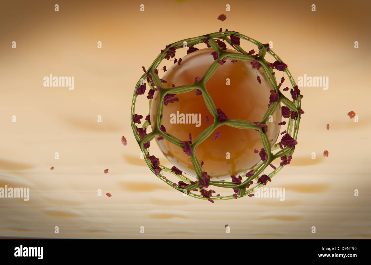 Clathrin Mediated Endocytosis Stock Photohttps://www.alamy.com/image-license-details/?v=1https://www.alamy.com/stock-photo-clathrin-mediated-endocytosis-57643068.html
RFD9NT90–Clathrin Mediated Endocytosis
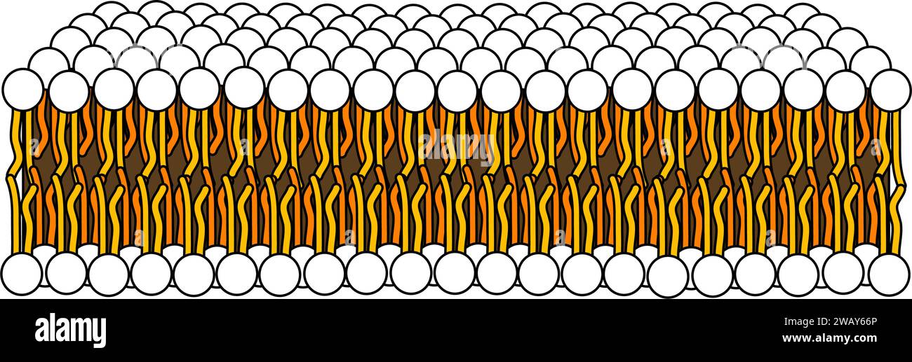 Structure of Phospholipid Molecule in Bilayer .Vector illustration. Stock Vectorhttps://www.alamy.com/image-license-details/?v=1https://www.alamy.com/structure-of-phospholipid-molecule-in-bilayer-vector-illustration-image591896670.html
RF2WAY66P–Structure of Phospholipid Molecule in Bilayer .Vector illustration.
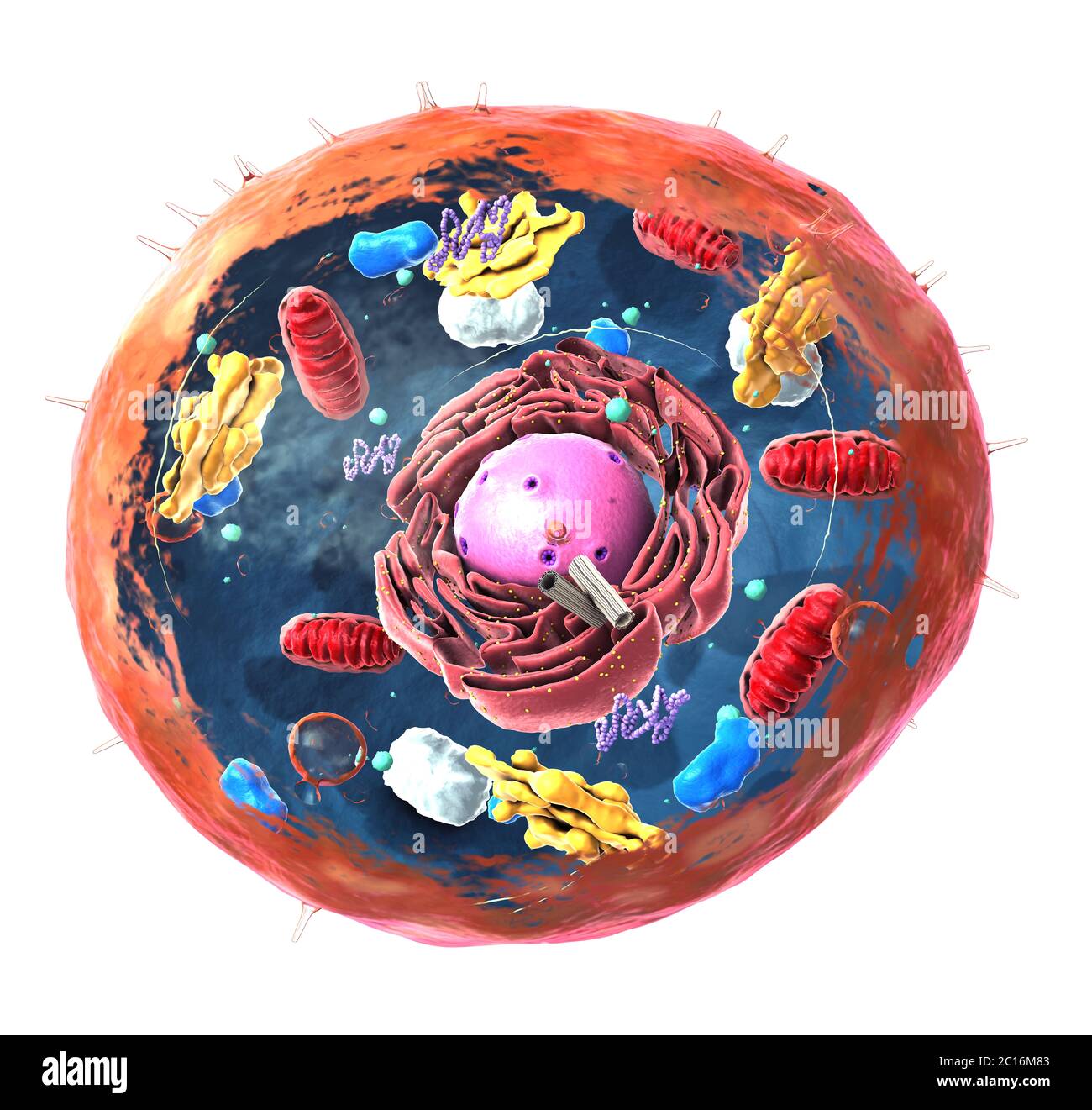 Components of Eukaryotic cell, nucleus and organelles and plasma membrane - 3d illustration Stock Photohttps://www.alamy.com/image-license-details/?v=1https://www.alamy.com/components-of-eukaryotic-cell-nucleus-and-organelles-and-plasma-membrane-3d-illustration-image362180003.html
RF2C16M83–Components of Eukaryotic cell, nucleus and organelles and plasma membrane - 3d illustration
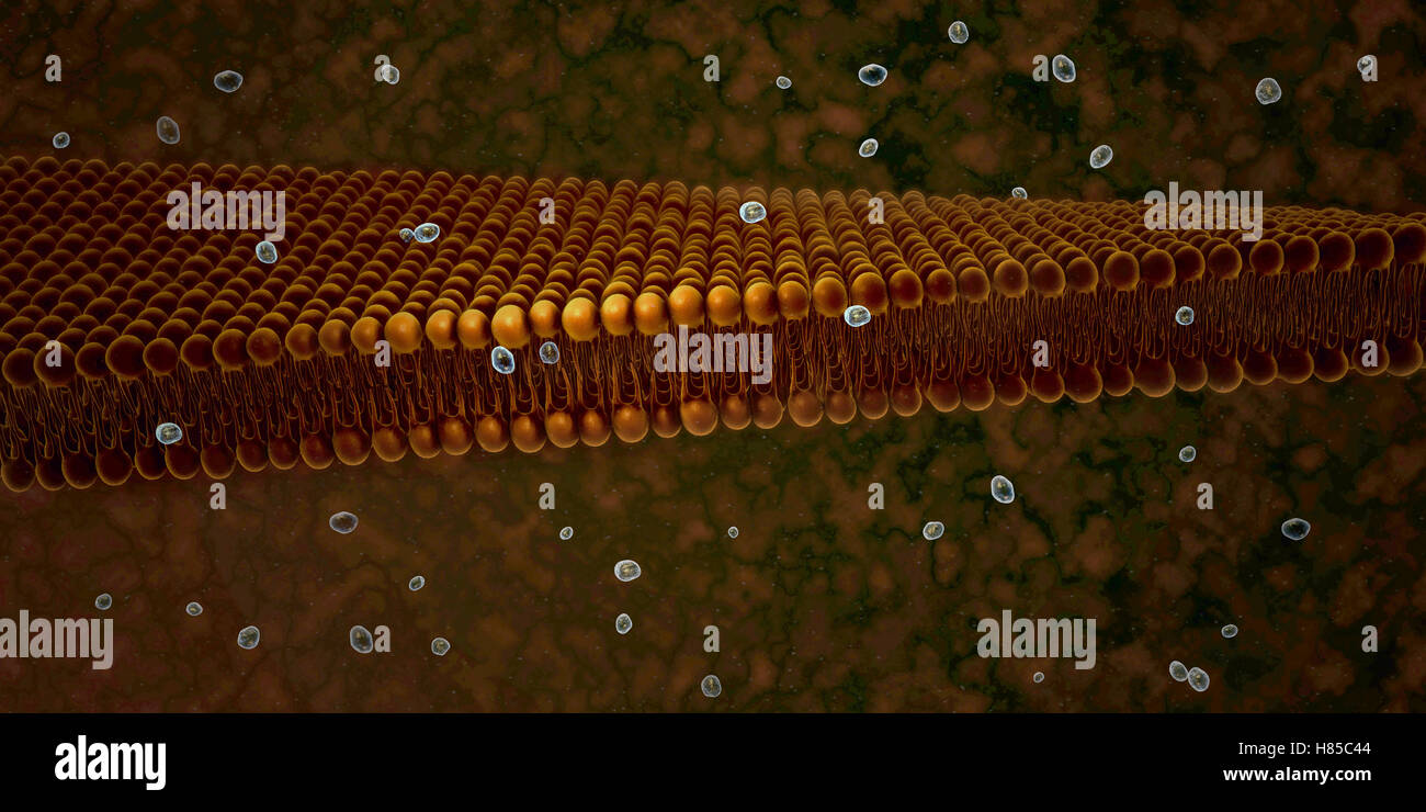 Plasma Membrane Of Cell With other molecules, 3d render Stock Photohttps://www.alamy.com/image-license-details/?v=1https://www.alamy.com/stock-photo-plasma-membrane-of-cell-with-other-molecules-3d-render-125509108.html
RFH85C44–Plasma Membrane Of Cell With other molecules, 3d render
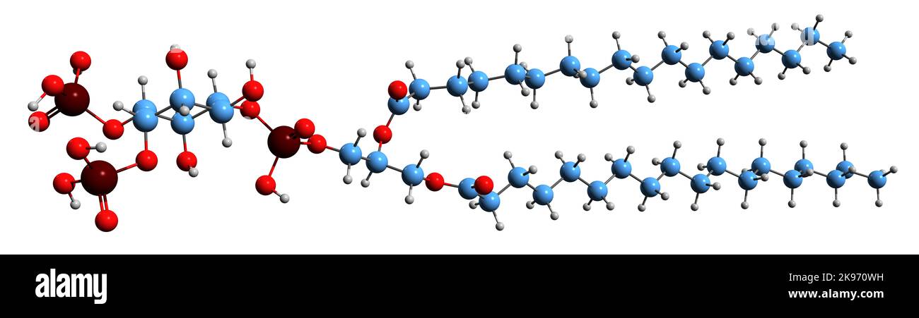 3D image of Phosphatidylinositol bisphosphate skeletal formula - molecular chemical structure of cell membranes phospholipid isolated on white backgr Stock Photohttps://www.alamy.com/image-license-details/?v=1https://www.alamy.com/3d-image-of-phosphatidylinositol-bisphosphate-skeletal-formula-molecular-chemical-structure-of-cell-membranes-phospholipid-isolated-on-white-backgr-image487576589.html
RF2K970WH–3D image of Phosphatidylinositol bisphosphate skeletal formula - molecular chemical structure of cell membranes phospholipid isolated on white backgr
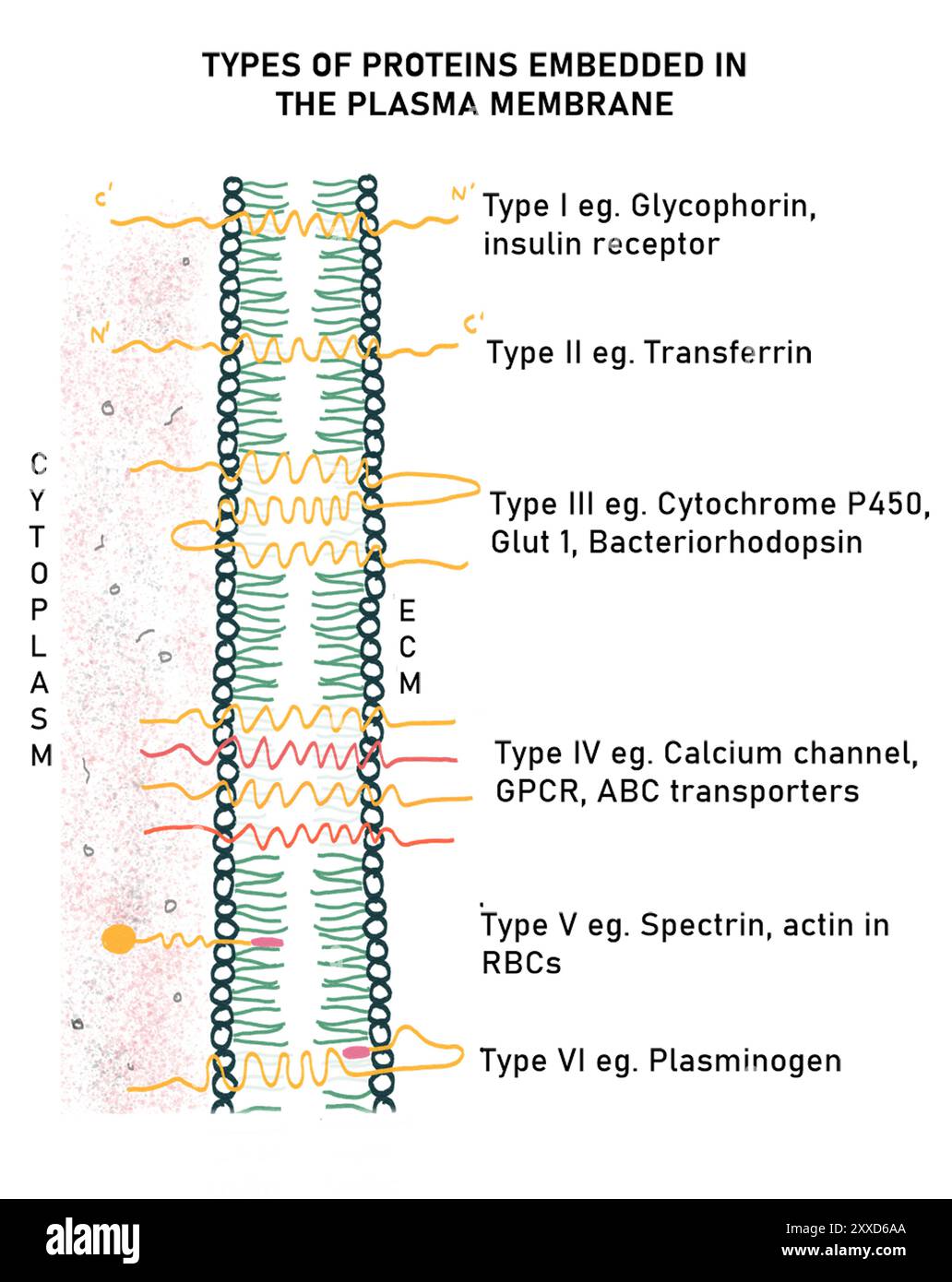 Types of protein in the plasma membrane, illustration. There are six categories of protein in the plasma membrane, on the basis of their arrangement. five are transmembrane proteins and one is a peripheral protein. Stock Photohttps://www.alamy.com/image-license-details/?v=1https://www.alamy.com/types-of-protein-in-the-plasma-membrane-illustration-there-are-six-categories-of-protein-in-the-plasma-membrane-on-the-basis-of-their-arrangement-five-are-transmembrane-proteins-and-one-is-a-peripheral-protein-image618634306.html
RF2XXD6AA–Types of protein in the plasma membrane, illustration. There are six categories of protein in the plasma membrane, on the basis of their arrangement. five are transmembrane proteins and one is a peripheral protein.
 Red science blood cell particles background. Life and biology, medicine scientific, molecular research. Virus or causative agent microscope view Stock Vectorhttps://www.alamy.com/image-license-details/?v=1https://www.alamy.com/red-science-blood-cell-particles-background-life-and-biology-medicine-scientific-molecular-research-virus-or-causative-agent-microscope-view-image412017843.html
RF2EX90XY–Red science blood cell particles background. Life and biology, medicine scientific, molecular research. Virus or causative agent microscope view
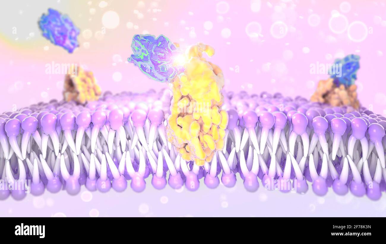 Structure of the plasma membrane of a cell. Lipids and fats viewed under a microscope. 3d render Stock Photohttps://www.alamy.com/image-license-details/?v=1https://www.alamy.com/structure-of-the-plasma-membrane-of-a-cell-lipids-and-fats-viewed-under-a-microscope-3d-render-image417542041.html
RF2F78K3N–Structure of the plasma membrane of a cell. Lipids and fats viewed under a microscope. 3d render
