SAVE UP TO 30% ON YOUR FIRST ORDER, APPLY CODE: HELLO30
Quick filters:
Microscopic image Stock Photos and Images
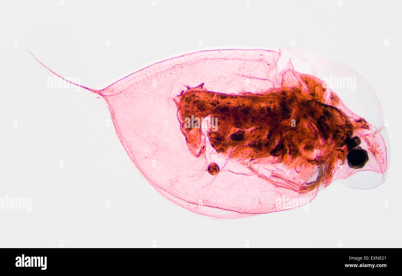 Microscopic Image of Pond Plankton Water Flea Daphina Stock Photohttps://www.alamy.com/licenses-and-pricing/?v=1https://www.alamy.com/stock-photo-microscopic-image-of-pond-plankton-water-flea-daphina-85294553.html
RFEXNE21–Microscopic Image of Pond Plankton Water Flea Daphina
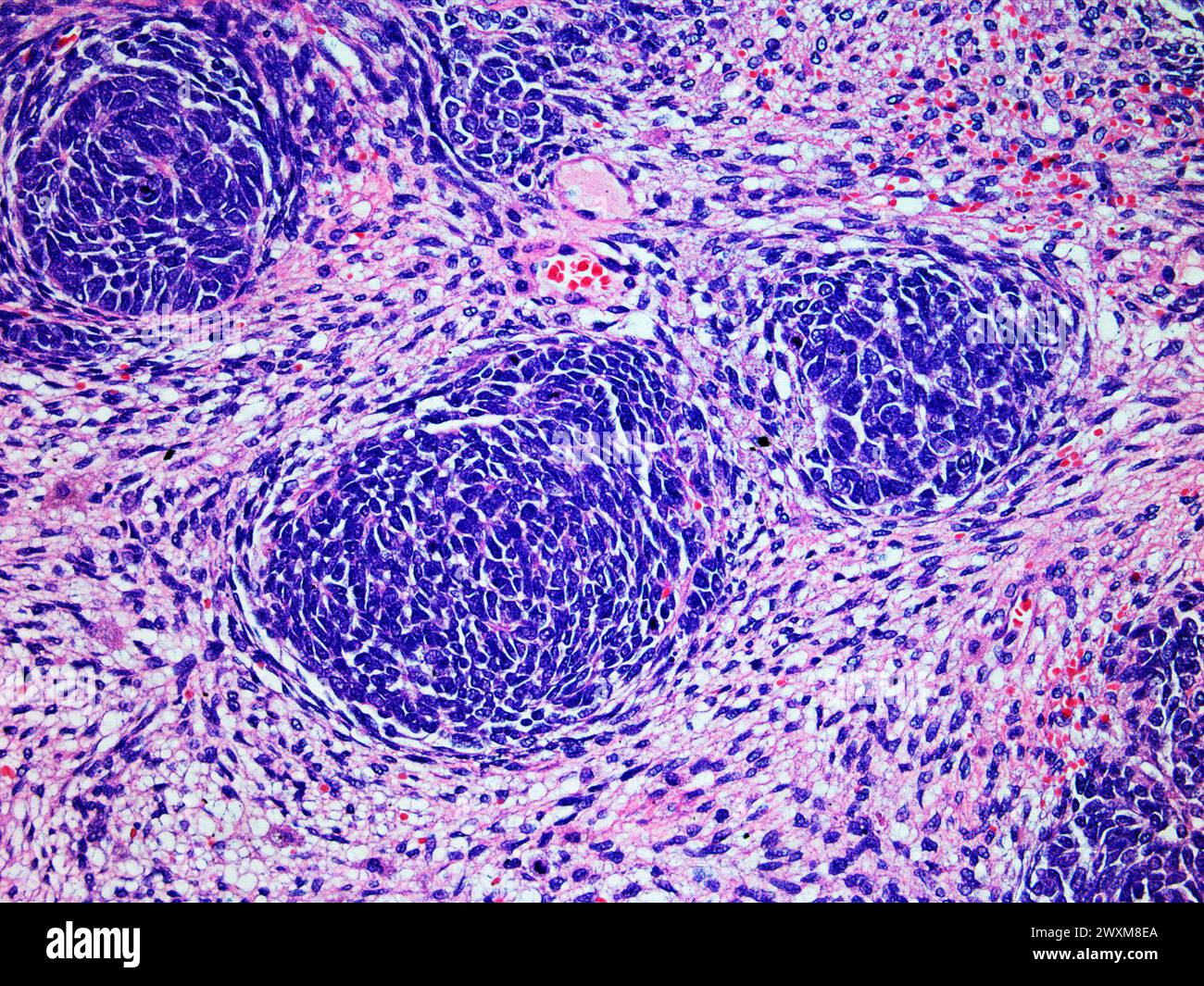 Microscopic Image of a Wilms Tumor or Nephroblastoma of a Childs Kidney Viewed at 200x Magnification with Hematoxylin and Eosin Staining One of the mo Stock Photohttps://www.alamy.com/licenses-and-pricing/?v=1https://www.alamy.com/microscopic-image-of-a-wilms-tumor-or-nephroblastoma-of-a-childs-kidney-viewed-at-200x-magnification-with-hematoxylin-and-eosin-staining-one-of-the-mo-image601579282.html
RF2WXM8EA–Microscopic Image of a Wilms Tumor or Nephroblastoma of a Childs Kidney Viewed at 200x Magnification with Hematoxylin and Eosin Staining One of the mo
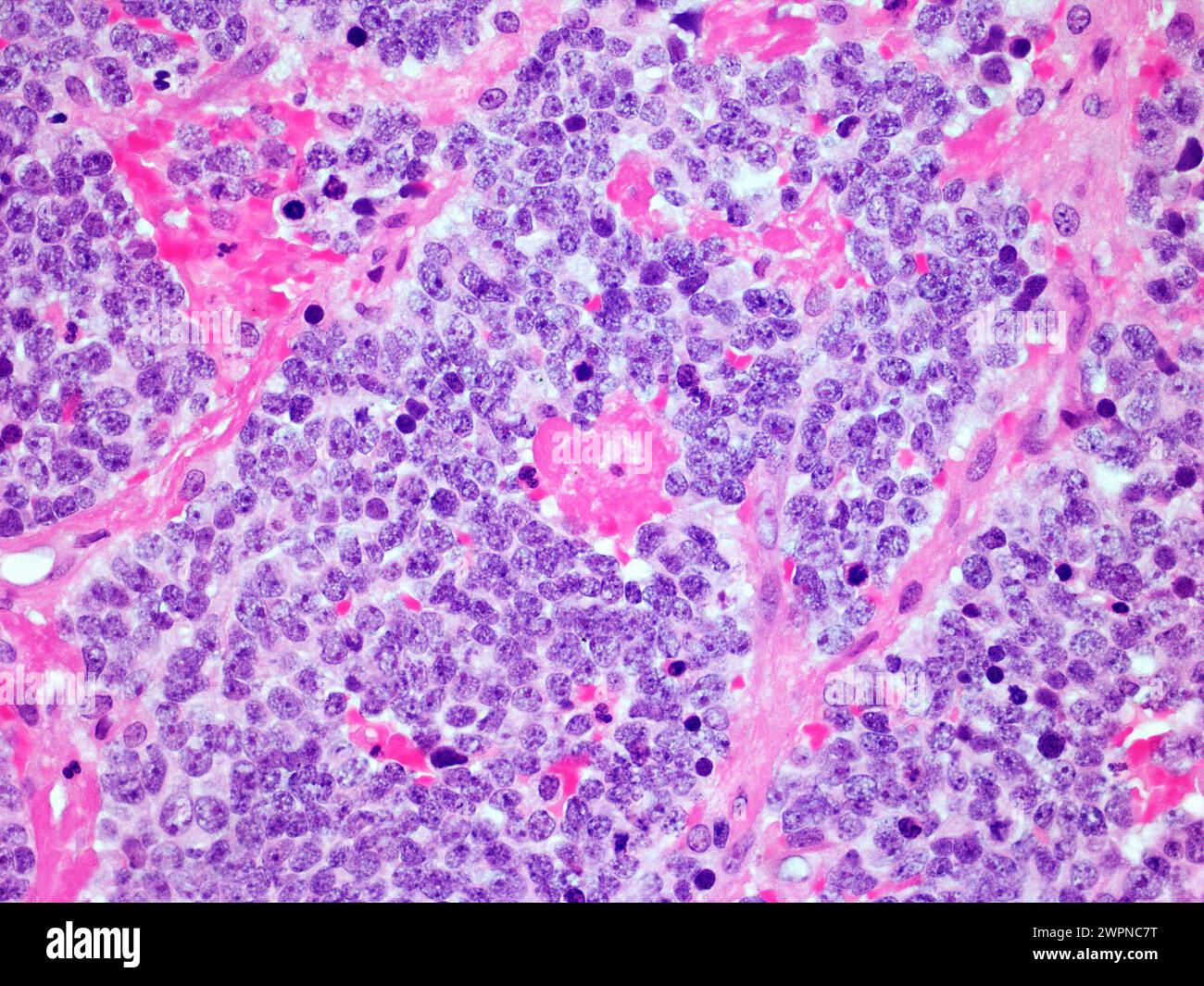 Microscopic Image of a Neuroblastoma Malignant Tumor of the Adrenal Gland Viewed at 300x Magnification with Haematoxylin and Eosin Staining Stock Photohttps://www.alamy.com/licenses-and-pricing/?v=1https://www.alamy.com/microscopic-image-of-a-neuroblastoma-malignant-tumor-of-the-adrenal-gland-viewed-at-300x-magnification-with-haematoxylin-and-eosin-staining-image599145564.html
RF2WPNC7T–Microscopic Image of a Neuroblastoma Malignant Tumor of the Adrenal Gland Viewed at 300x Magnification with Haematoxylin and Eosin Staining
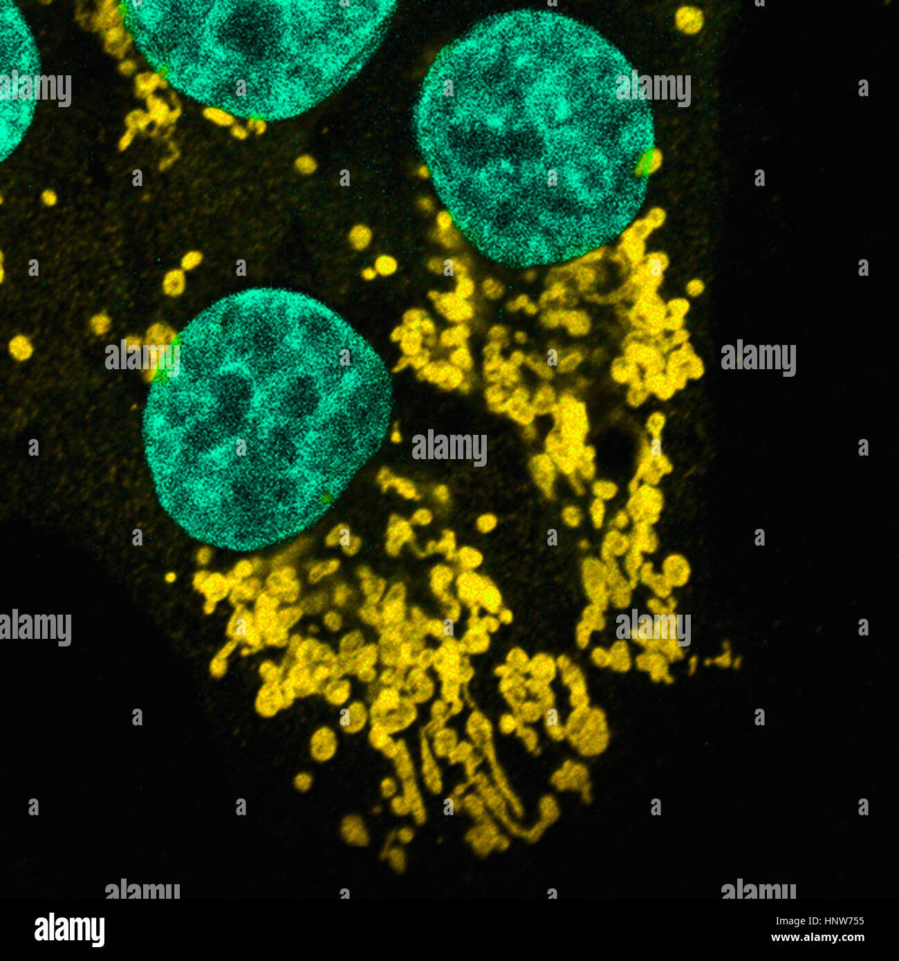 Microscopic image of mutated human epithelial cells Stock Photohttps://www.alamy.com/licenses-and-pricing/?v=1https://www.alamy.com/stock-photo-microscopic-image-of-mutated-human-epithelial-cells-133934785.html
RFHNW755–Microscopic image of mutated human epithelial cells
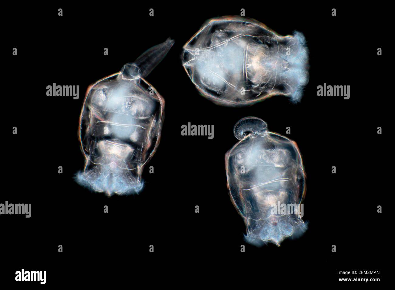 rotifers (Rotatoria), dark field microscopic image, magnification x80 related to 35 mm Stock Photohttps://www.alamy.com/licenses-and-pricing/?v=1https://www.alamy.com/rotifers-rotatoria-dark-field-microscopic-image-magnification-x80-related-to-35-mm-image408213421.html
RM2EM3MAN–rotifers (Rotatoria), dark field microscopic image, magnification x80 related to 35 mm
 a large background image of cells or bacteria under the microscope Stock Photohttps://www.alamy.com/licenses-and-pricing/?v=1https://www.alamy.com/stock-photo-a-large-background-image-of-cells-or-bacteria-under-the-microscope-15109757.html
RFAM0M5J–a large background image of cells or bacteria under the microscope
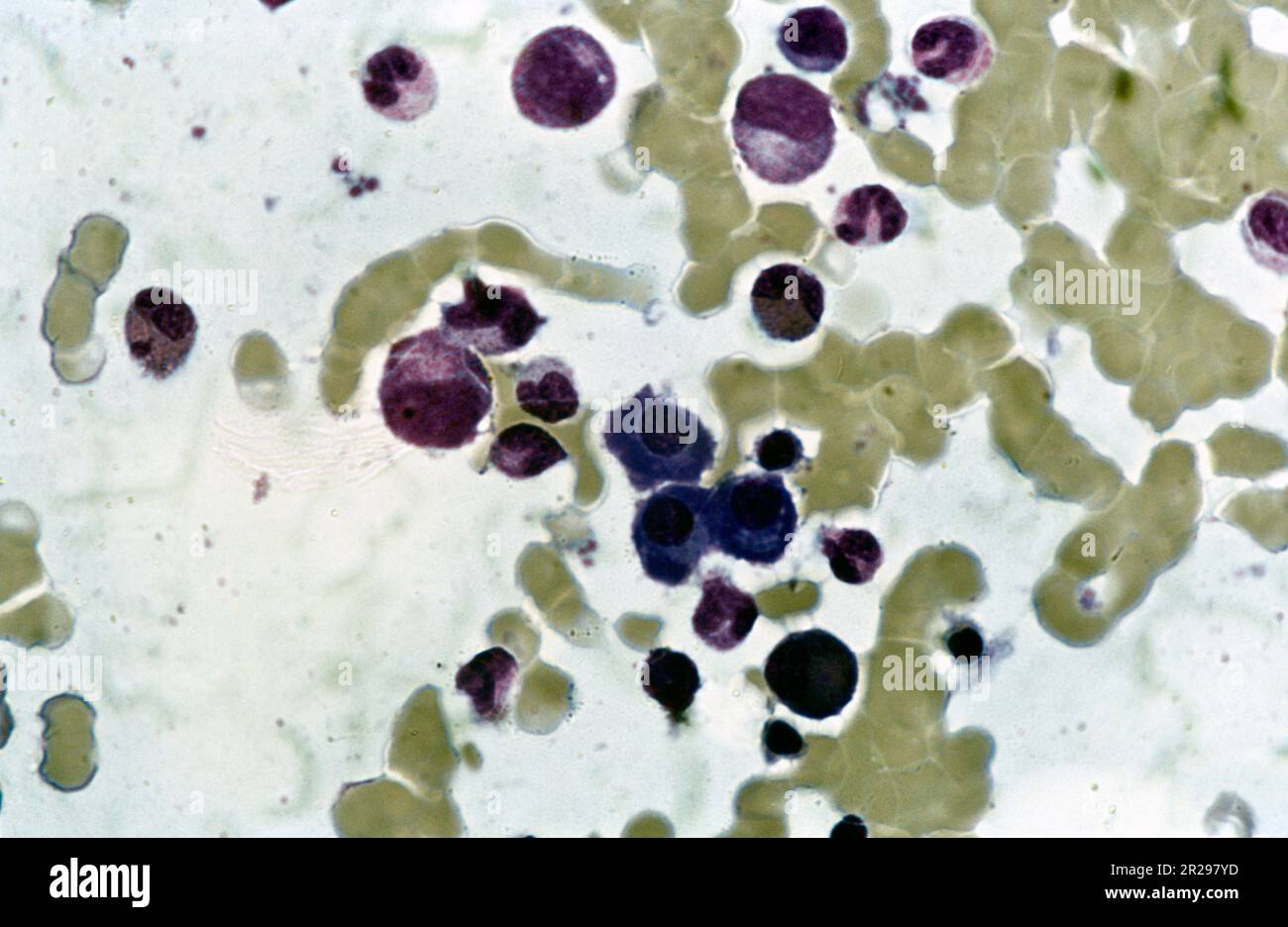 Microscopic Image of Dividing Blood Cells Stock Photohttps://www.alamy.com/licenses-and-pricing/?v=1https://www.alamy.com/microscopic-image-of-dividing-blood-cells-image552164913.html
RM2R297YD–Microscopic Image of Dividing Blood Cells
 a large rendered image of bacteria or cells under a microscope Stock Photohttps://www.alamy.com/licenses-and-pricing/?v=1https://www.alamy.com/stock-photo-a-large-rendered-image-of-bacteria-or-cells-under-a-microscope-14558755.html
RFAHX48M–a large rendered image of bacteria or cells under a microscope
 Microscopic image of skin tissue. Stock Photohttps://www.alamy.com/licenses-and-pricing/?v=1https://www.alamy.com/stock-photo-microscopic-image-of-skin-tissue-101776726.html
RFFWG972–Microscopic image of skin tissue.
 Science students looking at microscopic image on computer Stock Photohttps://www.alamy.com/licenses-and-pricing/?v=1https://www.alamy.com/stock-photo-science-students-looking-at-microscopic-image-on-computer-78168744.html
RFEF4W0T–Science students looking at microscopic image on computer
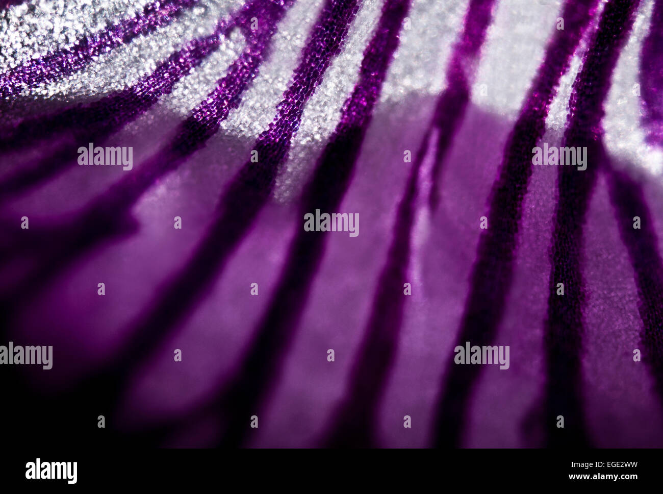 microscopic image of a flower petal Stock Photohttps://www.alamy.com/licenses-and-pricing/?v=1https://www.alamy.com/stock-photo-microscopic-image-of-a-flower-petal-78985589.html
RMEGE2WW–microscopic image of a flower petal
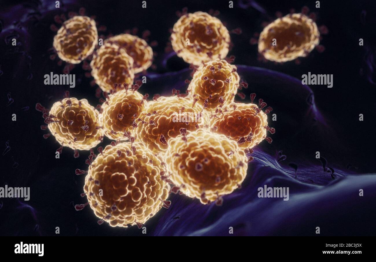 Details of Coronavirus COVID-19 on human cells, 3D illustration as a microscopic image inside the human body based on SEM SARS photos Stock Photohttps://www.alamy.com/licenses-and-pricing/?v=1https://www.alamy.com/details-of-coronavirus-covid-19-on-human-cells-3d-illustration-as-a-microscopic-image-inside-the-human-body-based-on-sem-sars-photos-image351663366.html
RF2BC3J5X–Details of Coronavirus COVID-19 on human cells, 3D illustration as a microscopic image inside the human body based on SEM SARS photos
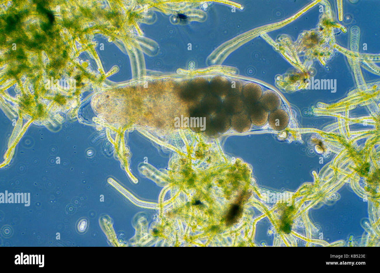 Water Bear (Hypsibius sp) microscopic image, animal is less than one mm in length, can enter cryptobiosis to withstand temperature and moisture extremes Stock Photohttps://www.alamy.com/licenses-and-pricing/?v=1https://www.alamy.com/stock-image-water-bear-hypsibius-sp-microscopic-image-animal-is-less-than-one-161765954.html
RMKB523E–Water Bear (Hypsibius sp) microscopic image, animal is less than one mm in length, can enter cryptobiosis to withstand temperature and moisture extremes
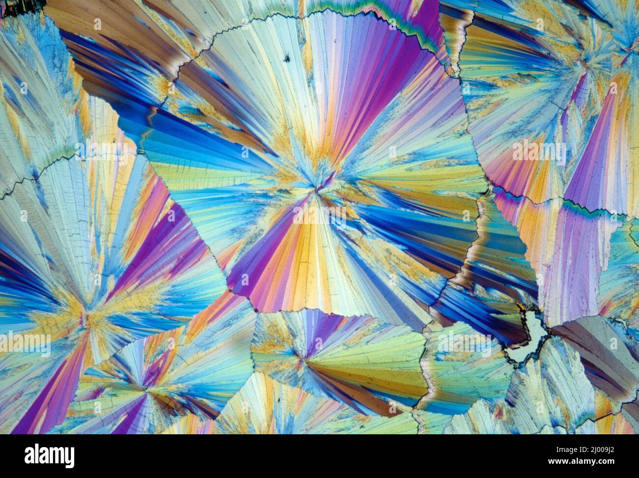 Still life. Microscopic image of chemical crystals. Sugar. Stock Photohttps://www.alamy.com/licenses-and-pricing/?v=1https://www.alamy.com/still-life-microscopic-image-of-chemical-crystals-sugar-image464687498.html
RM2J009J2–Still life. Microscopic image of chemical crystals. Sugar.
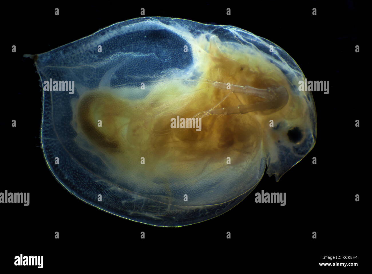 Microscopic image of daphnia, dark field technique Stock Photohttps://www.alamy.com/licenses-and-pricing/?v=1https://www.alamy.com/stock-image-microscopic-image-of-daphnia-dark-field-technique-162697728.html
RFKCKEH4–Microscopic image of daphnia, dark field technique
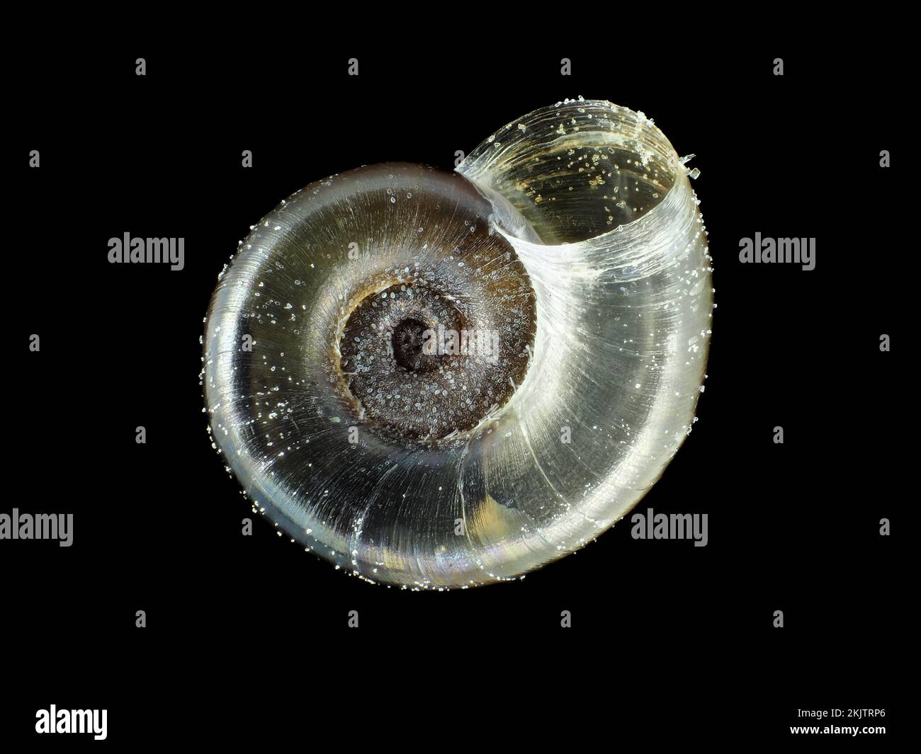 Tiny fungivore snail shell under the microscope Stock Photohttps://www.alamy.com/licenses-and-pricing/?v=1https://www.alamy.com/tiny-fungivore-snail-shell-under-the-microscope-image493499614.html
RM2KJTRP6–Tiny fungivore snail shell under the microscope
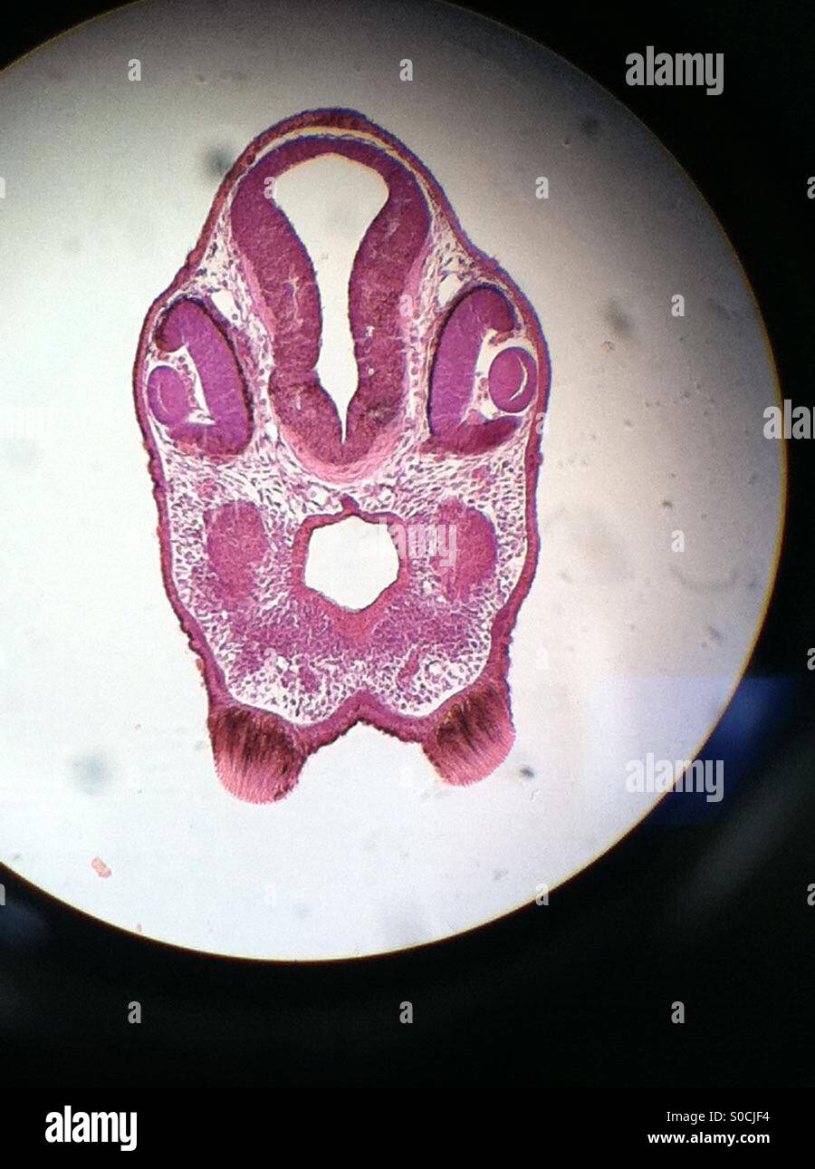 Chick embryo microscopic slide image. Image taken from the microscope. Stock Photohttps://www.alamy.com/licenses-and-pricing/?v=1https://www.alamy.com/stock-photo-chick-embryo-microscopic-slide-image-image-taken-from-the-microscope-310064584.html
RFS0CJF4–Chick embryo microscopic slide image. Image taken from the microscope.
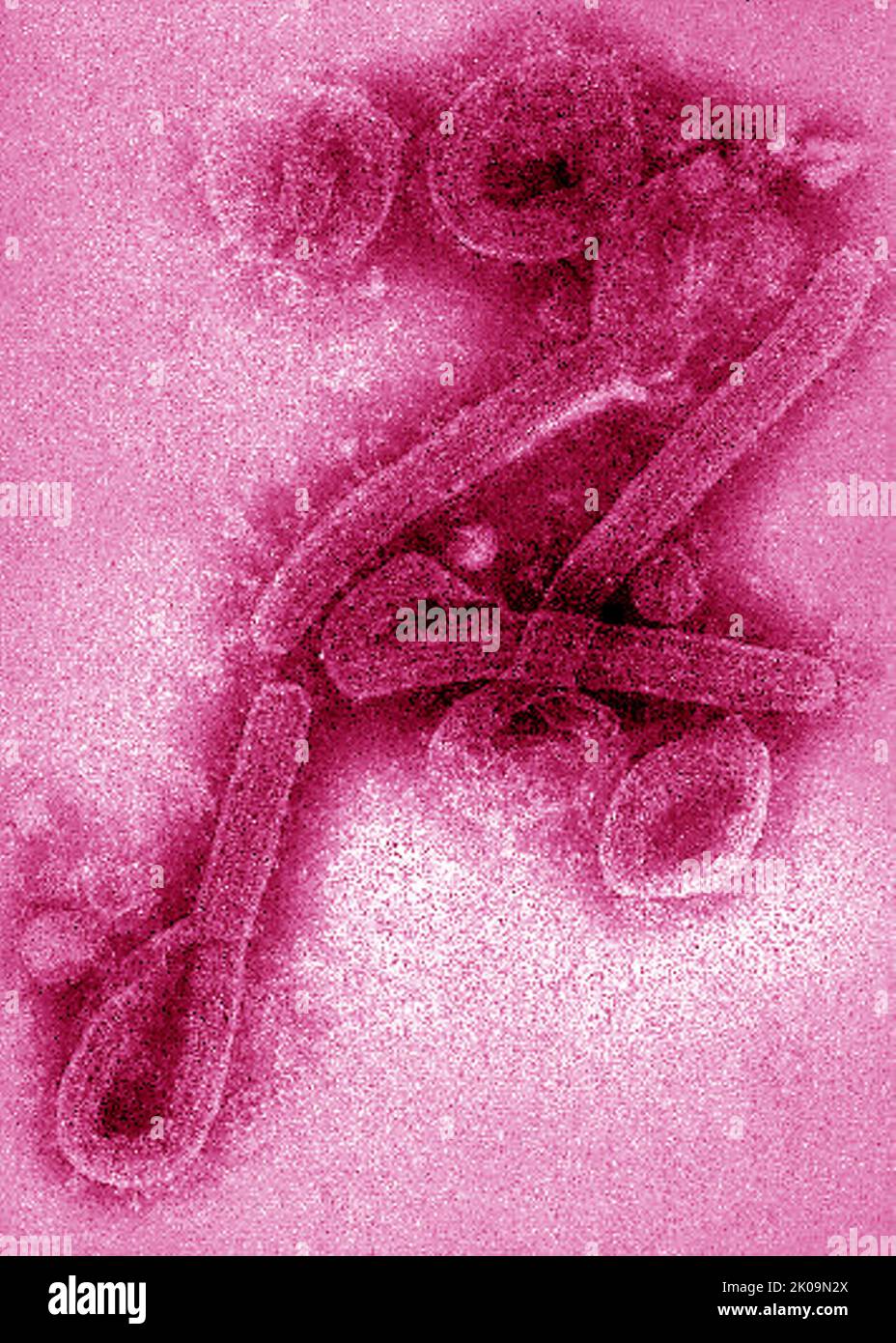 Transmission electron microscopic (TEM) image of Marburg virus virions. Stock Photohttps://www.alamy.com/licenses-and-pricing/?v=1https://www.alamy.com/transmission-electron-microscopic-tem-image-of-marburg-virus-virions-image482104418.html
RM2K09N2X–Transmission electron microscopic (TEM) image of Marburg virus virions.
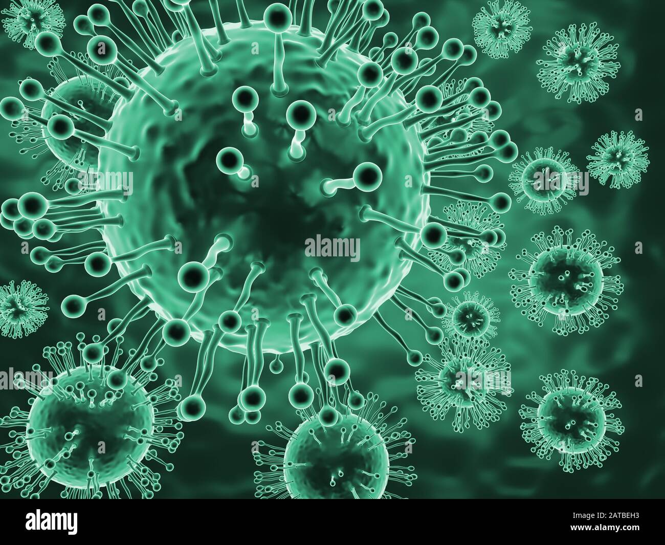 Microscopic image of deadly coronavirus particles Stock Photohttps://www.alamy.com/licenses-and-pricing/?v=1https://www.alamy.com/microscopic-image-of-deadly-coronavirus-particles-image342001663.html
RF2ATBEH3–Microscopic image of deadly coronavirus particles
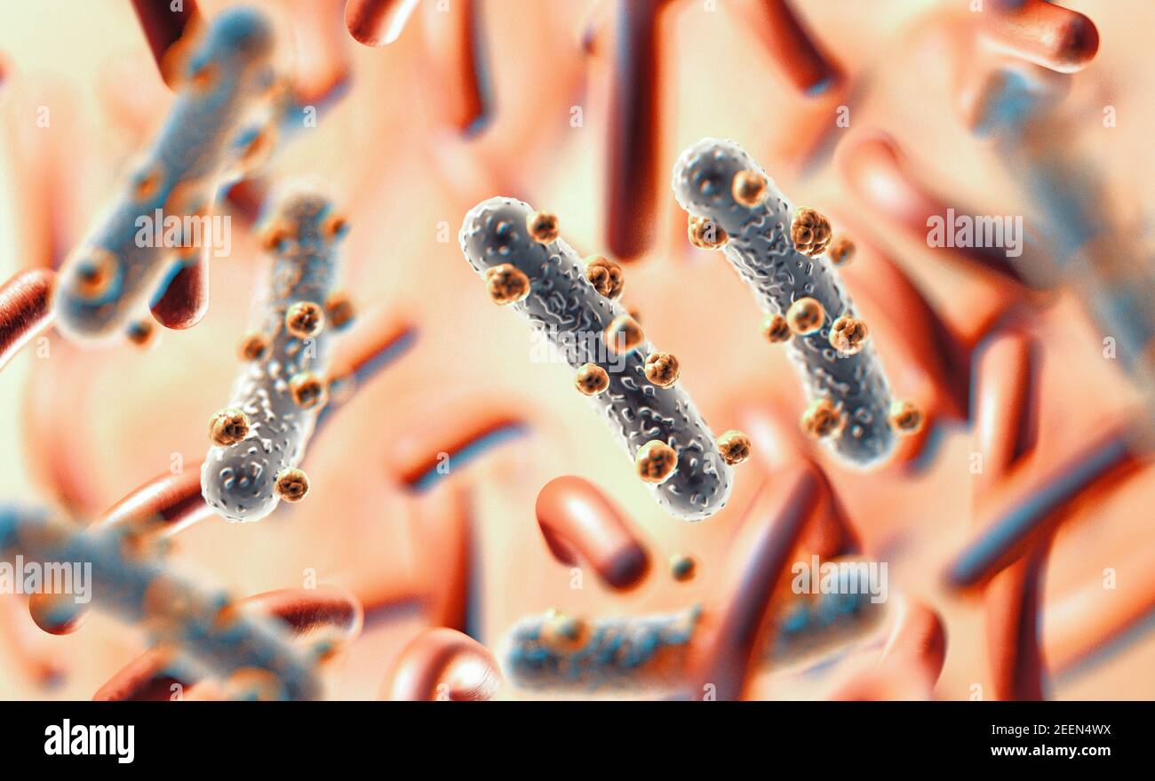 3d illustration of microscopic image of a virus or infectious cell.Microbacteria and bacterial organisms.biology and science background Stock Photohttps://www.alamy.com/licenses-and-pricing/?v=1https://www.alamy.com/3d-illustration-of-microscopic-image-of-a-virus-or-infectious-cellmicrobacteria-and-bacterial-organismsbiology-and-science-background-image404908502.html
RF2EEN4WX–3d illustration of microscopic image of a virus or infectious cell.Microbacteria and bacterial organisms.biology and science background
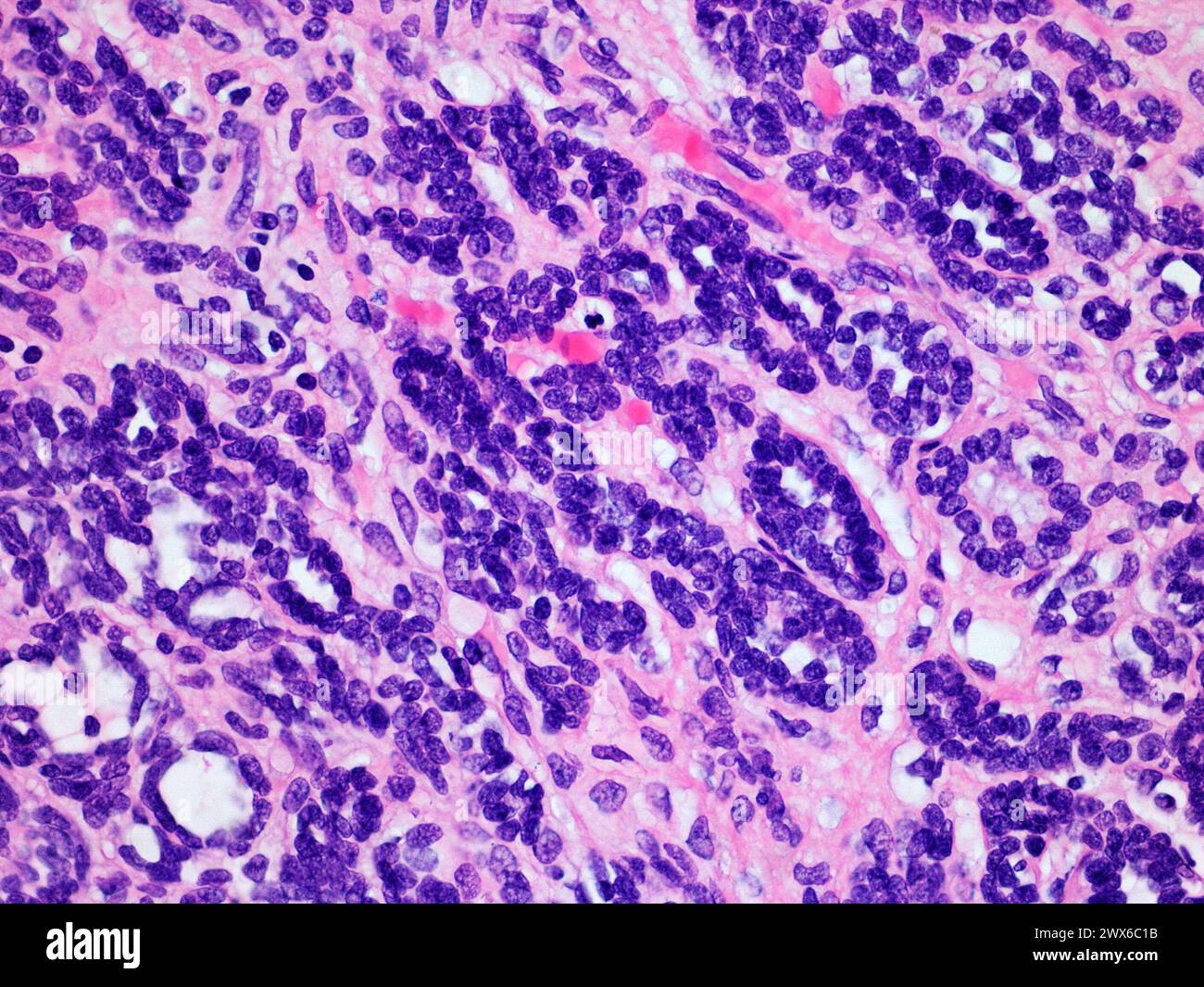 Microscopic Image of a Wilms Tumor or Nephroblastoma of a Childs Kidney Viewed at 400x Magnification with Hematoxylin and Eosin Staining Stock Photohttps://www.alamy.com/licenses-and-pricing/?v=1https://www.alamy.com/microscopic-image-of-a-wilms-tumor-or-nephroblastoma-of-a-childs-kidney-viewed-at-400x-magnification-with-hematoxylin-and-eosin-staining-image601274727.html
RF2WX6C1B–Microscopic Image of a Wilms Tumor or Nephroblastoma of a Childs Kidney Viewed at 400x Magnification with Hematoxylin and Eosin Staining
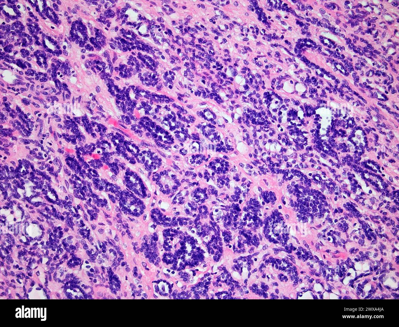 Wilms Tumor or Nephroblastoma of a Childs Kidney Viewed at 200x Magnification with Hematoxylin and Eosin Staining One of the most Common Cancers Affec Stock Photohttps://www.alamy.com/licenses-and-pricing/?v=1https://www.alamy.com/wilms-tumor-or-nephroblastoma-of-a-childs-kidney-viewed-at-200x-magnification-with-hematoxylin-and-eosin-staining-one-of-the-most-common-cancers-affec-image601356738.html
RF2WXA4JA–Wilms Tumor or Nephroblastoma of a Childs Kidney Viewed at 200x Magnification with Hematoxylin and Eosin Staining One of the most Common Cancers Affec
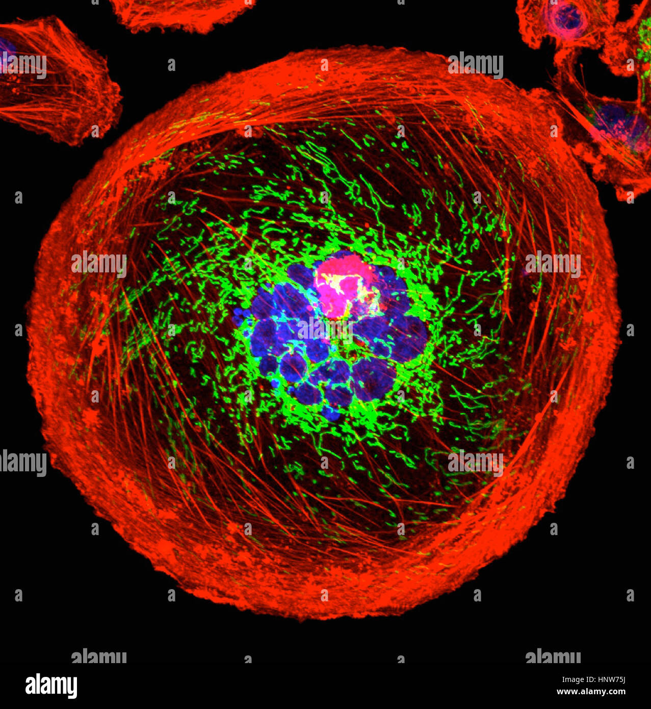 Microscopic image of polyploid giant cancer cell Stock Photohttps://www.alamy.com/licenses-and-pricing/?v=1https://www.alamy.com/stock-photo-microscopic-image-of-polyploid-giant-cancer-cell-133934798.html
RFHNW75J–Microscopic image of polyploid giant cancer cell
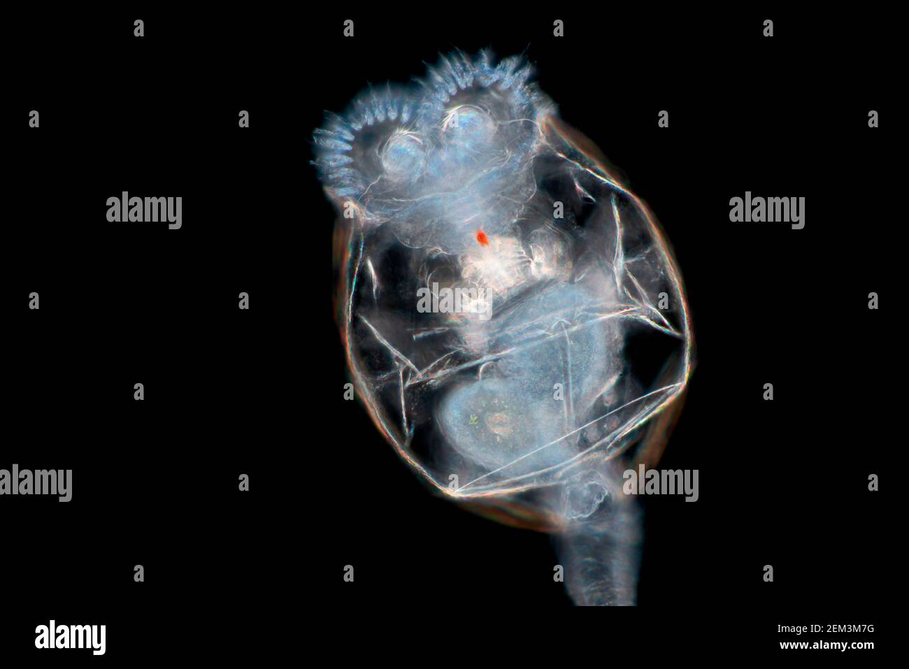 rotifers (Rotatoria), dark field microscopic image, magnification x100 related to 35 mm Stock Photohttps://www.alamy.com/licenses-and-pricing/?v=1https://www.alamy.com/rotifers-rotatoria-dark-field-microscopic-image-magnification-x100-related-to-35-mm-image408213332.html
RM2EM3M7G–rotifers (Rotatoria), dark field microscopic image, magnification x100 related to 35 mm
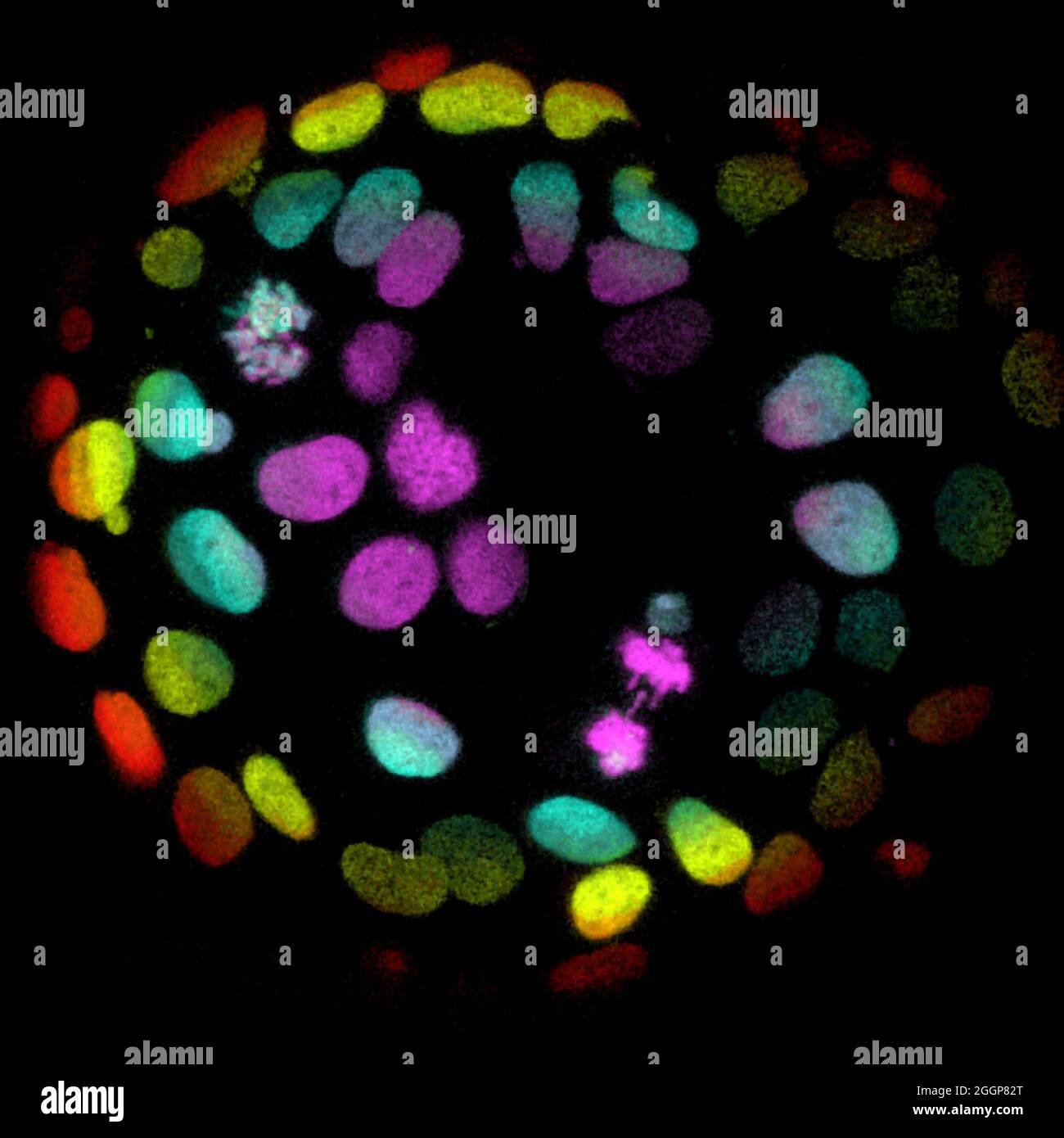 Microscopic image of colorectal cells grown into organoids. Stock Photohttps://www.alamy.com/licenses-and-pricing/?v=1https://www.alamy.com/microscopic-image-of-colorectal-cells-grown-into-organoids-image440582992.html
RM2GGP82T–Microscopic image of colorectal cells grown into organoids.
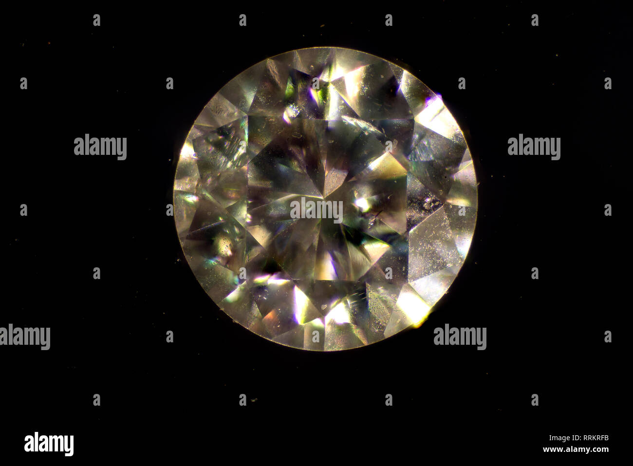 MIcroscopic image. Diamond is a solid form of the element carbon with its atoms arranged in a crystal structure called diamond cubic. Stock Photohttps://www.alamy.com/licenses-and-pricing/?v=1https://www.alamy.com/microscopic-image-diamond-is-a-solid-form-of-the-element-carbon-with-its-atoms-arranged-in-a-crystal-structure-called-diamond-cubic-image238307423.html
RMRRKRFB–MIcroscopic image. Diamond is a solid form of the element carbon with its atoms arranged in a crystal structure called diamond cubic.
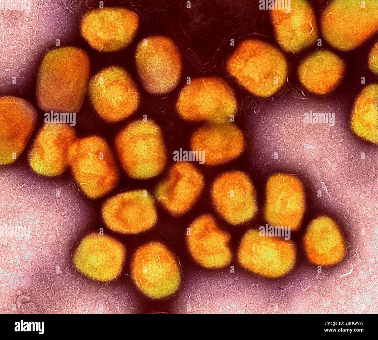 Fort Detrick, United States. 26th July, 2022. A colorized transmission electron micrograph of monkeypox virus particles (gold) cultivated and purified from cell culture captured at the NIAID Integrated Research Facility released July 26, 2022, in Fort Detrick, Maryland. Credit: NIAID/NIAID/Alamy Live News Stock Photohttps://www.alamy.com/licenses-and-pricing/?v=1https://www.alamy.com/fort-detrick-united-states-26th-july-2022-a-colorized-transmission-electron-micrograph-of-monkeypox-virus-particles-gold-cultivated-and-purified-from-cell-culture-captured-at-the-niaid-integrated-research-facility-released-july-26-2022-in-fort-detrick-maryland-credit-niaidniaidalamy-live-news-image476130197.html
RM2JJHGWW–Fort Detrick, United States. 26th July, 2022. A colorized transmission electron micrograph of monkeypox virus particles (gold) cultivated and purified from cell culture captured at the NIAID Integrated Research Facility released July 26, 2022, in Fort Detrick, Maryland. Credit: NIAID/NIAID/Alamy Live News
 Transmission electron microscopic image of an isolate from the first U.S. case of COVID-19, formerly known as 2019-nCoV. The spherical viral particles, colorized blue, contain cross-sections through the viral genome, seen as black dots. Credit: UPI/Alamy Live News Stock Photohttps://www.alamy.com/licenses-and-pricing/?v=1https://www.alamy.com/transmission-electron-microscopic-image-of-an-isolate-from-the-first-us-case-of-covid-19-formerly-known-as-2019-ncov-the-spherical-viral-particles-colorized-blue-contain-cross-sections-through-the-viral-genome-seen-as-black-dots-credit-upialamy-live-news-image348816489.html
RM2B7DXYN–Transmission electron microscopic image of an isolate from the first U.S. case of COVID-19, formerly known as 2019-nCoV. The spherical viral particles, colorized blue, contain cross-sections through the viral genome, seen as black dots. Credit: UPI/Alamy Live News
 Science students looking at microscopic image on computer Stock Photohttps://www.alamy.com/licenses-and-pricing/?v=1https://www.alamy.com/stock-photo-science-students-looking-at-microscopic-image-on-computer-78168741.html
RFEF4W0N–Science students looking at microscopic image on computer
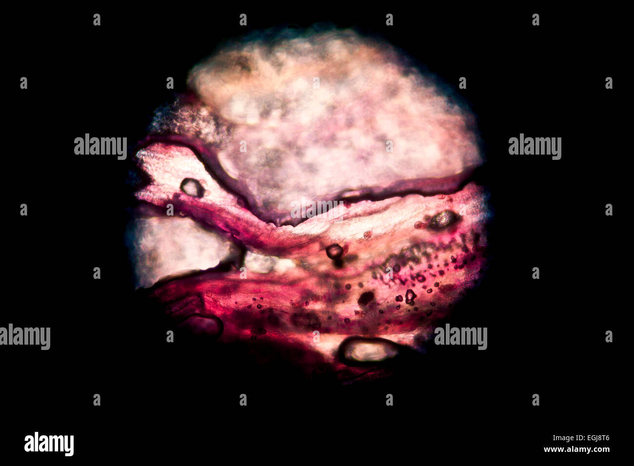 Circular microscopic image of a pink flower petal Stock Photohttps://www.alamy.com/licenses-and-pricing/?v=1https://www.alamy.com/stock-photo-circular-microscopic-image-of-a-pink-flower-petal-79078054.html
RMEGJ8T6–Circular microscopic image of a pink flower petal
 Details of Coronavirus COVID-19 on human cells, 3D illustration as a microscopic image inside the human body based on SEM SARS photos Stock Photohttps://www.alamy.com/licenses-and-pricing/?v=1https://www.alamy.com/details-of-coronavirus-covid-19-on-human-cells-3d-illustration-as-a-microscopic-image-inside-the-human-body-based-on-sem-sars-photos-image351663470.html
RF2BC3J9J–Details of Coronavirus COVID-19 on human cells, 3D illustration as a microscopic image inside the human body based on SEM SARS photos
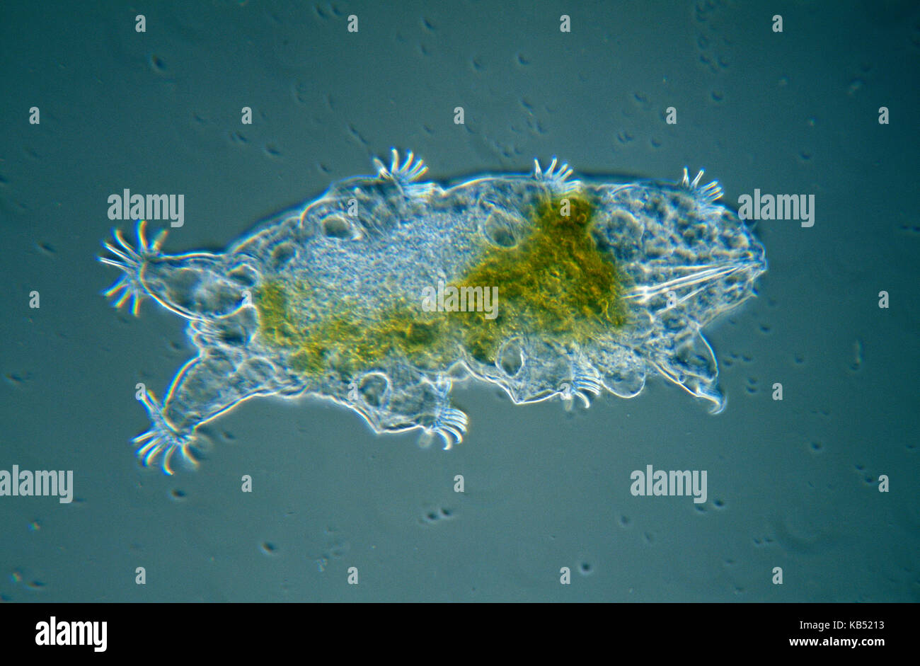 Tardigrade (Echiniscoides sigismundi) microscopic image, animal is less than one mm in length, can enter cryptobiosis to withstand temperature and moisture extremes Stock Photohttps://www.alamy.com/licenses-and-pricing/?v=1https://www.alamy.com/stock-image-tardigrade-echiniscoides-sigismundi-microscopic-image-animal-is-less-161765887.html
RMKB5213–Tardigrade (Echiniscoides sigismundi) microscopic image, animal is less than one mm in length, can enter cryptobiosis to withstand temperature and moisture extremes
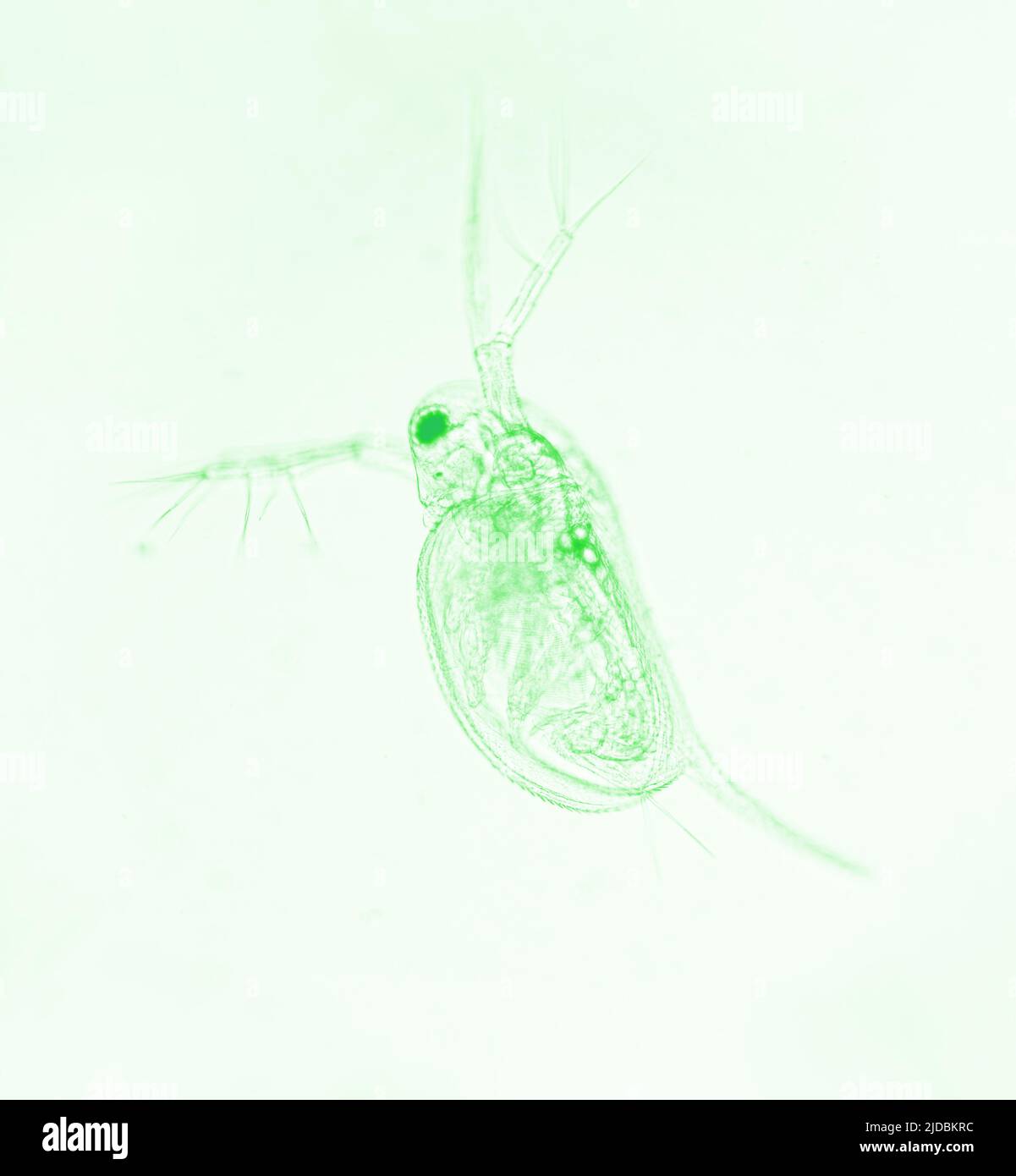 Zooplankton Water Flea Daphnia, microscopic image of crustacea, green effect Stock Photohttps://www.alamy.com/licenses-and-pricing/?v=1https://www.alamy.com/zooplankton-water-flea-daphnia-microscopic-image-of-crustacea-green-effect-image472927488.html
RF2JDBKRC–Zooplankton Water Flea Daphnia, microscopic image of crustacea, green effect
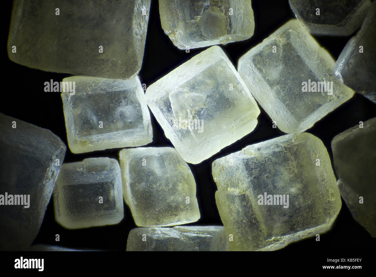 Brown sugar on black background microscopic image, dark field technique, magnification x 40 Stock Photohttps://www.alamy.com/licenses-and-pricing/?v=1https://www.alamy.com/stock-image-brown-sugar-on-black-background-microscopic-image-dark-field-technique-161776467.html
RFKB5FEY–Brown sugar on black background microscopic image, dark field technique, magnification x 40
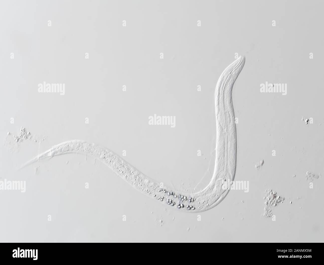 Microscopic free-living nematode worm, possibly Panagrellus sp., horizontal field of view is approximately 235 micrometers Stock Photohttps://www.alamy.com/licenses-and-pricing/?v=1https://www.alamy.com/microscopic-free-living-nematode-worm-possibly-panagrellus-sp-horizontal-field-of-view-is-approximately-235-micrometers-image340364352.html
RM2ANMX5M–Microscopic free-living nematode worm, possibly Panagrellus sp., horizontal field of view is approximately 235 micrometers
 Microscopic image of brain neurons Stock Photohttps://www.alamy.com/licenses-and-pricing/?v=1https://www.alamy.com/microscopic-image-of-brain-neurons-image330240451.html
RM2A57N1R–Microscopic image of brain neurons
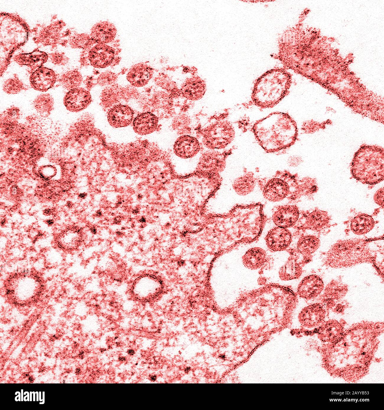 Transmission electron microscopic image of an isolate from the first U.S. case of COVID-19, formerly known as 2019-nCoV. The spherical extracellular viral particles contain cross-sections through the viral genome, seen as red dots. Stock Photohttps://www.alamy.com/licenses-and-pricing/?v=1https://www.alamy.com/transmission-electron-microscopic-image-of-an-isolate-from-the-first-us-case-of-covid-19-formerly-known-as-2019-ncov-the-spherical-extracellular-viral-particles-contain-cross-sections-through-the-viral-genome-seen-as-red-dots-image344194175.html
RF2AYYB53–Transmission electron microscopic image of an isolate from the first U.S. case of COVID-19, formerly known as 2019-nCoV. The spherical extracellular viral particles contain cross-sections through the viral genome, seen as red dots.
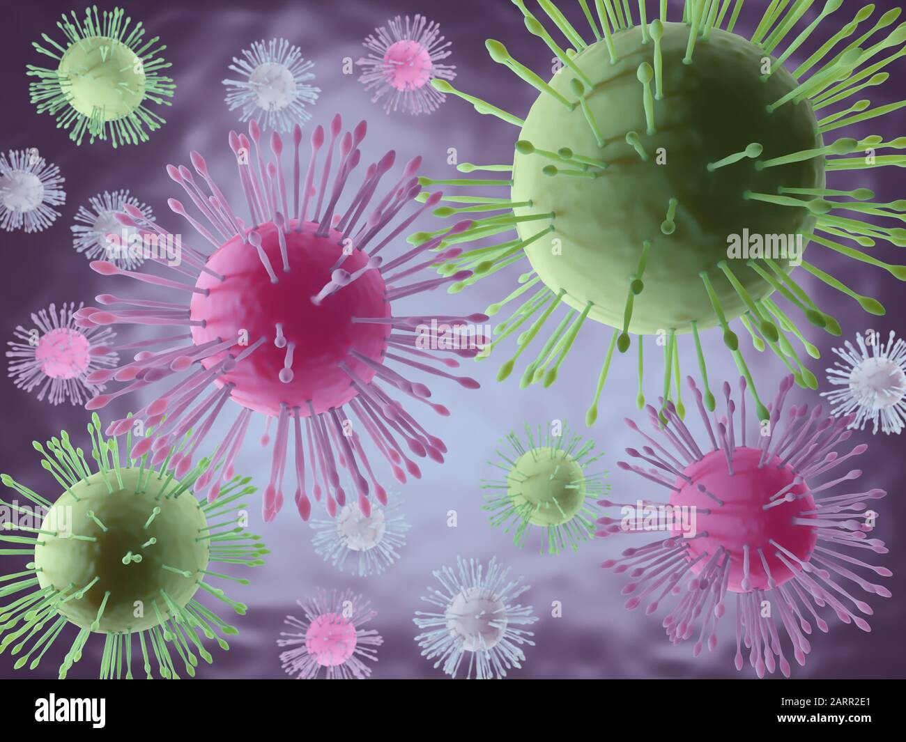 3D rendering of microscopic image of deadly coronavirus particles Stock Photohttps://www.alamy.com/licenses-and-pricing/?v=1https://www.alamy.com/3d-rendering-of-microscopic-image-of-deadly-coronavirus-particles-image341640937.html
RF2ARR2E1–3D rendering of microscopic image of deadly coronavirus particles
 3d illustration of microscopic image of a virus or infectious cell.Microbacteria and bacterial organisms.biology and science background. Stock Photohttps://www.alamy.com/licenses-and-pricing/?v=1https://www.alamy.com/3d-illustration-of-microscopic-image-of-a-virus-or-infectious-cellmicrobacteria-and-bacterial-organismsbiology-and-science-background-image523143778.html
RF2NB376A–3d illustration of microscopic image of a virus or infectious cell.Microbacteria and bacterial organisms.biology and science background.
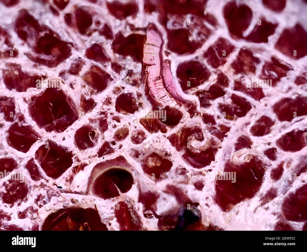 A microscopic image of stomach tissue Stock Photohttps://www.alamy.com/licenses-and-pricing/?v=1https://www.alamy.com/a-microscopic-image-of-stomach-tissue-image473728222.html
RM2JEM552–A microscopic image of stomach tissue
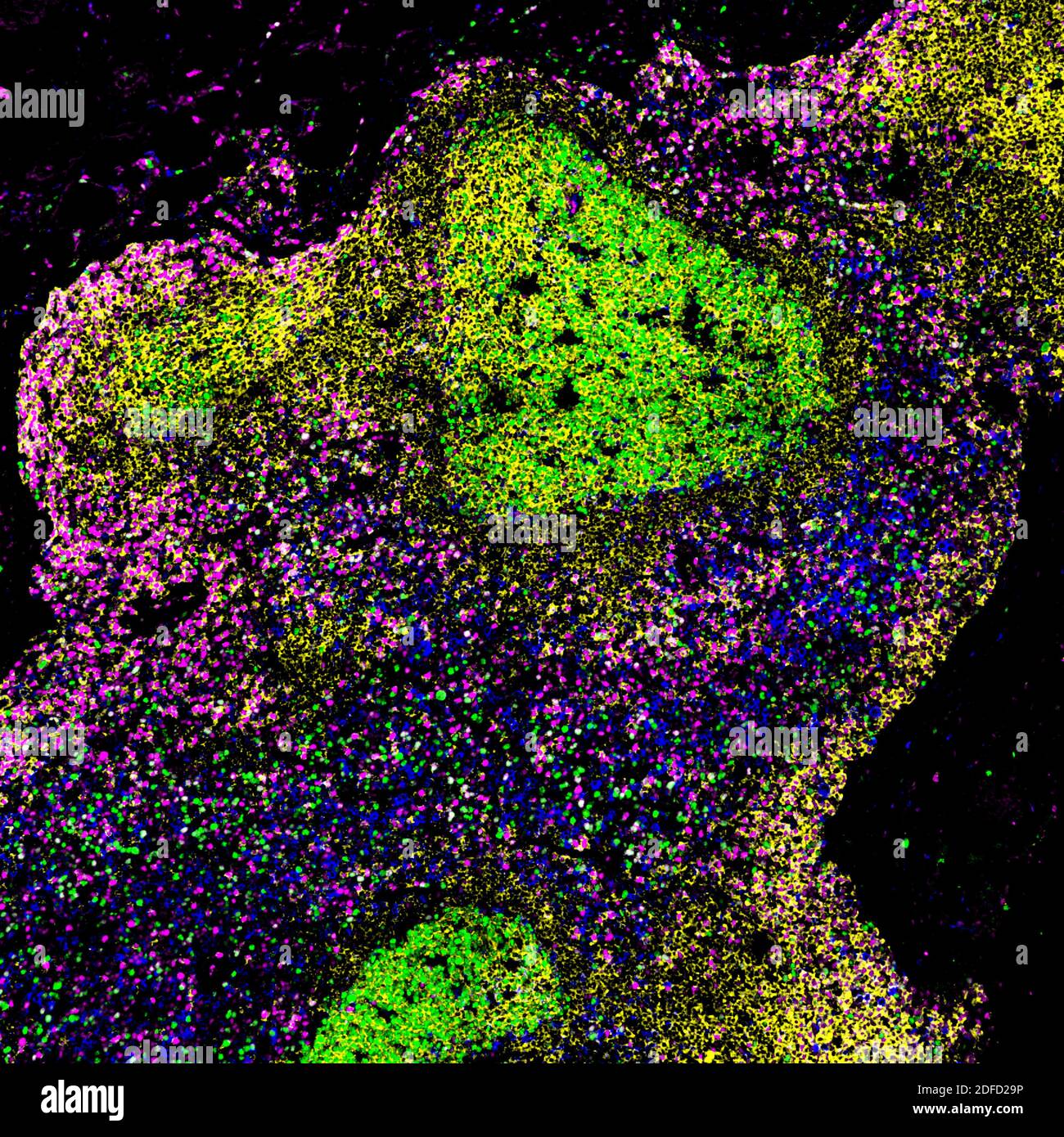 A microscopic image of a biopsied lymph node of a person with untreated HIV, showing large germinal centers containing abnormal proliferating B cells Stock Photohttps://www.alamy.com/licenses-and-pricing/?v=1https://www.alamy.com/a-microscopic-image-of-a-biopsied-lymph-node-of-a-person-with-untreated-hiv-showing-large-germinal-centers-containing-abnormal-proliferating-b-cells-image388135154.html
RF2DFD29P–A microscopic image of a biopsied lymph node of a person with untreated HIV, showing large germinal centers containing abnormal proliferating B cells
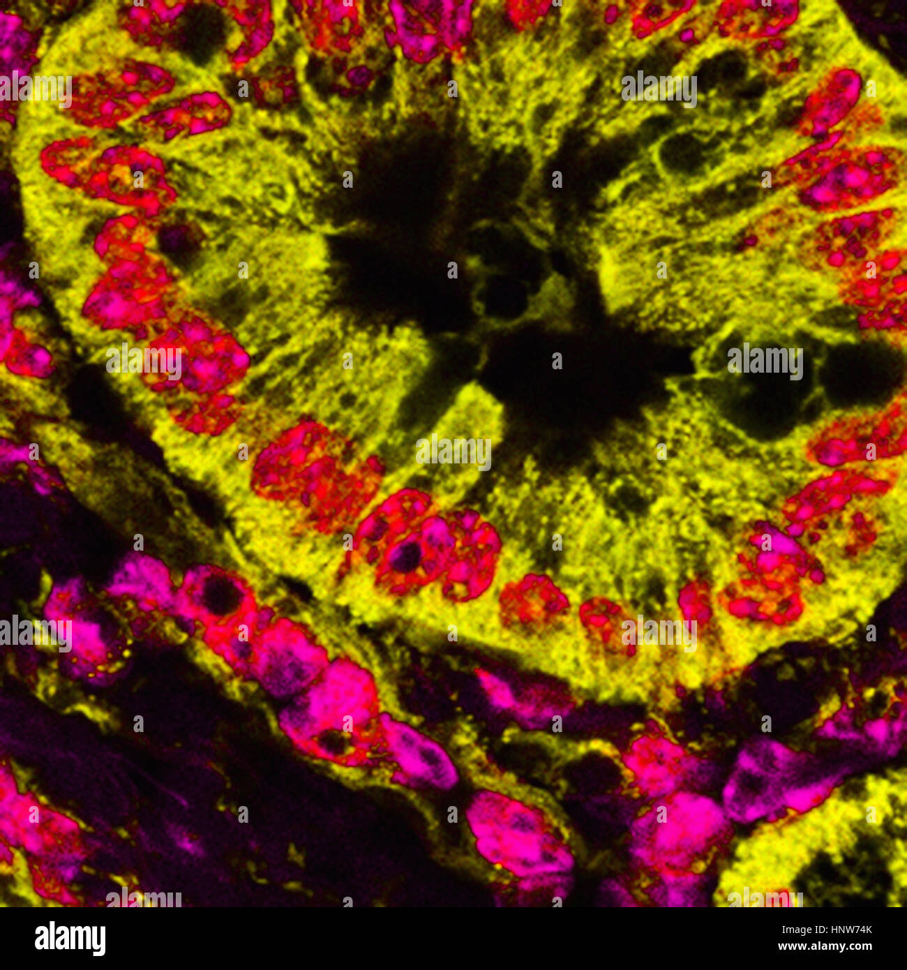 Microscopic image of mitochondrial stained pancreatic cancer cells Stock Photohttps://www.alamy.com/licenses-and-pricing/?v=1https://www.alamy.com/stock-photo-microscopic-image-of-mitochondrial-stained-pancreatic-cancer-cells-133934771.html
RFHNW74K–Microscopic image of mitochondrial stained pancreatic cancer cells
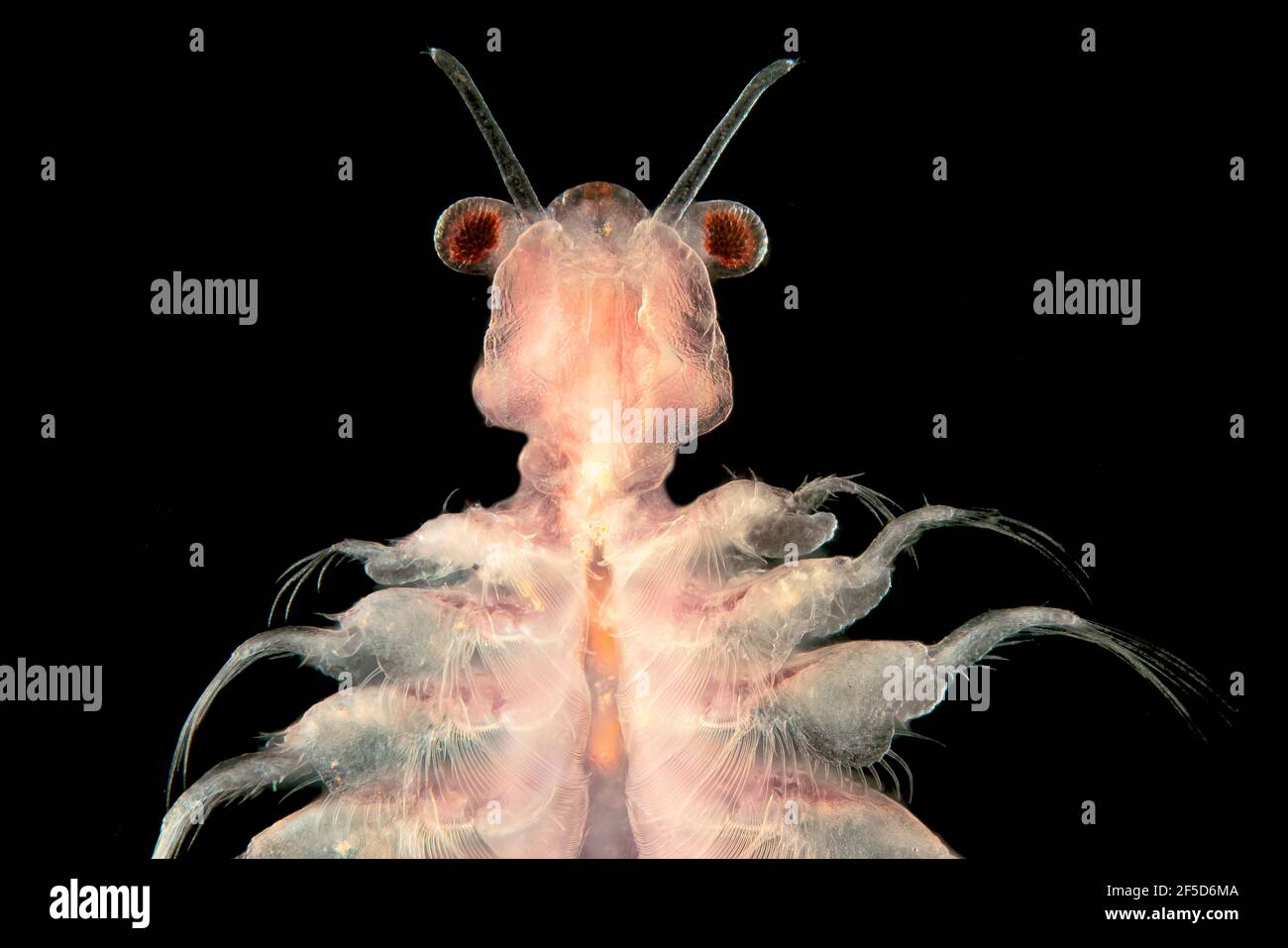 brine shrimp (Artemia salina), dark field microscopic image, magnification x20 related to 35 mm Stock Photohttps://www.alamy.com/licenses-and-pricing/?v=1https://www.alamy.com/brine-shrimp-artemia-salina-dark-field-microscopic-image-magnification-x20-related-to-35-mm-image416412762.html
RM2F5D6MA–brine shrimp (Artemia salina), dark field microscopic image, magnification x20 related to 35 mm
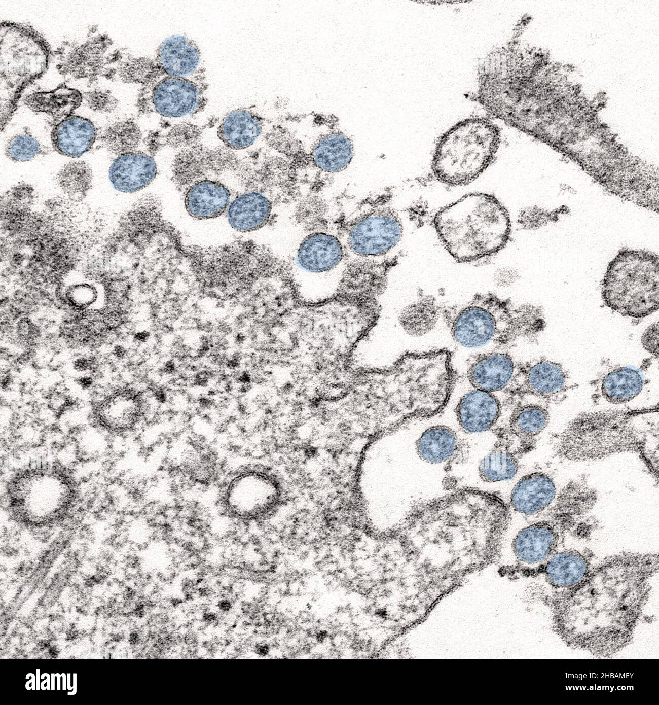 Transmission electron microscopic image of an isolate from the first U.S. case of COVID-19, formerly known as 2019-nCoV. The spherical viral particles, colourised blue, contain cross-sections through the viral genome, seen as black dots. An optimised and enhanced version of an image produced by the US Centers for Disease Control and Prevention / Credit: CDC / C.S. Goldsmith & A. Tamin Stock Photohttps://www.alamy.com/licenses-and-pricing/?v=1https://www.alamy.com/transmission-electron-microscopic-image-of-an-isolate-from-the-first-us-case-of-covid-19-formerly-known-as-2019-ncov-the-spherical-viral-particles-colourised-blue-contain-cross-sections-through-the-viral-genome-seen-as-black-dots-an-optimised-and-enhanced-version-of-an-image-produced-by-the-us-centers-for-disease-control-and-prevention-credit-cdc-cs-goldsmith-a-tamin-image454466403.html
RM2HBAMEY–Transmission electron microscopic image of an isolate from the first U.S. case of COVID-19, formerly known as 2019-nCoV. The spherical viral particles, colourised blue, contain cross-sections through the viral genome, seen as black dots. An optimised and enhanced version of an image produced by the US Centers for Disease Control and Prevention / Credit: CDC / C.S. Goldsmith & A. Tamin
 Microscopic image Stock Photohttps://www.alamy.com/licenses-and-pricing/?v=1https://www.alamy.com/stock-photo-microscopic-image-95593682.html
RMFFEJM2–Microscopic image
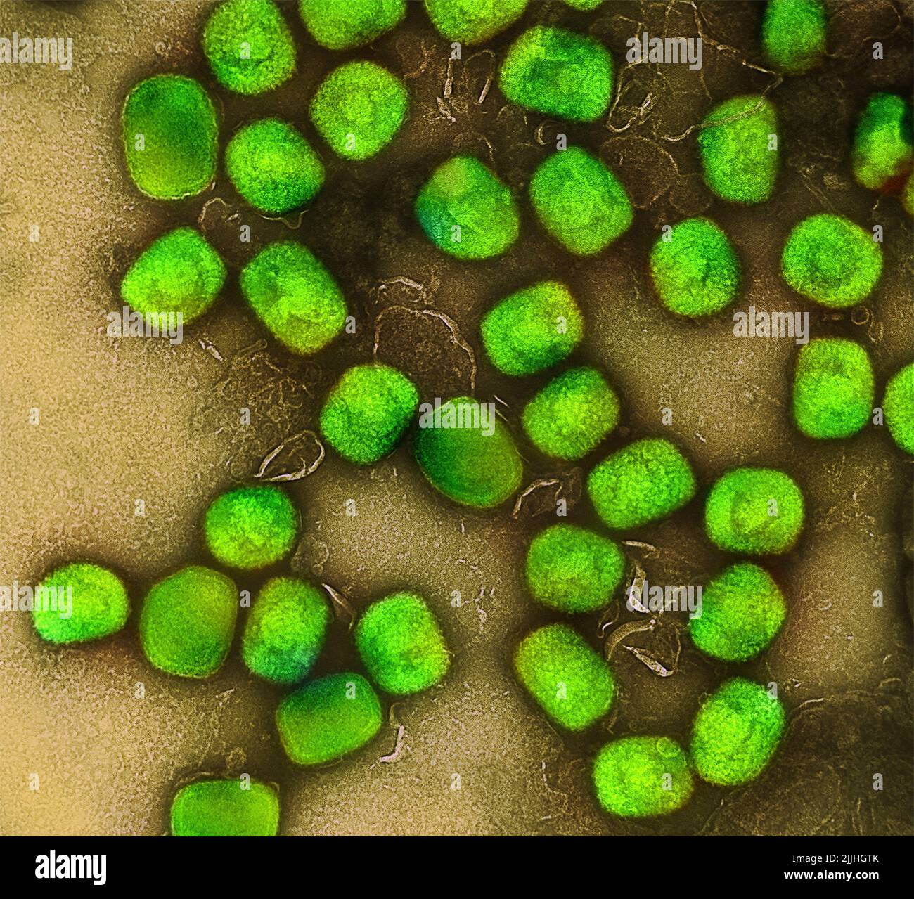 Fort Detrick, United States. 26th July, 2022. A colorized transmission electron micrograph of monkeypox virus particles (green) cultivated and purified from cell culture captured at the NIAID Integrated Research Facility released July 26, 2022, in Fort Detrick, Maryland. Credit: NIAID/NIAID/Alamy Live News Stock Photohttps://www.alamy.com/licenses-and-pricing/?v=1https://www.alamy.com/fort-detrick-united-states-26th-july-2022-a-colorized-transmission-electron-micrograph-of-monkeypox-virus-particles-green-cultivated-and-purified-from-cell-culture-captured-at-the-niaid-integrated-research-facility-released-july-26-2022-in-fort-detrick-maryland-credit-niaidniaidalamy-live-news-image476130163.html
RM2JJHGTK–Fort Detrick, United States. 26th July, 2022. A colorized transmission electron micrograph of monkeypox virus particles (green) cultivated and purified from cell culture captured at the NIAID Integrated Research Facility released July 26, 2022, in Fort Detrick, Maryland. Credit: NIAID/NIAID/Alamy Live News
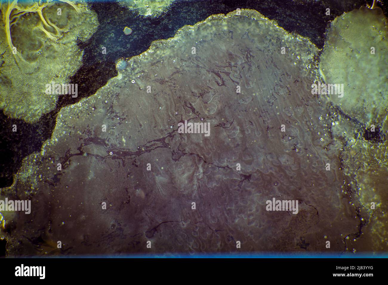 Fungal growth on biological material microscopic image Stock Photohttps://www.alamy.com/licenses-and-pricing/?v=1https://www.alamy.com/fungal-growth-on-biological-material-microscopic-image-image469684980.html
RF2J83YYG–Fungal growth on biological material microscopic image
 Science students looking at microscopic image on computer Stock Photohttps://www.alamy.com/licenses-and-pricing/?v=1https://www.alamy.com/stock-photo-science-students-looking-at-microscopic-image-on-computer-78168743.html
RFEF4W0R–Science students looking at microscopic image on computer
 Microscopic image of a water droplet on a leaf Stock Photohttps://www.alamy.com/licenses-and-pricing/?v=1https://www.alamy.com/stock-photo-microscopic-image-of-a-water-droplet-on-a-leaf-79078048.html
RMEGJ8T0–Microscopic image of a water droplet on a leaf
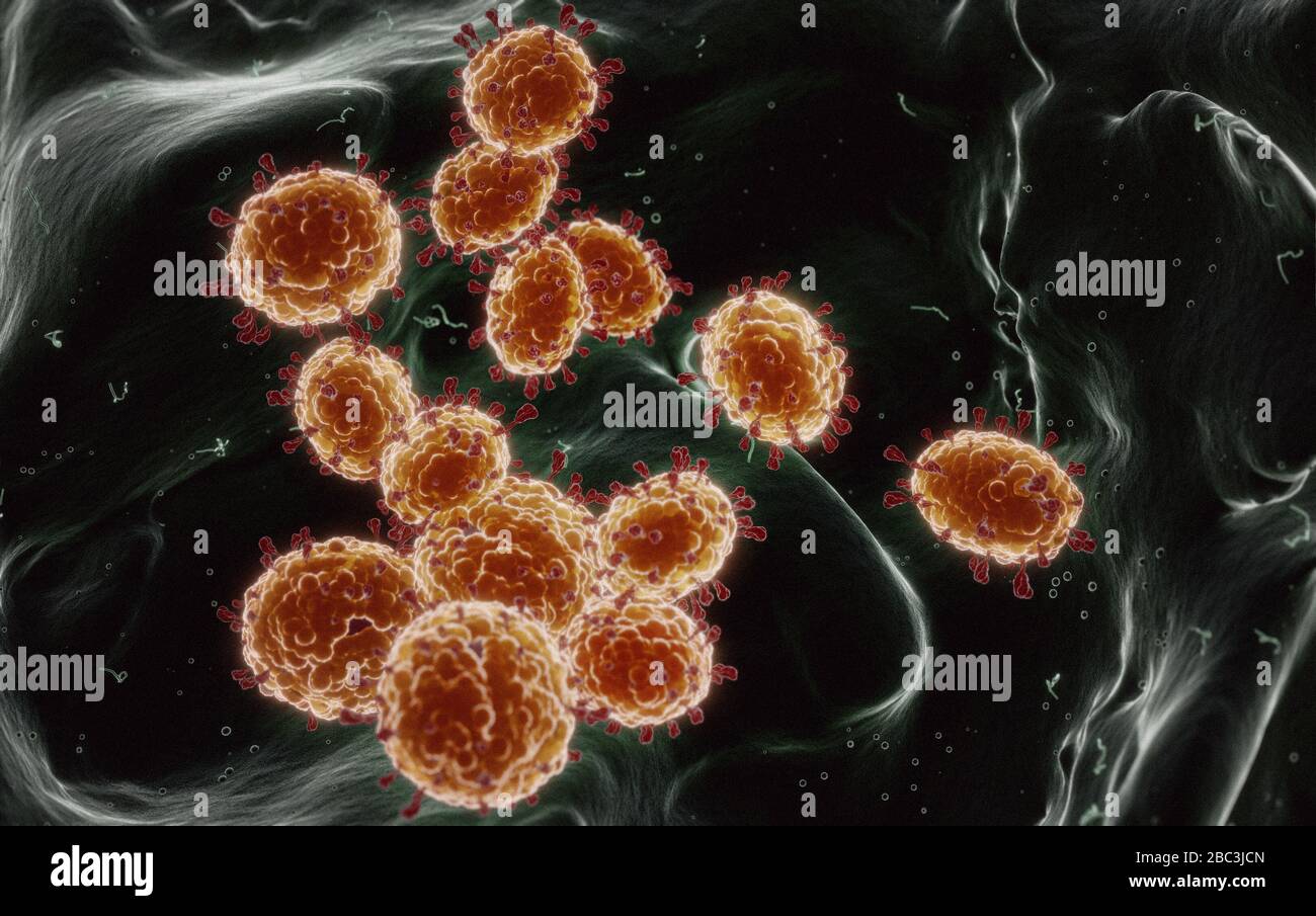 Details of Coronavirus COVID-19 on human cells, 3D illustration as a microscopic image inside the human body based on SEM SARS photos Stock Photohttps://www.alamy.com/licenses-and-pricing/?v=1https://www.alamy.com/details-of-coronavirus-covid-19-on-human-cells-3d-illustration-as-a-microscopic-image-inside-the-human-body-based-on-sem-sars-photos-image351663557.html
RF2BC3JCN–Details of Coronavirus COVID-19 on human cells, 3D illustration as a microscopic image inside the human body based on SEM SARS photos
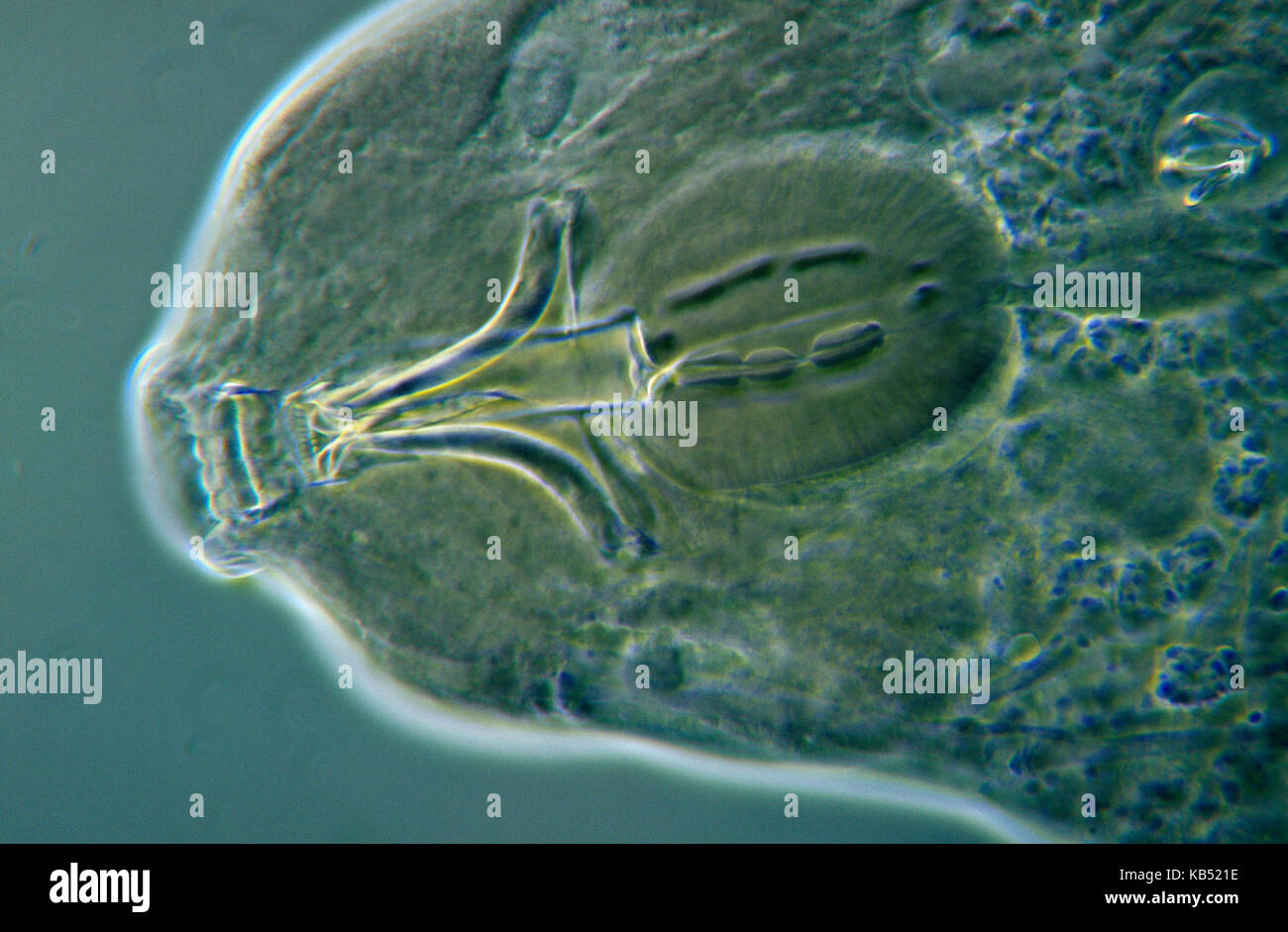 Water Bear (Macrobiotus richtersi) microscopic image, animal is less than one mm in length, can enter cryptobiosis to withstand temperature and moisture extremes Stock Photohttps://www.alamy.com/licenses-and-pricing/?v=1https://www.alamy.com/stock-image-water-bear-macrobiotus-richtersi-microscopic-image-animal-is-less-161765898.html
RMKB521E–Water Bear (Macrobiotus richtersi) microscopic image, animal is less than one mm in length, can enter cryptobiosis to withstand temperature and moisture extremes
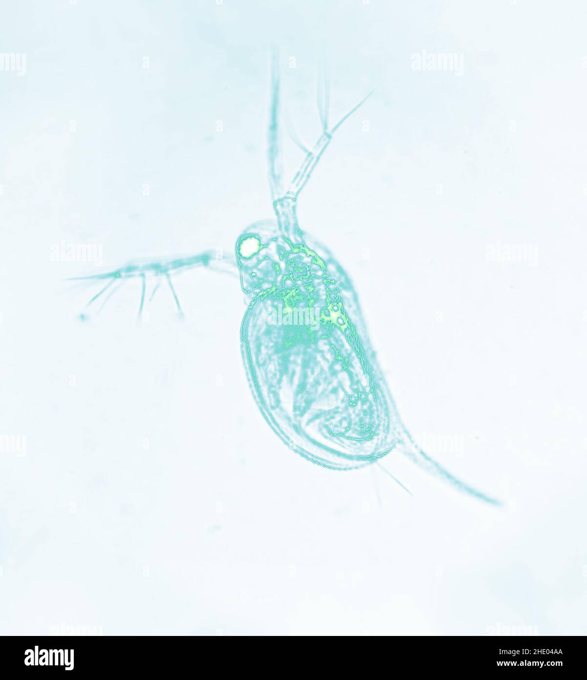 Microscopic image of zooplankton Water Flea Daphnia Stock Photohttps://www.alamy.com/licenses-and-pricing/?v=1https://www.alamy.com/microscopic-image-of-zooplankton-water-flea-daphnia-image456078178.html
RF2HE04AA–Microscopic image of zooplankton Water Flea Daphnia
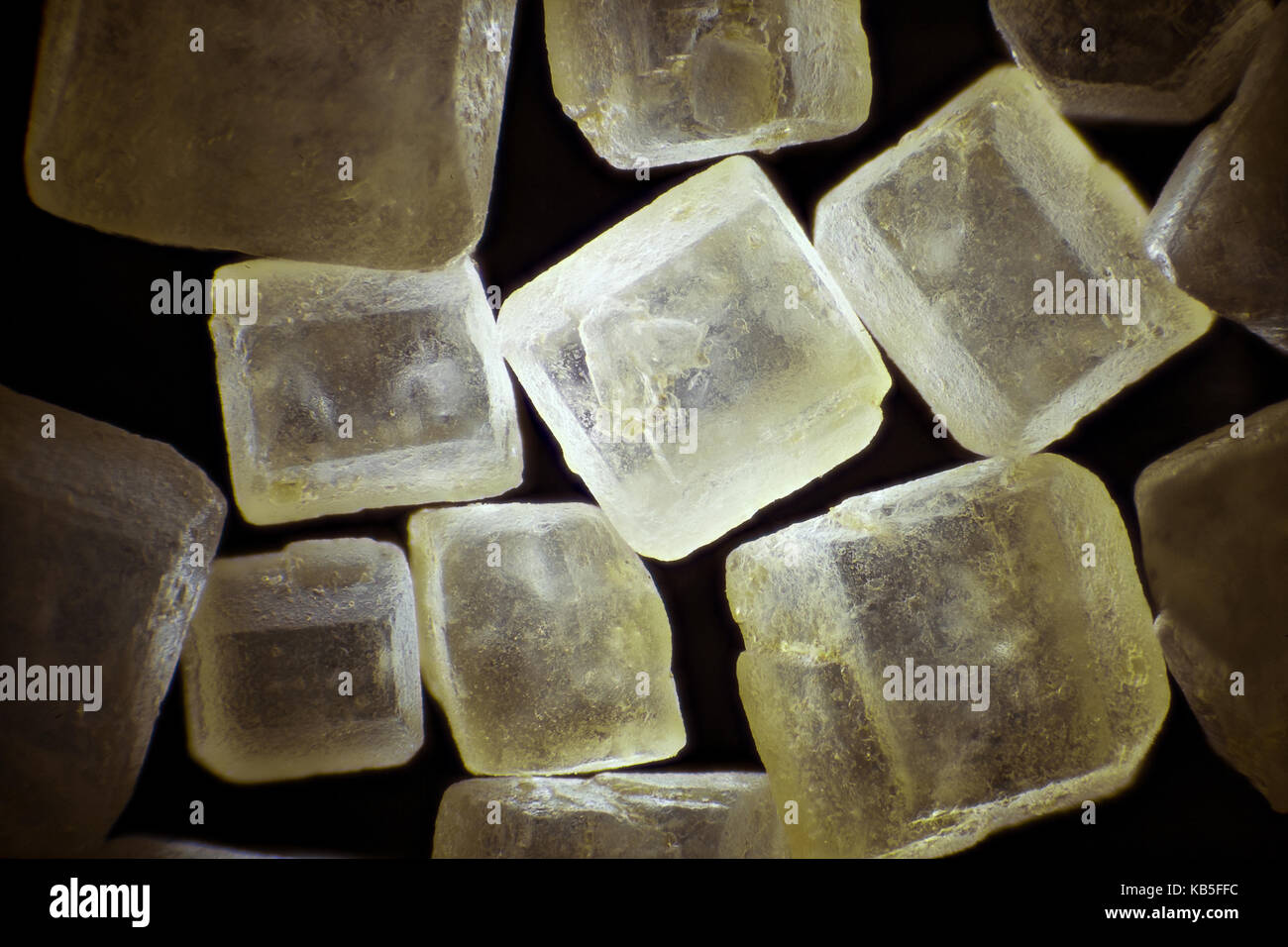 Brown sugar on black background microscopic image, dark field technique, magnification x 40 Stock Photohttps://www.alamy.com/licenses-and-pricing/?v=1https://www.alamy.com/stock-image-brown-sugar-on-black-background-microscopic-image-dark-field-technique-161776480.html
RFKB5FFC–Brown sugar on black background microscopic image, dark field technique, magnification x 40
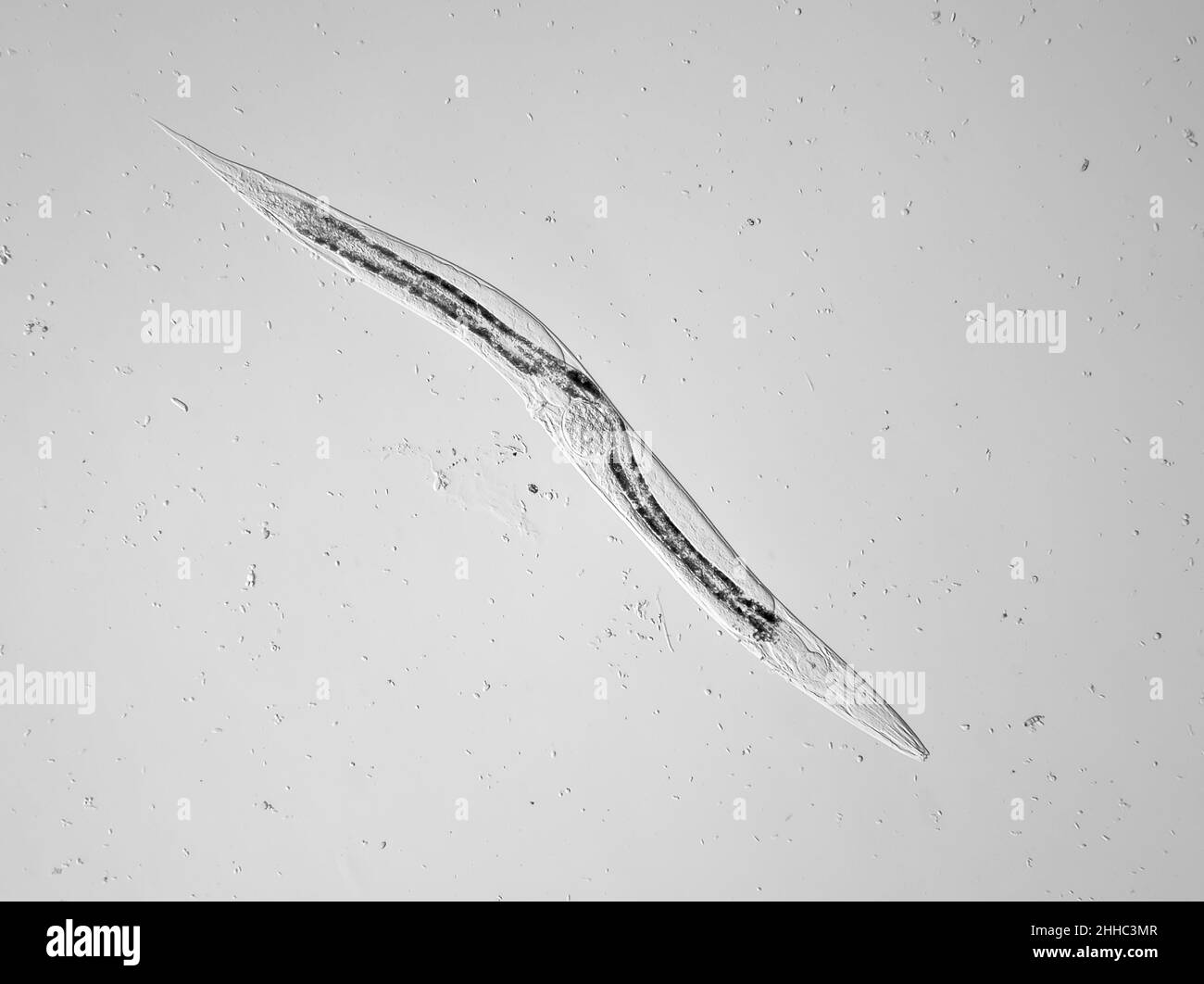 Microscopic free-living nematode worm (with an egg inside) from garden soil, possibly Panagrellus sp., horizontal field of view is about 1.1mm Stock Photohttps://www.alamy.com/licenses-and-pricing/?v=1https://www.alamy.com/microscopic-free-living-nematode-worm-with-an-egg-inside-from-garden-soil-possibly-panagrellus-sp-horizontal-field-of-view-is-about-11mm-image458185079.html
RM2HHC3MR–Microscopic free-living nematode worm (with an egg inside) from garden soil, possibly Panagrellus sp., horizontal field of view is about 1.1mm
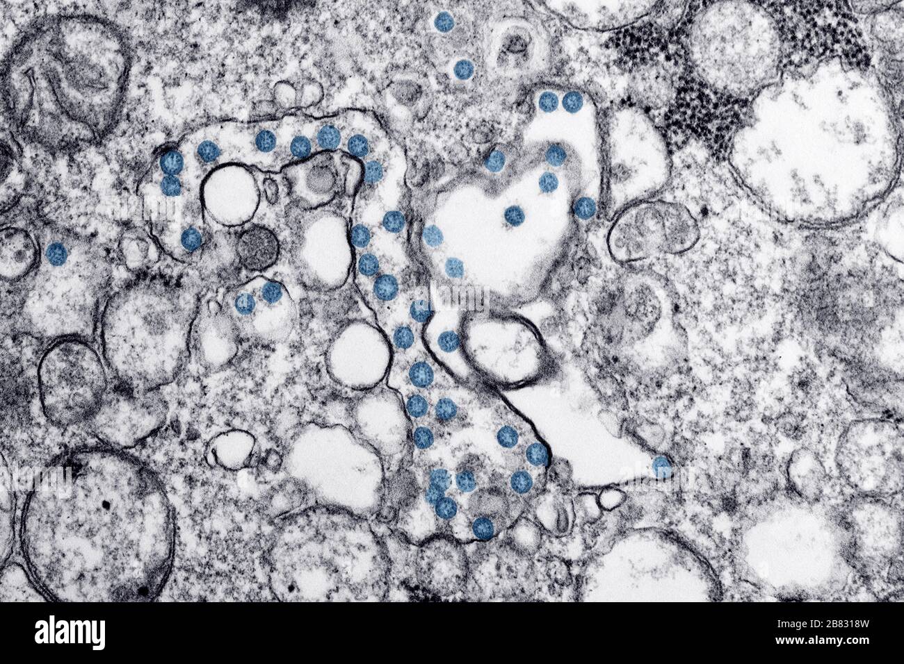 Transmission electron microscopic image of an isolate from the first US case of COVID-19, formerly known as 2019-nCoV, a coronavirus, March, 2020. The spherical viral particles, colorized blue, contain cross-sections through the viral genome, seen as black dots. Image courtesy CDC/ Hannah A Bullock, Azaibi Tamin. () Stock Photohttps://www.alamy.com/licenses-and-pricing/?v=1https://www.alamy.com/transmission-electron-microscopic-image-of-an-isolate-from-the-first-us-case-of-covid-19-formerly-known-as-2019-ncov-a-coronavirus-march-2020-the-spherical-viral-particles-colorized-blue-contain-cross-sections-through-the-viral-genome-seen-as-black-dots-image-courtesy-cdc-hannah-a-bullock-azaibi-tamin-image349191497.html
RM2B8318W–Transmission electron microscopic image of an isolate from the first US case of COVID-19, formerly known as 2019-nCoV, a coronavirus, March, 2020. The spherical viral particles, colorized blue, contain cross-sections through the viral genome, seen as black dots. Image courtesy CDC/ Hannah A Bullock, Azaibi Tamin. ()
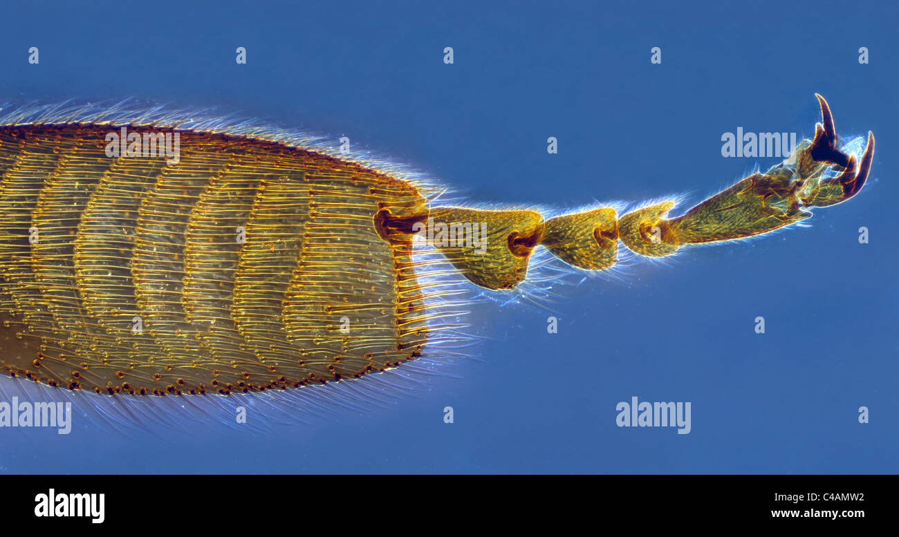 Microscopic image of bee leg isolated on blue background Stock Photohttps://www.alamy.com/licenses-and-pricing/?v=1https://www.alamy.com/stock-photo-microscopic-image-of-bee-leg-isolated-on-blue-background-37115262.html
RFC4AMW2–Microscopic image of bee leg isolated on blue background
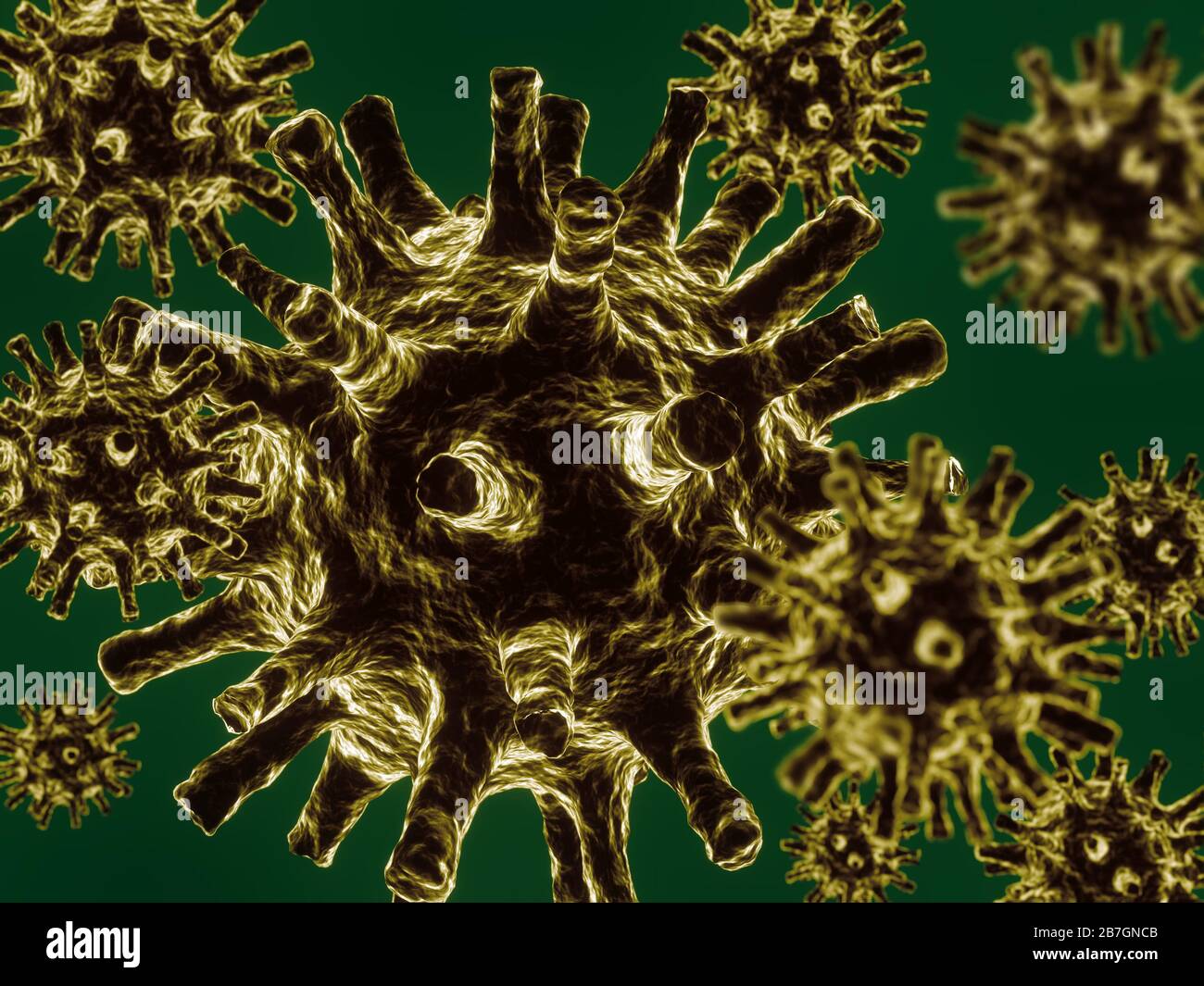 3D rendering og microscopic image of deadly coronavirus particles Stock Photohttps://www.alamy.com/licenses-and-pricing/?v=1https://www.alamy.com/3d-rendering-og-microscopic-image-of-deadly-coronavirus-particles-image348877995.html
RF2B7GNCB–3D rendering og microscopic image of deadly coronavirus particles
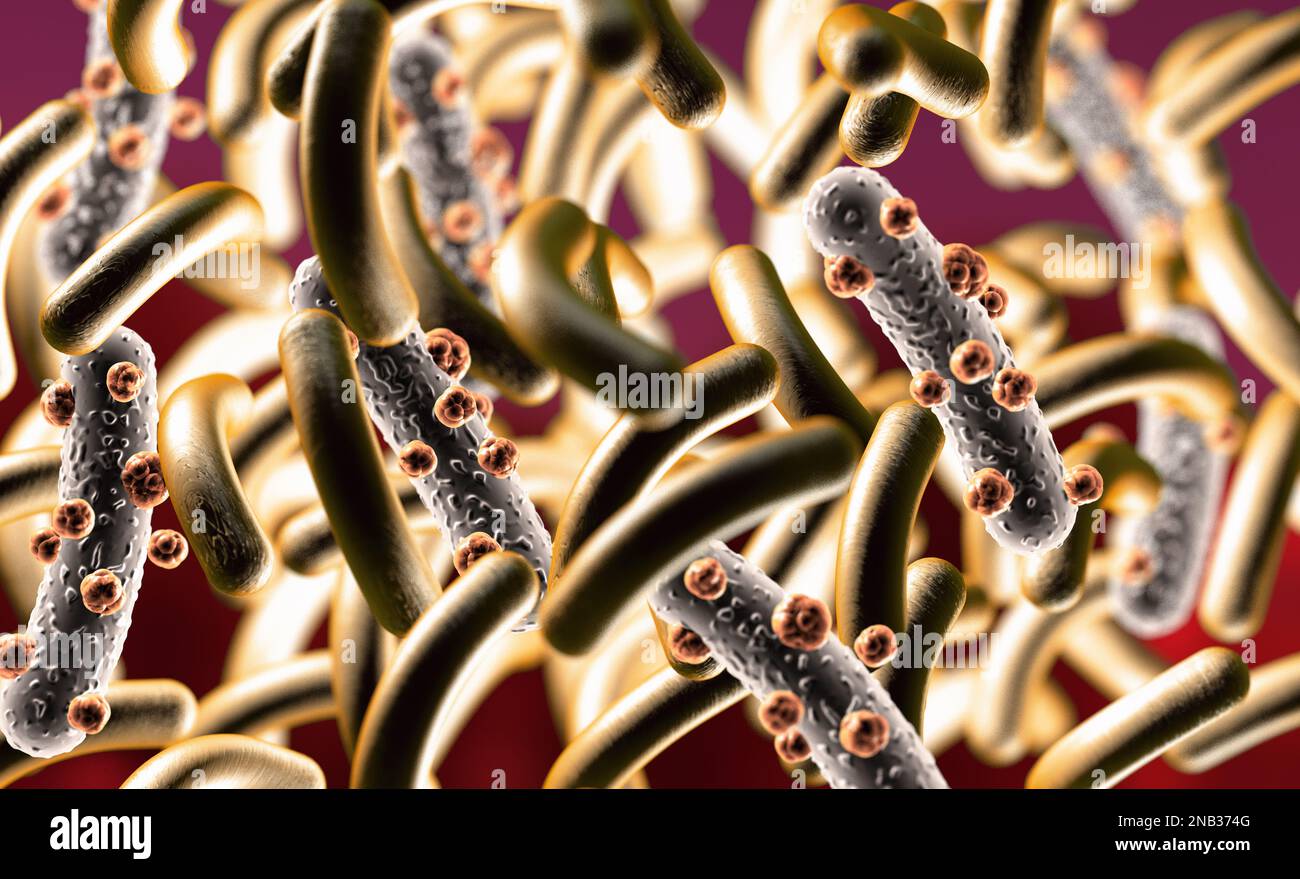 3d illustration of microscopic image of a virus or infectious cell.Microbacteria and bacterial organisms.biology and science background. Stock Photohttps://www.alamy.com/licenses-and-pricing/?v=1https://www.alamy.com/3d-illustration-of-microscopic-image-of-a-virus-or-infectious-cellmicrobacteria-and-bacterial-organismsbiology-and-science-background-image523143728.html
RF2NB374G–3d illustration of microscopic image of a virus or infectious cell.Microbacteria and bacterial organisms.biology and science background.
 An intense and continuing interest in all aspects of the plot to assassinate Abraham Lincoln resulted in curiosities like this, even though it was not published until 1867, two years after the event. A magnified image of John WIlkes Booth within a bullet representing the one which he killed President Lincoln with on April 14, 1865. By an unidentified artist. Stock Photohttps://www.alamy.com/licenses-and-pricing/?v=1https://www.alamy.com/an-intense-and-continuing-interest-in-all-aspects-of-the-plot-to-assassinate-abraham-lincoln-resulted-in-curiosities-like-this-even-though-it-was-not-published-until-1867-two-years-after-the-event-a-magnified-image-of-john-wilkes-booth-within-a-bullet-representing-the-one-which-he-killed-president-lincoln-with-on-april-14-1865-by-an-unidentified-artist-image461420853.html
RM2HPKF05–An intense and continuing interest in all aspects of the plot to assassinate Abraham Lincoln resulted in curiosities like this, even though it was not published until 1867, two years after the event. A magnified image of John WIlkes Booth within a bullet representing the one which he killed President Lincoln with on April 14, 1865. By an unidentified artist.
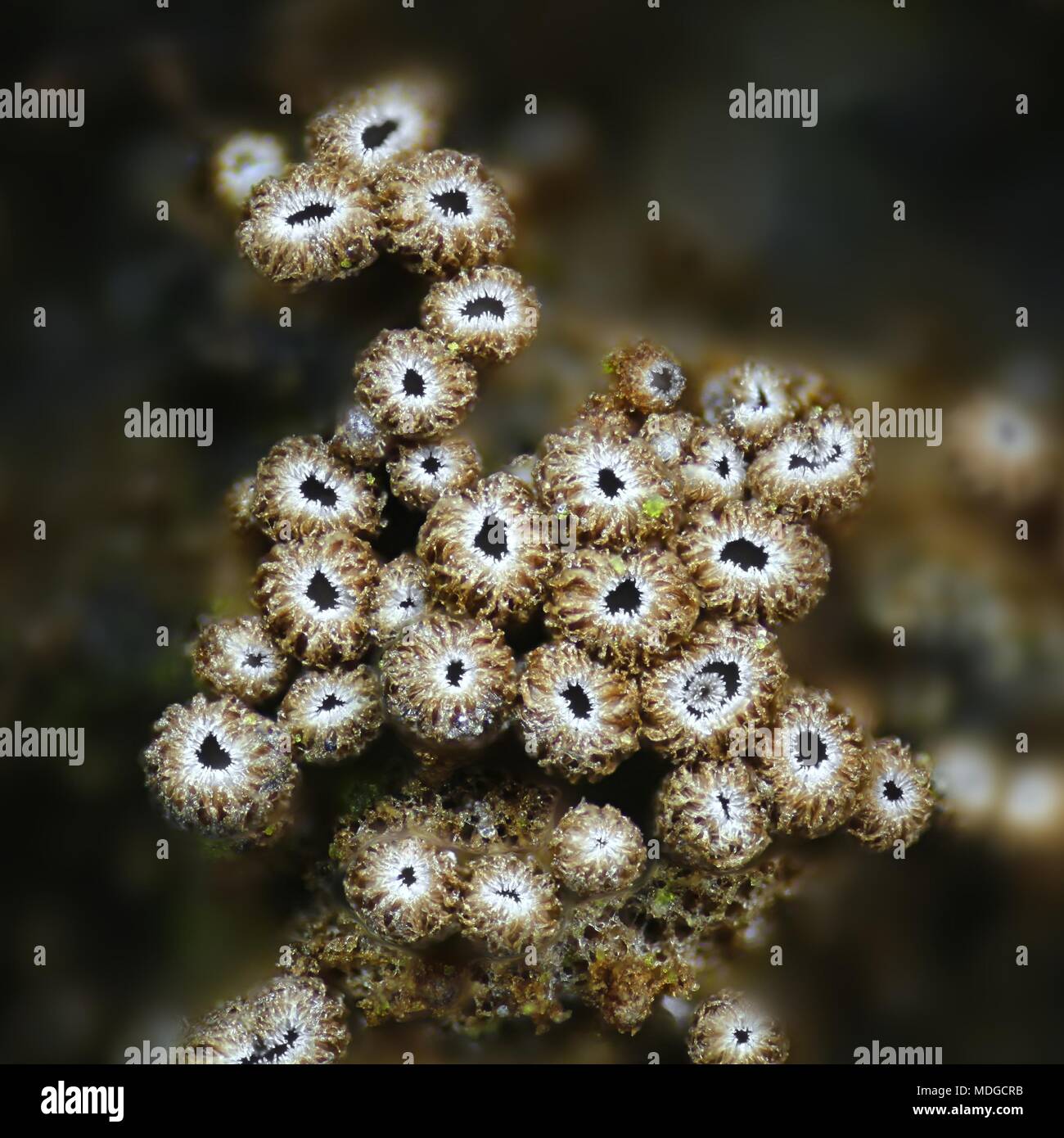 Microscopic fungus, Merismodes anomala, a microscope image Stock Photohttps://www.alamy.com/licenses-and-pricing/?v=1https://www.alamy.com/microscopic-fungus-merismodes-anomala-a-microscope-image-image180455503.html
RFMDGCRB–Microscopic fungus, Merismodes anomala, a microscope image
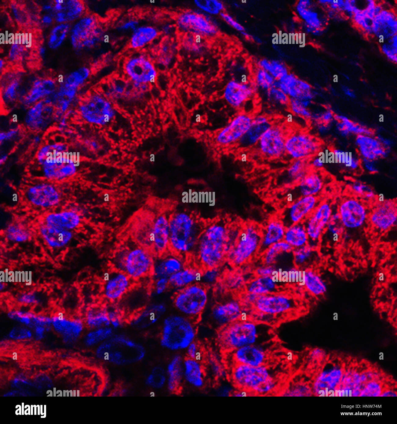 Microscopic image of mitochondrial stained pancreatic cancer cells Stock Photohttps://www.alamy.com/licenses-and-pricing/?v=1https://www.alamy.com/stock-photo-microscopic-image-of-mitochondrial-stained-pancreatic-cancer-cells-133934772.html
RFHNW74M–Microscopic image of mitochondrial stained pancreatic cancer cells
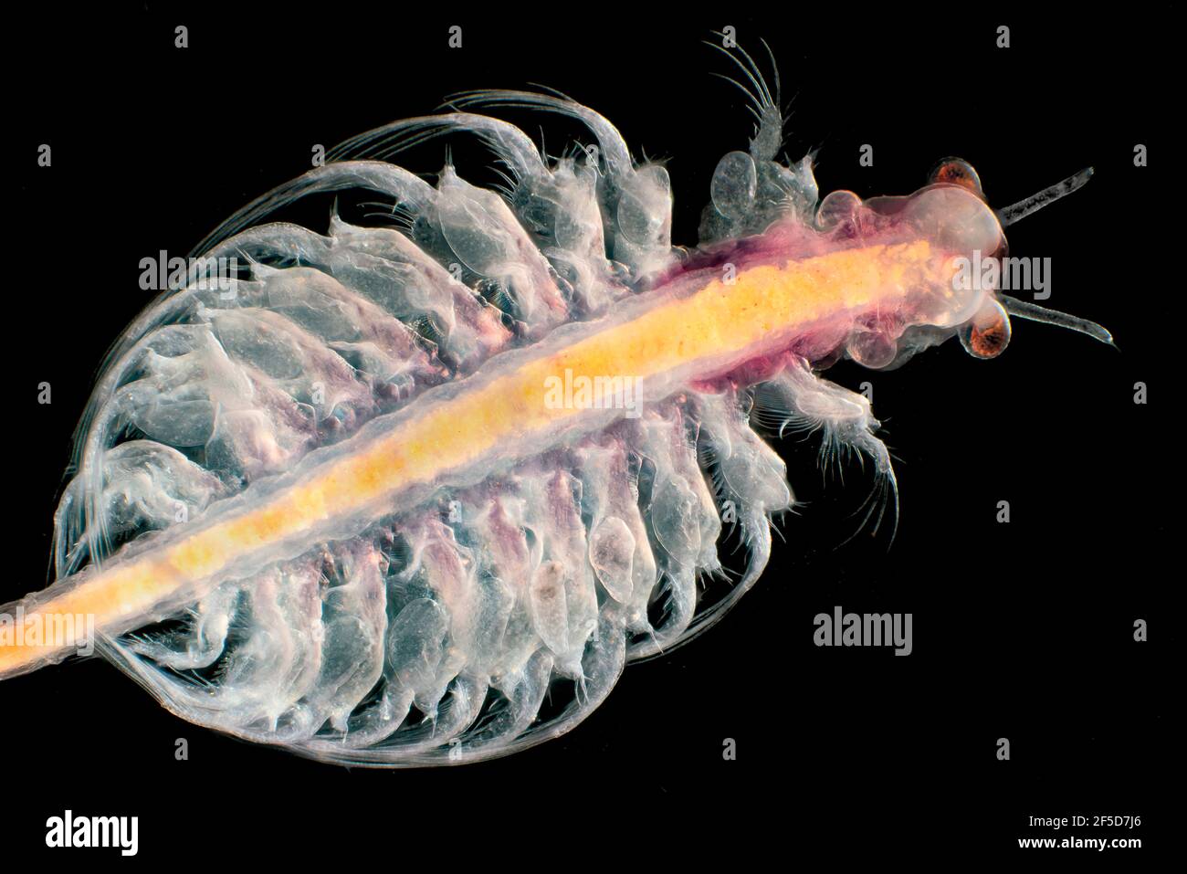 brine shrimp (Artemia salina), dark field microscopic image, magnification x20 related to 35 mm Stock Photohttps://www.alamy.com/licenses-and-pricing/?v=1https://www.alamy.com/brine-shrimp-artemia-salina-dark-field-microscopic-image-magnification-x20-related-to-35-mm-image416413486.html
RM2F5D7J6–brine shrimp (Artemia salina), dark field microscopic image, magnification x20 related to 35 mm
 Transmission electron microscopic image of an isolate from the first U.S. case of COVID-19, formerly known as 2019-nCoV. The spherical viral particles, colourised blue, contain cross-sections through the viral genome, seen as black dots. An optimised and enhanced version of an image produced by the US Centers for Disease Control and Prevention / Credit: CDC / C.S. Goldsmith & A. Tamin Stock Photohttps://www.alamy.com/licenses-and-pricing/?v=1https://www.alamy.com/transmission-electron-microscopic-image-of-an-isolate-from-the-first-us-case-of-covid-19-formerly-known-as-2019-ncov-the-spherical-viral-particles-colourised-blue-contain-cross-sections-through-the-viral-genome-seen-as-black-dots-an-optimised-and-enhanced-version-of-an-image-produced-by-the-us-centers-for-disease-control-and-prevention-credit-cdc-cs-goldsmith-a-tamin-image454466402.html
RM2HBAMEX–Transmission electron microscopic image of an isolate from the first U.S. case of COVID-19, formerly known as 2019-nCoV. The spherical viral particles, colourised blue, contain cross-sections through the viral genome, seen as black dots. An optimised and enhanced version of an image produced by the US Centers for Disease Control and Prevention / Credit: CDC / C.S. Goldsmith & A. Tamin
 Microscopic image Stock Photohttps://www.alamy.com/licenses-and-pricing/?v=1https://www.alamy.com/stock-photo-microscopic-image-95594729.html
RMFFEM1D–Microscopic image
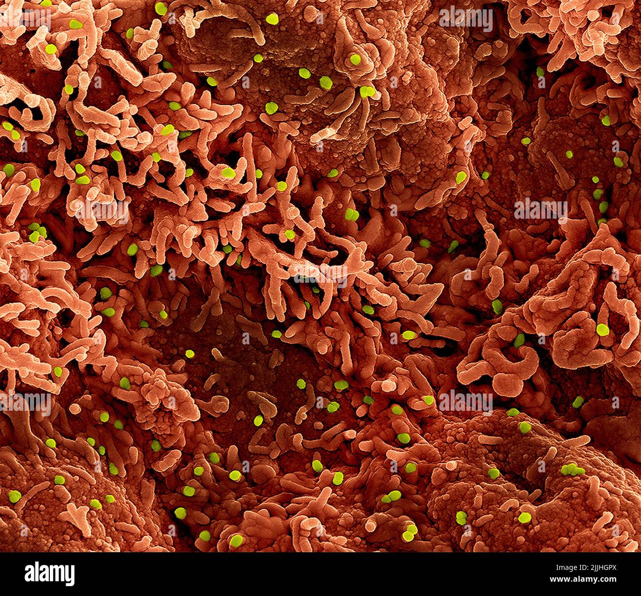 Fort Detrick, United States. 26th July, 2022. A colorized scanning electron micrograph of monkeypox virus (lime) on the surface of infected VERO E6 cells (orange) captured at the NIAID Integrated Research Facility released July 26, 2022, in Fort Detrick, Maryland. Credit: NIAID/NIAID/Alamy Live News Stock Photohttps://www.alamy.com/licenses-and-pricing/?v=1https://www.alamy.com/fort-detrick-united-states-26th-july-2022-a-colorized-scanning-electron-micrograph-of-monkeypox-virus-lime-on-the-surface-of-infected-vero-e6-cells-orange-captured-at-the-niaid-integrated-research-facility-released-july-26-2022-in-fort-detrick-maryland-credit-niaidniaidalamy-live-news-image476130114.html
RM2JJHGPX–Fort Detrick, United States. 26th July, 2022. A colorized scanning electron micrograph of monkeypox virus (lime) on the surface of infected VERO E6 cells (orange) captured at the NIAID Integrated Research Facility released July 26, 2022, in Fort Detrick, Maryland. Credit: NIAID/NIAID/Alamy Live News
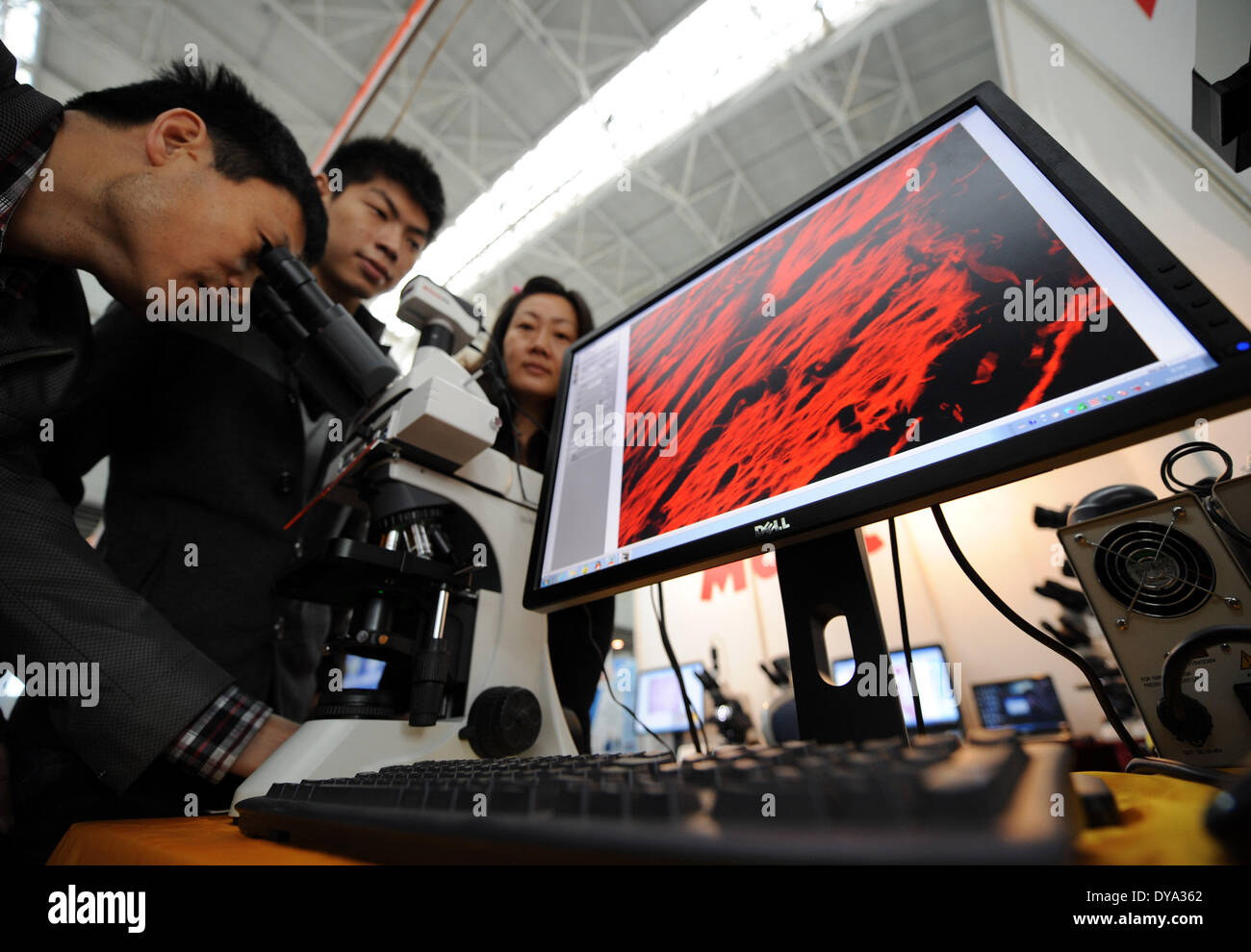 Nanjing, China . 11th Apr, 2014. Visitors experience a microscopic image processing system during the 2014 China International Education Equipment & Science Exhibition in Nanjing, capital of east China's Jiangsu Province, April 11, 2014. The exhibiton, with the participation of 430 exhibitors from more than 20 countries and regions, is held in Nanjing through April 11 to 13. Credit: Xinhua/Alamy Live News Stock Photohttps://www.alamy.com/licenses-and-pricing/?v=1https://www.alamy.com/nanjing-china-11th-apr-2014-visitors-experience-a-microscopic-image-image68448858.html
RMDYA362–Nanjing, China . 11th Apr, 2014. Visitors experience a microscopic image processing system during the 2014 China International Education Equipment & Science Exhibition in Nanjing, capital of east China's Jiangsu Province, April 11, 2014. The exhibiton, with the participation of 430 exhibitors from more than 20 countries and regions, is held in Nanjing through April 11 to 13. Credit: Xinhua/Alamy Live News
 Science students looking at microscopic image on computer with lecturer Stock Photohttps://www.alamy.com/licenses-and-pricing/?v=1https://www.alamy.com/stock-photo-science-students-looking-at-microscopic-image-on-computer-with-lecturer-78164850.html
RFEF4M1P–Science students looking at microscopic image on computer with lecturer
 Abstract microscopic image. Dust and pollen in pond water. Stock Photohttps://www.alamy.com/licenses-and-pricing/?v=1https://www.alamy.com/stock-photo-abstract-microscopic-image-dust-and-pollen-in-pond-water-79078466.html
RMEGJ9AX–Abstract microscopic image. Dust and pollen in pond water.
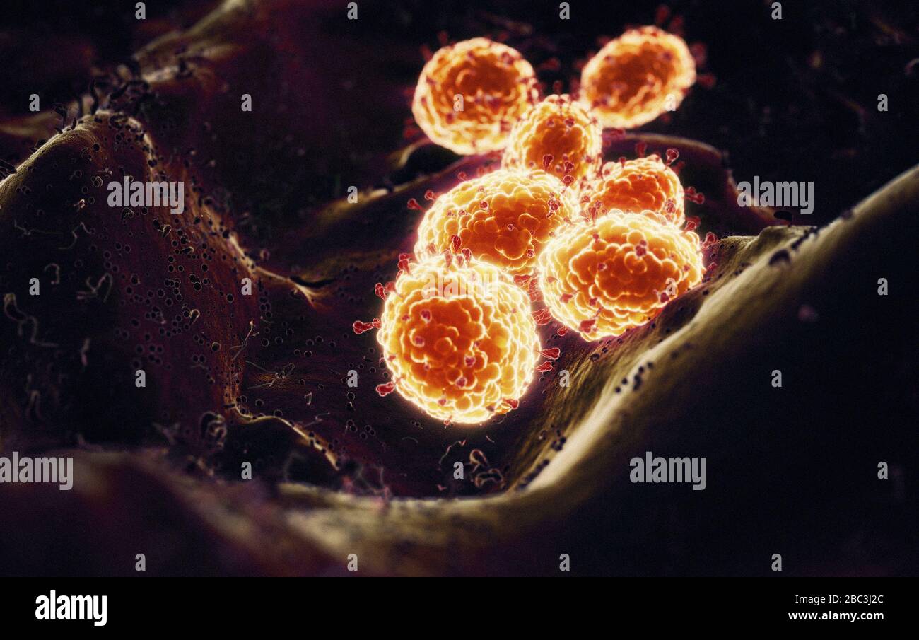 Details of Coronavirus COVID-19 on human cells, 3D illustration as a microscopic image inside the human body based on SEM SARS photos Stock Photohttps://www.alamy.com/licenses-and-pricing/?v=1https://www.alamy.com/details-of-coronavirus-covid-19-on-human-cells-3d-illustration-as-a-microscopic-image-inside-the-human-body-based-on-sem-sars-photos-image351663268.html
RF2BC3J2C–Details of Coronavirus COVID-19 on human cells, 3D illustration as a microscopic image inside the human body based on SEM SARS photos
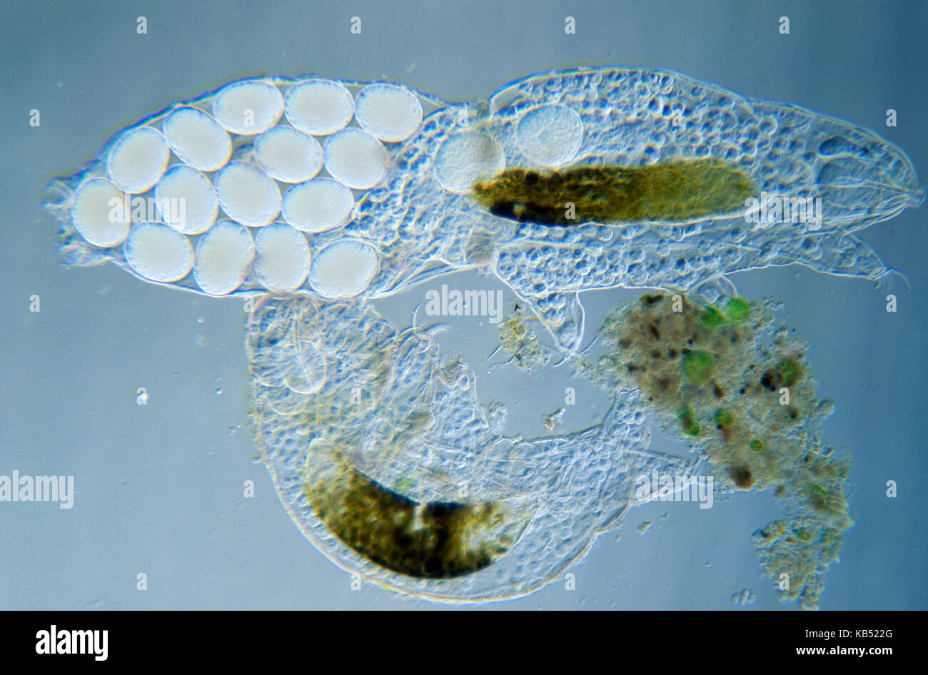 Tardigrade (Pseudobiotus megalonyx) microscopic image, animal can reach more than one mm in length, lives in fresh and brackish water in Europe Stock Photohttps://www.alamy.com/licenses-and-pricing/?v=1https://www.alamy.com/stock-image-tardigrade-pseudobiotus-megalonyx-microscopic-image-animal-can-reach-161765928.html
RMKB522G–Tardigrade (Pseudobiotus megalonyx) microscopic image, animal can reach more than one mm in length, lives in fresh and brackish water in Europe
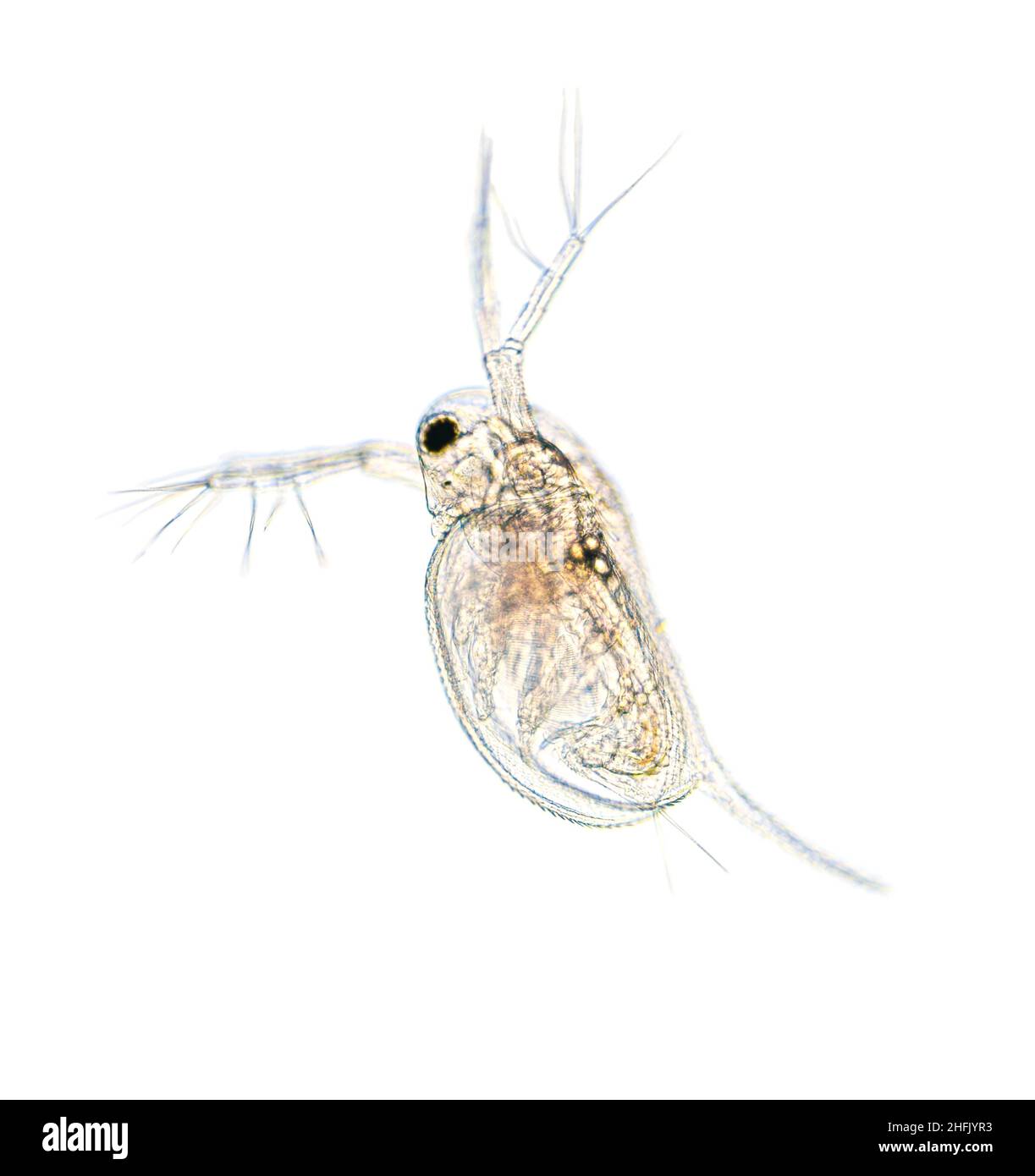 Microscopic image of zooplankton Water Flea Daphnia Stock Photohttps://www.alamy.com/licenses-and-pricing/?v=1https://www.alamy.com/microscopic-image-of-zooplankton-water-flea-daphnia-image457106359.html
RF2HFJYR3–Microscopic image of zooplankton Water Flea Daphnia
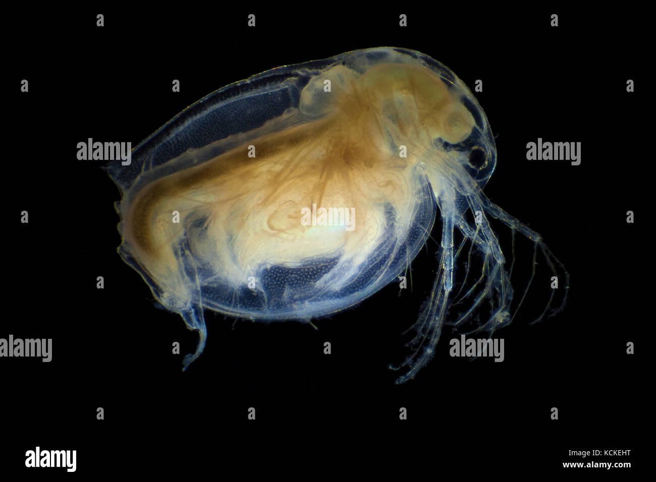 Microscopic image of daphnia, dark field technique Stock Photohttps://www.alamy.com/licenses-and-pricing/?v=1https://www.alamy.com/stock-image-microscopic-image-of-daphnia-dark-field-technique-162697748.html
RFKCKEHT–Microscopic image of daphnia, dark field technique
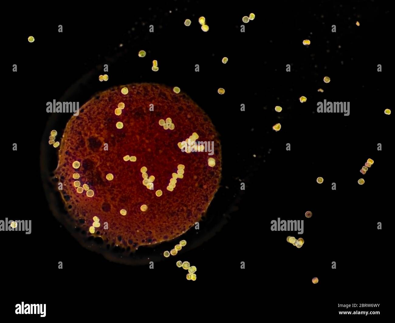 Spores of unidentified mold (found on peach), stained with lactophenol cotton blue, inverted bright field micrograph, 50x microscope objective Stock Photohttps://www.alamy.com/licenses-and-pricing/?v=1https://www.alamy.com/spores-of-unidentified-mold-found-on-peach-stained-with-lactophenol-cotton-blue-inverted-bright-field-micrograph-50x-microscope-objective-image358898679.html
RM2BRW6WY–Spores of unidentified mold (found on peach), stained with lactophenol cotton blue, inverted bright field micrograph, 50x microscope objective
 microscopic image of the patterns made by a pigeon feather Stock Photohttps://www.alamy.com/licenses-and-pricing/?v=1https://www.alamy.com/stock-photo-microscopic-image-of-the-patterns-made-by-a-pigeon-feather-27637079.html
RMBGXYAF–microscopic image of the patterns made by a pigeon feather
 Fungal growth on biological material microscopic image Stock Photohttps://www.alamy.com/licenses-and-pricing/?v=1https://www.alamy.com/fungal-growth-on-biological-material-microscopic-image-image432463406.html
RF2G3GBDJ–Fungal growth on biological material microscopic image
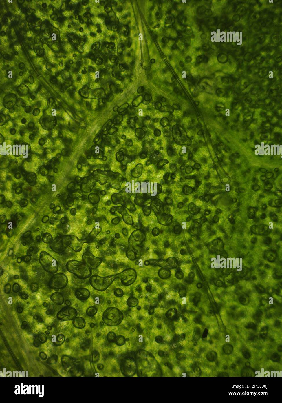 Pontic rhodendron, Pontic alpine rose, Common pontic rhododendron (Rhododendron ponticum) microscopic image of cells Stock Photohttps://www.alamy.com/licenses-and-pricing/?v=1https://www.alamy.com/pontic-rhodendron-pontic-alpine-rose-common-pontic-rhododendron-rhododendron-ponticum-microscopic-image-of-cells-image543363202.html
RM2PG098J–Pontic rhodendron, Pontic alpine rose, Common pontic rhododendron (Rhododendron ponticum) microscopic image of cells
 3d illustration of microscopic image of a virus or infectious cell.Microbacteria and bacterial organisms.biology and science background. Stock Photohttps://www.alamy.com/licenses-and-pricing/?v=1https://www.alamy.com/3d-illustration-of-microscopic-image-of-a-virus-or-infectious-cellmicrobacteria-and-bacterial-organismsbiology-and-science-background-image523143720.html
RF2NB3748–3d illustration of microscopic image of a virus or infectious cell.Microbacteria and bacterial organisms.biology and science background.
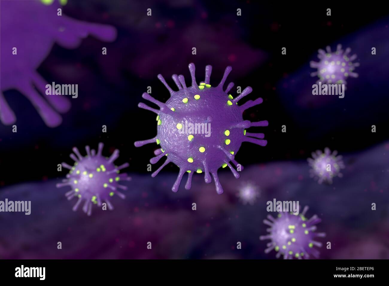 Microscopic image of Coronavirus 2019 (COVID-19), an infectious disease caused by severe acute respiratory syndrome coronavirus 2 (SARS-CoV-2). Stock Photohttps://www.alamy.com/licenses-and-pricing/?v=1https://www.alamy.com/microscopic-image-of-coronavirus-2019-covid-19-an-infectious-disease-caused-by-severe-acute-respiratory-syndrome-coronavirus-2-sars-cov-2-image353350990.html
RF2BETEP6–Microscopic image of Coronavirus 2019 (COVID-19), an infectious disease caused by severe acute respiratory syndrome coronavirus 2 (SARS-CoV-2).
 In this photo illustration a medical syringe is seen in front of a microscopic image of coronavirus. Stock Photohttps://www.alamy.com/licenses-and-pricing/?v=1https://www.alamy.com/in-this-photo-illustration-a-medical-syringe-is-seen-in-front-of-a-microscopic-image-of-coronavirus-image425297401.html
RM2FKWY4W–In this photo illustration a medical syringe is seen in front of a microscopic image of coronavirus.
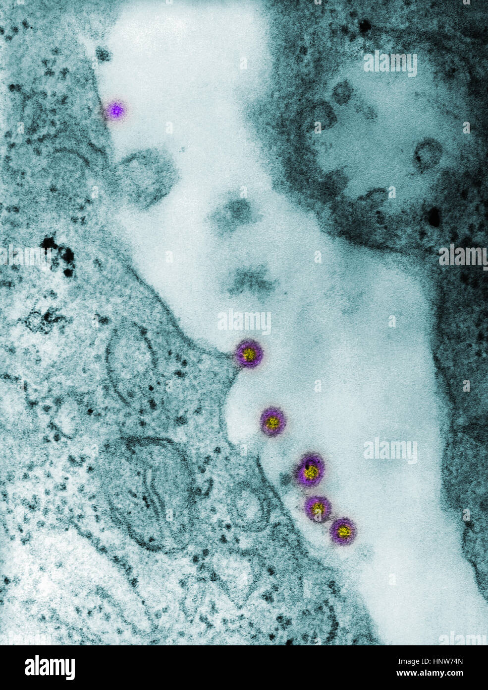 Full frame microscopic image of rubella virus virions budding from the host cell surface to be freed into the host’s system Stock Photohttps://www.alamy.com/licenses-and-pricing/?v=1https://www.alamy.com/stock-photo-full-frame-microscopic-image-of-rubella-virus-virions-budding-from-133934773.html
RFHNW74N–Full frame microscopic image of rubella virus virions budding from the host cell surface to be freed into the host’s system
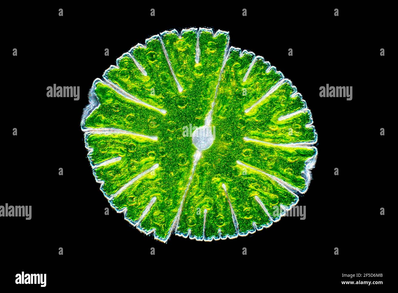 green alga (Micrasterias rotata), dark field microscopic image, magnification x100 related to 35 mm, Germany Stock Photohttps://www.alamy.com/licenses-and-pricing/?v=1https://www.alamy.com/green-alga-micrasterias-rotata-dark-field-microscopic-image-magnification-x100-related-to-35-mm-germany-image416412763.html
RM2F5D6MB–green alga (Micrasterias rotata), dark field microscopic image, magnification x100 related to 35 mm, Germany
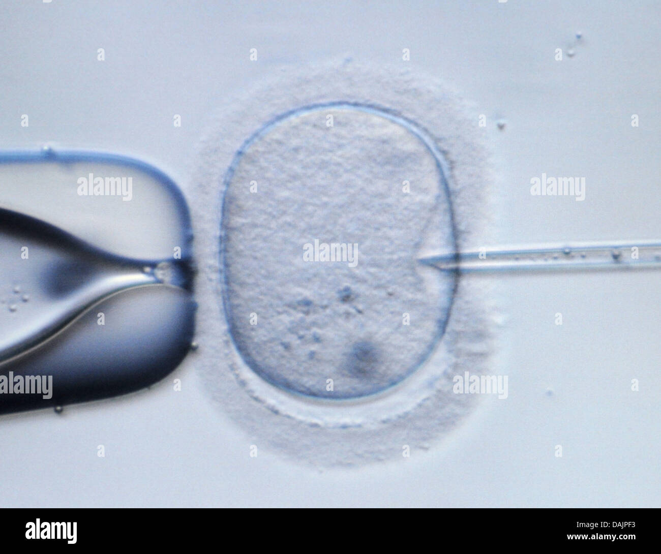 (file) - A dpa file picture dated 09 February 2009 shows a microscopic image of a human ovum being punctured by an injection needle at a laboratory in Dresden, Germany. Preimplantation genetic diagnosis is a controversial topic in Germany. Photo: Ralf Hirschberger Stock Photohttps://www.alamy.com/licenses-and-pricing/?v=1https://www.alamy.com/stock-photo-file-a-dpa-file-picture-dated-09-february-2009-shows-a-microscopic-58190471.html
RMDAJPF3–(file) - A dpa file picture dated 09 February 2009 shows a microscopic image of a human ovum being punctured by an injection needle at a laboratory in Dresden, Germany. Preimplantation genetic diagnosis is a controversial topic in Germany. Photo: Ralf Hirschberger
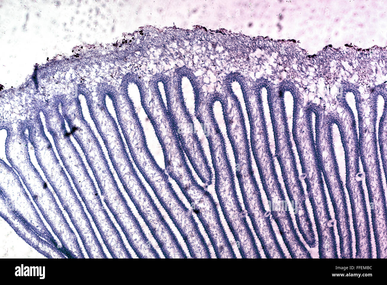 Microscopic image Stock Photohttps://www.alamy.com/licenses-and-pricing/?v=1https://www.alamy.com/stock-photo-microscopic-image-95595008.html
RFFFEMBC–Microscopic image
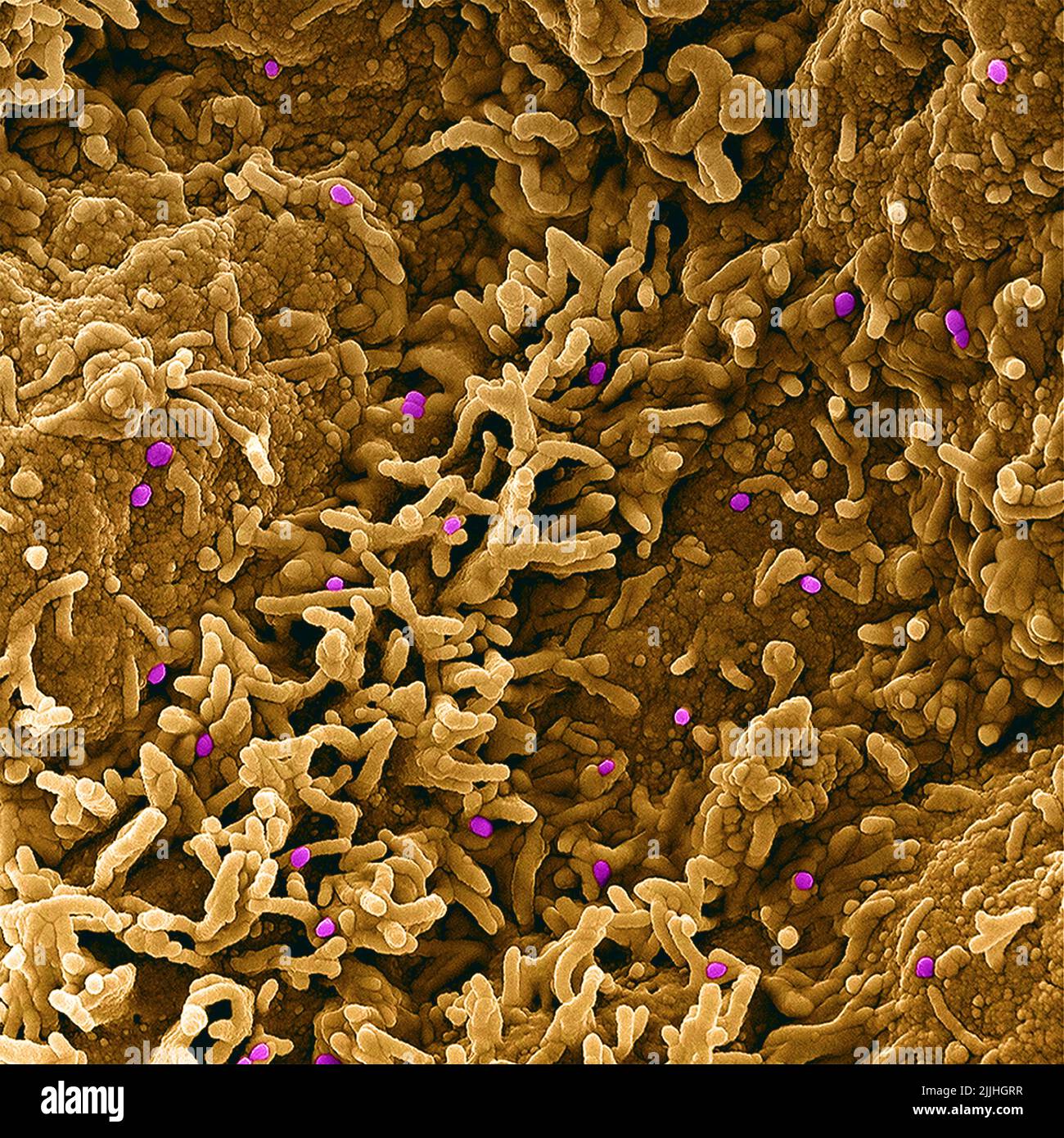 Fort Detrick, United States. 26th July, 2022. A colorized scanning electron micrograph of monkeypox virus (purple) on the surface of infected VERO E6 cells (tan) captured at the NIAID Integrated Research Facility released July 26, 2022, in Fort Detrick, Maryland. Credit: NIAID/NIAID/Alamy Live News Stock Photohttps://www.alamy.com/licenses-and-pricing/?v=1https://www.alamy.com/fort-detrick-united-states-26th-july-2022-a-colorized-scanning-electron-micrograph-of-monkeypox-virus-purple-on-the-surface-of-infected-vero-e6-cells-tan-captured-at-the-niaid-integrated-research-facility-released-july-26-2022-in-fort-detrick-maryland-credit-niaidniaidalamy-live-news-image476130139.html
RM2JJHGRR–Fort Detrick, United States. 26th July, 2022. A colorized scanning electron micrograph of monkeypox virus (purple) on the surface of infected VERO E6 cells (tan) captured at the NIAID Integrated Research Facility released July 26, 2022, in Fort Detrick, Maryland. Credit: NIAID/NIAID/Alamy Live News
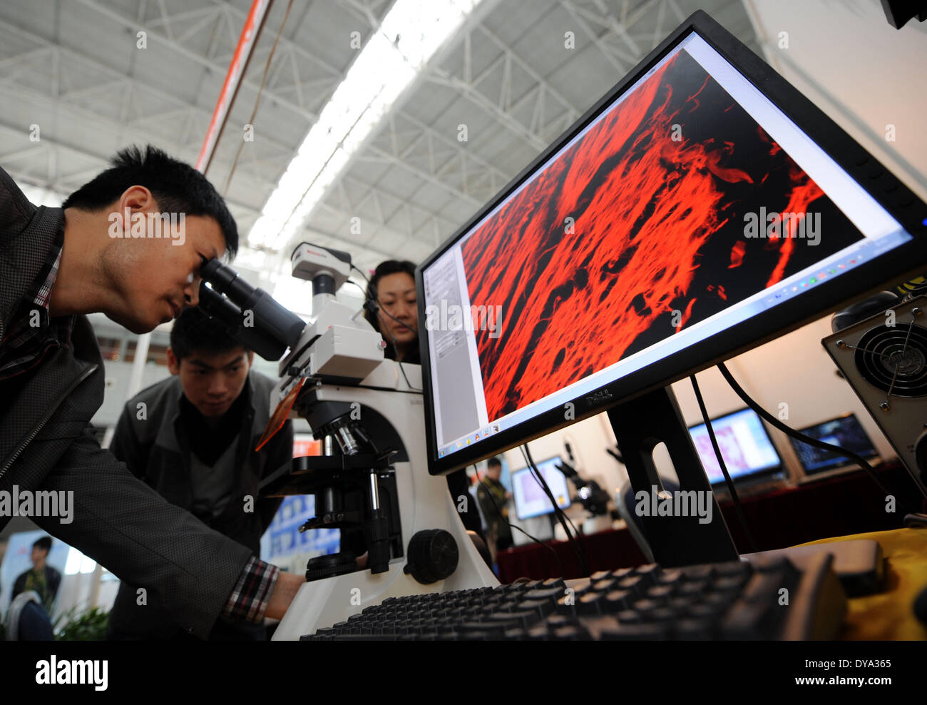 Nanjing, China . 11th Apr, 2014. Visitors experience a microscopic image processing system during the 2014 China International Education Equipment & Science Exhibition in Nanjing, capital of east China's Jiangsu Province, April 11, 2014. The exhibiton, with the participation of 430 exhibitors from more than 20 countries and regions, is held in Nanjing through April 11 to 13. Credit: Xinhua/Alamy Live News Stock Photohttps://www.alamy.com/licenses-and-pricing/?v=1https://www.alamy.com/nanjing-china-11th-apr-2014-visitors-experience-a-microscopic-image-image68448861.html
RMDYA365–Nanjing, China . 11th Apr, 2014. Visitors experience a microscopic image processing system during the 2014 China International Education Equipment & Science Exhibition in Nanjing, capital of east China's Jiangsu Province, April 11, 2014. The exhibiton, with the participation of 430 exhibitors from more than 20 countries and regions, is held in Nanjing through April 11 to 13. Credit: Xinhua/Alamy Live News
 Science students looking at microscopic image on computer Stock Photohttps://www.alamy.com/licenses-and-pricing/?v=1https://www.alamy.com/stock-photo-science-students-looking-at-microscopic-image-on-computer-78168753.html
RFEF4W15–Science students looking at microscopic image on computer
 Pink glitter Stock Photohttps://www.alamy.com/licenses-and-pricing/?v=1https://www.alamy.com/stock-photo-pink-glitter-78985576.html
RMEGE2WC–Pink glitter
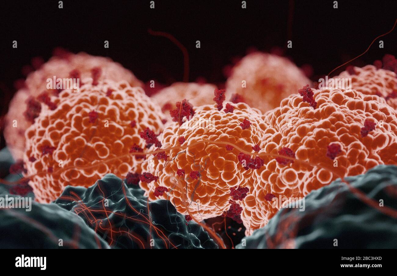 Coronavirus COVID-19 microscopy attacking human cells, 3D render as a microscopic image inside the human body based in SEM SARS photos Stock Photohttps://www.alamy.com/licenses-and-pricing/?v=1https://www.alamy.com/coronavirus-covid-19-microscopy-attacking-human-cells-3d-render-as-a-microscopic-image-inside-the-human-body-based-in-sem-sars-photos-image351663157.html
RF2BC3HXD–Coronavirus COVID-19 microscopy attacking human cells, 3D render as a microscopic image inside the human body based in SEM SARS photos
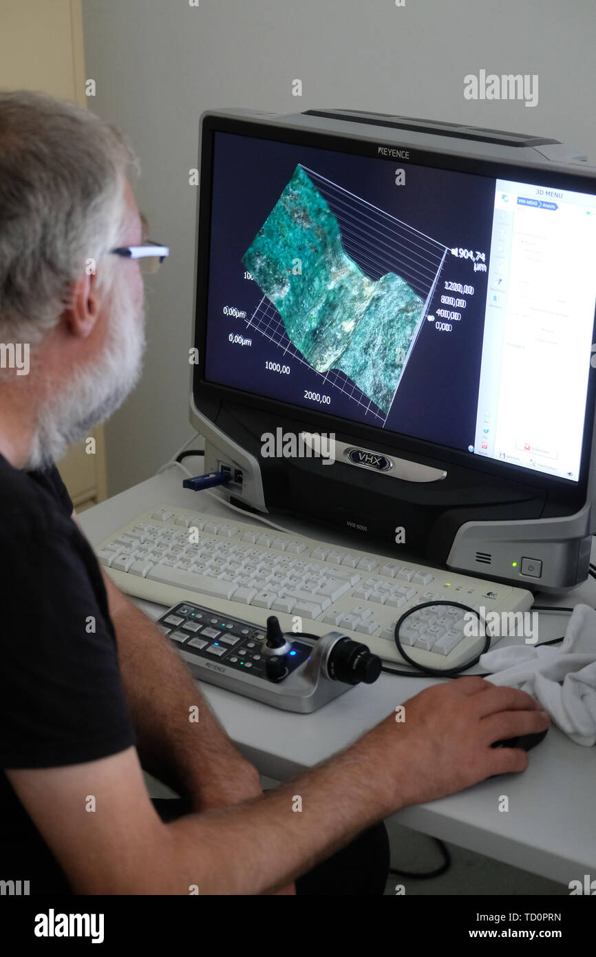 Halle, Germany. 03rd June, 2019. Christian Heinrich Wunderlich from the State Office for Monument Preservation and Archaeology Saxony-Anhalt sits in the workshop of the institution at a monitor on which a microscopic image of the Nebra Sky Disk is shown. (to dpa 'Researchers discover new gold traces on sky disk of Nebra') Credit: Sebastian Willnow/dpa-Zentralbild/dpa/Alamy Live News Stock Photohttps://www.alamy.com/licenses-and-pricing/?v=1https://www.alamy.com/halle-germany-03rd-june-2019-christian-heinrich-wunderlich-from-the-state-office-for-monument-preservation-and-archaeology-saxony-anhalt-sits-in-the-workshop-of-the-institution-at-a-monitor-on-which-a-microscopic-image-of-the-nebra-sky-disk-is-shown-to-dpa-researchers-discover-new-gold-traces-on-sky-disk-of-nebra-credit-sebastian-willnowdpa-zentralbilddpaalamy-live-news-image248953593.html
RMTD0PRN–Halle, Germany. 03rd June, 2019. Christian Heinrich Wunderlich from the State Office for Monument Preservation and Archaeology Saxony-Anhalt sits in the workshop of the institution at a monitor on which a microscopic image of the Nebra Sky Disk is shown. (to dpa 'Researchers discover new gold traces on sky disk of Nebra') Credit: Sebastian Willnow/dpa-Zentralbild/dpa/Alamy Live News
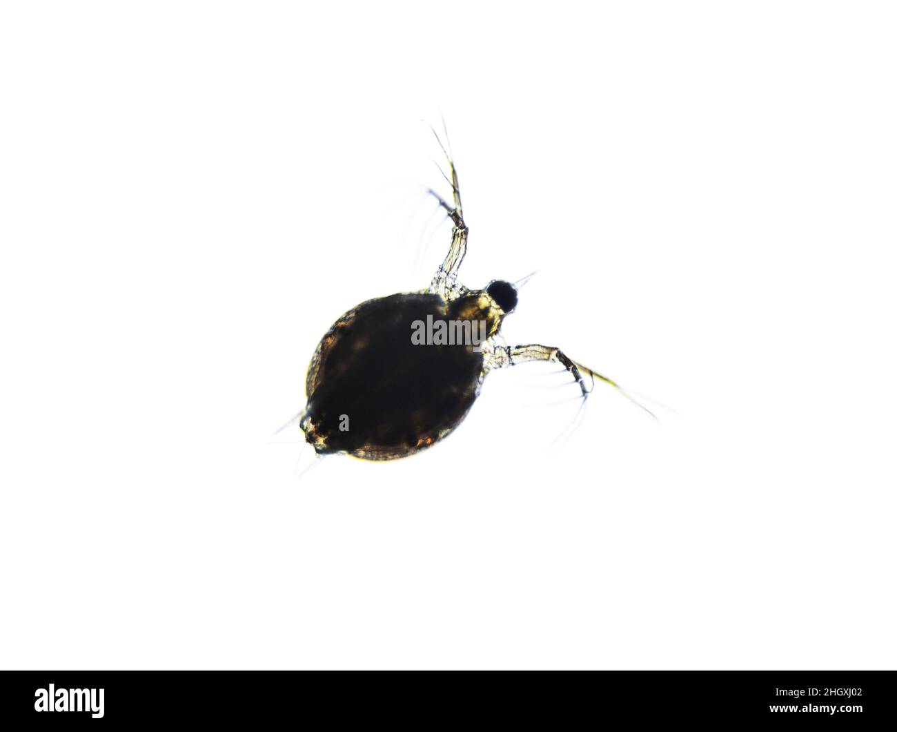 Microscopic image of zooplankton Water Flea Daphnia Stock Photohttps://www.alamy.com/licenses-and-pricing/?v=1https://www.alamy.com/microscopic-image-of-zooplankton-water-flea-daphnia-image457888930.html
RF2HGXJ02–Microscopic image of zooplankton Water Flea Daphnia
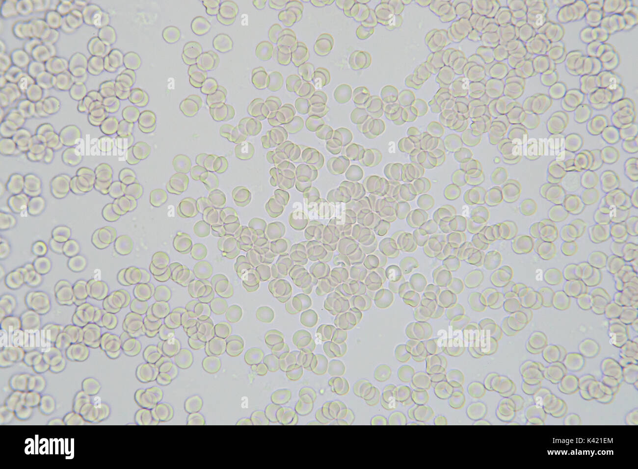 Blood cells microscopic image magnification x 400 Stock Photohttps://www.alamy.com/licenses-and-pricing/?v=1https://www.alamy.com/blood-cells-microscopic-image-magnification-x-400-image157397036.html
RFK421EM–Blood cells microscopic image magnification x 400
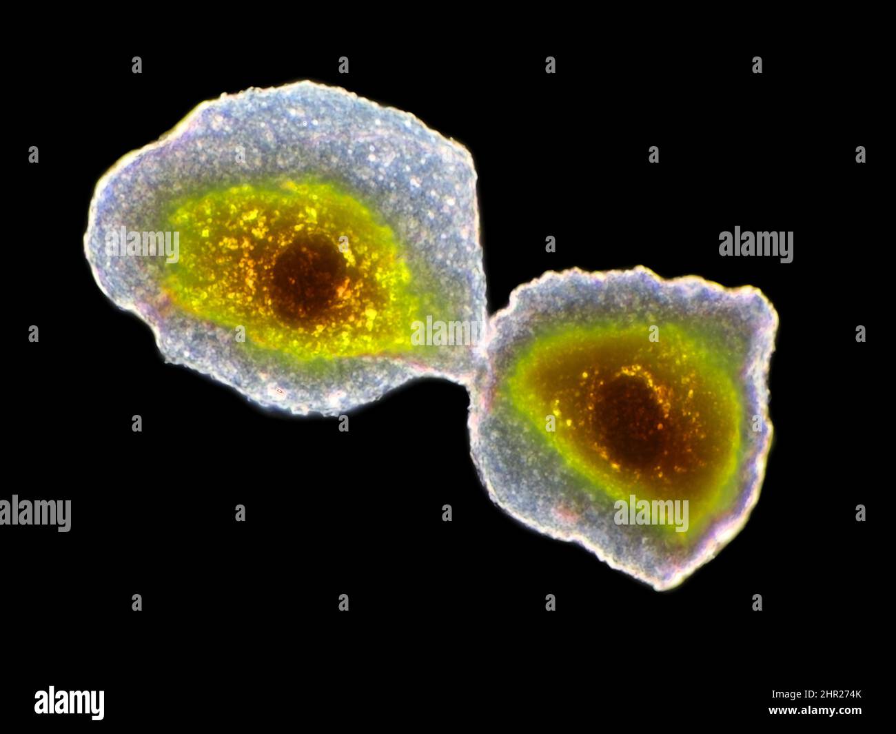 Human cheek epithelial cells under the microscope, horizontal field of view is about 125 micrometers Stock Photohttps://www.alamy.com/licenses-and-pricing/?v=1https://www.alamy.com/human-cheek-epithelial-cells-under-the-microscope-horizontal-field-of-view-is-about-125-micrometers-image461656179.html
RM2HR274K–Human cheek epithelial cells under the microscope, horizontal field of view is about 125 micrometers
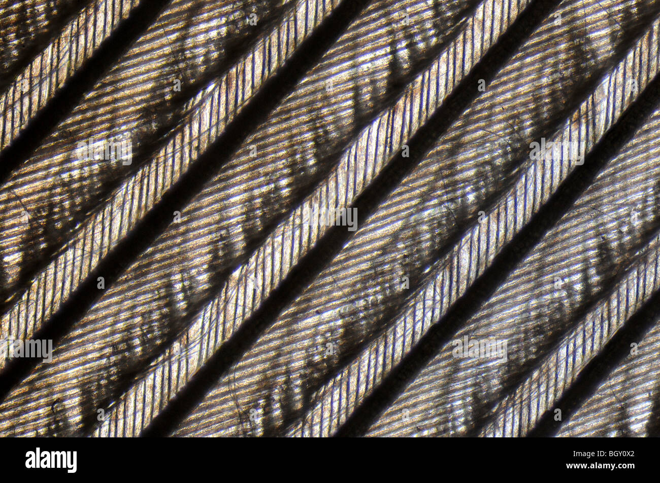 microscopic image of the patterns made by a pigeon feather Stock Photohttps://www.alamy.com/licenses-and-pricing/?v=1https://www.alamy.com/stock-photo-microscopic-image-of-the-patterns-made-by-a-pigeon-feather-27638298.html
RMBGY0X2–microscopic image of the patterns made by a pigeon feather
 3D illustration. Background image, abstract concept of microscopic life, virus and bacteria. Stock Photohttps://www.alamy.com/licenses-and-pricing/?v=1https://www.alamy.com/3d-illustration-background-image-abstract-concept-of-microscopic-life-virus-and-bacteria-image228122941.html
RFR73W3W–3D illustration. Background image, abstract concept of microscopic life, virus and bacteria.
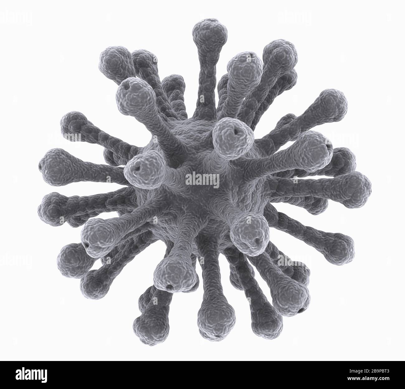 isolated microscopic image of virus Stock Photohttps://www.alamy.com/licenses-and-pricing/?v=1https://www.alamy.com/isolated-microscopic-image-of-virus-image350231507.html
RF2B9PBT3–isolated microscopic image of virus
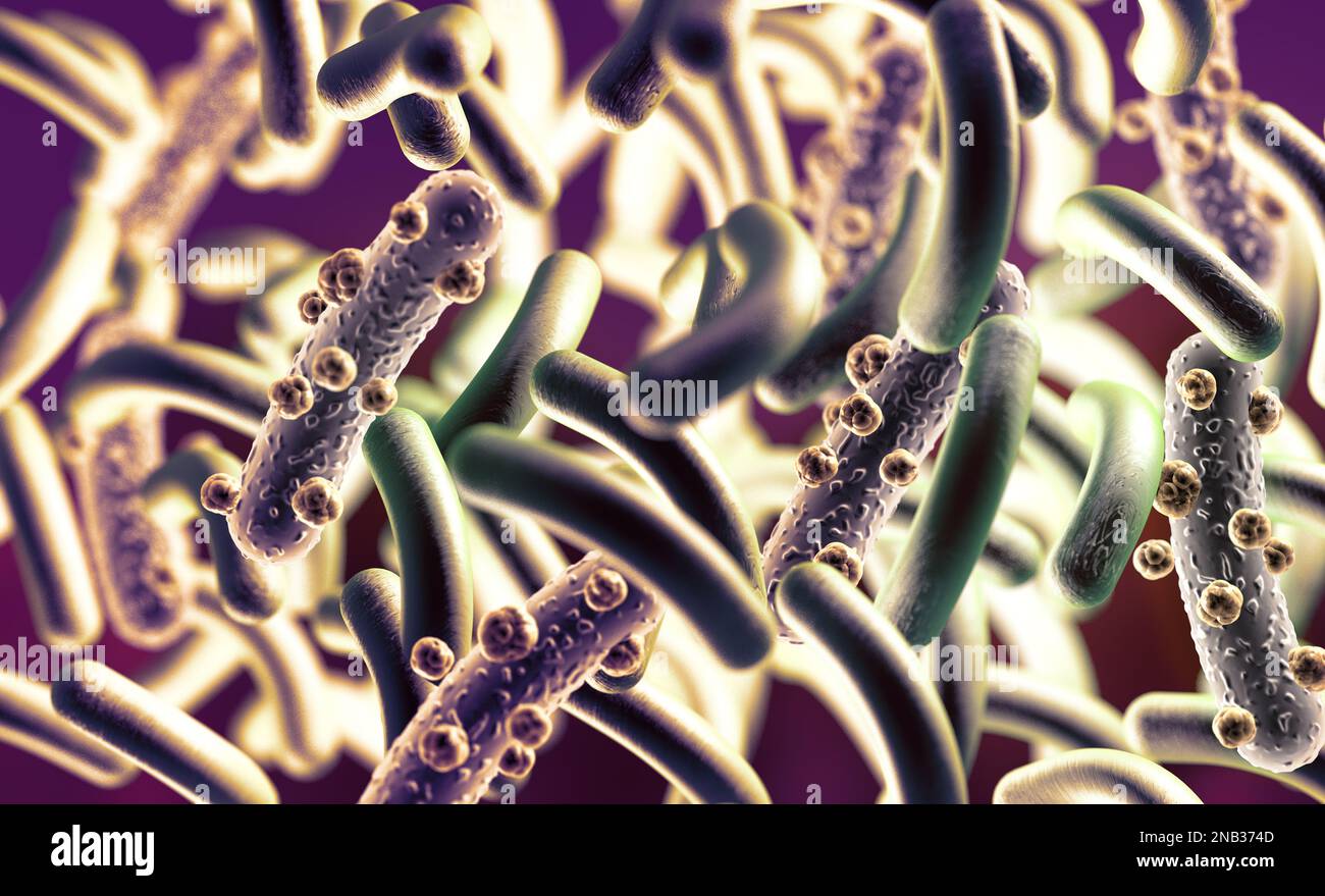 3d illustration of microscopic image of a virus or infectious cell.Microbacteria and bacterial organisms.biology and science background. Stock Photohttps://www.alamy.com/licenses-and-pricing/?v=1https://www.alamy.com/3d-illustration-of-microscopic-image-of-a-virus-or-infectious-cellmicrobacteria-and-bacterial-organismsbiology-and-science-background-image523143725.html
RF2NB374D–3d illustration of microscopic image of a virus or infectious cell.Microbacteria and bacterial organisms.biology and science background.
 Composite image of microscopic cells on abstract screen Stock Photohttps://www.alamy.com/licenses-and-pricing/?v=1https://www.alamy.com/composite-image-of-microscopic-cells-on-abstract-screen-image66697326.html
RFDTE93A–Composite image of microscopic cells on abstract screen
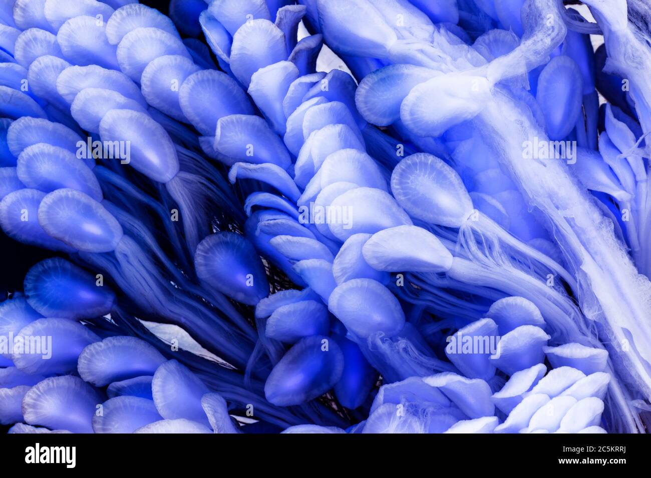 Microscopic Image of Surreal Blue Milkweed Seeds Stock Photohttps://www.alamy.com/licenses-and-pricing/?v=1https://www.alamy.com/microscopic-image-of-surreal-blue-milkweed-seeds-image364926790.html
RF2C5KRRJ–Microscopic Image of Surreal Blue Milkweed Seeds
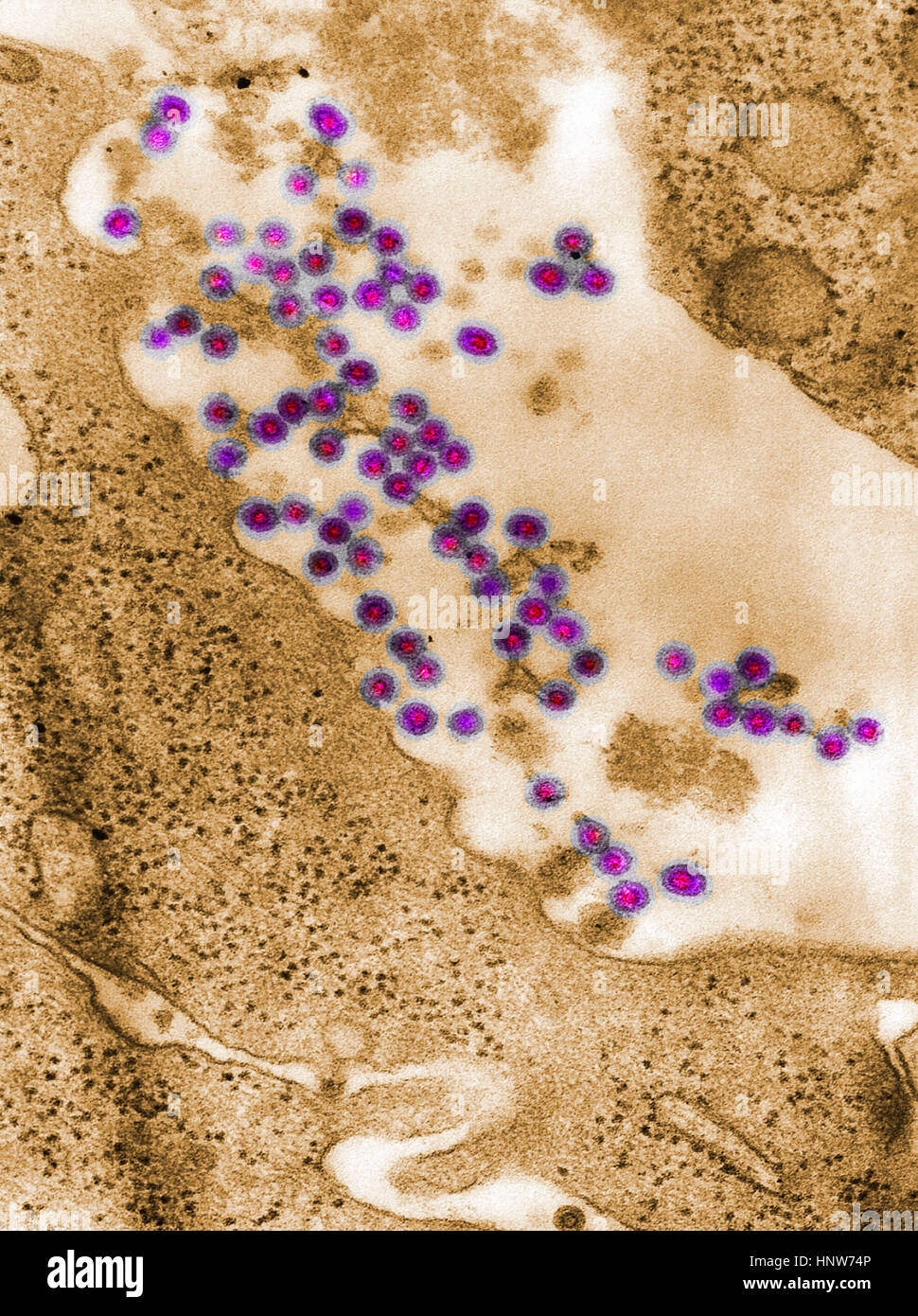 Full frame microscopic image of rubella virus virions budding from the host cell surface to be freed into the host’s system Stock Photohttps://www.alamy.com/licenses-and-pricing/?v=1https://www.alamy.com/stock-photo-full-frame-microscopic-image-of-rubella-virus-virions-budding-from-133934774.html
RFHNW74P–Full frame microscopic image of rubella virus virions budding from the host cell surface to be freed into the host’s system
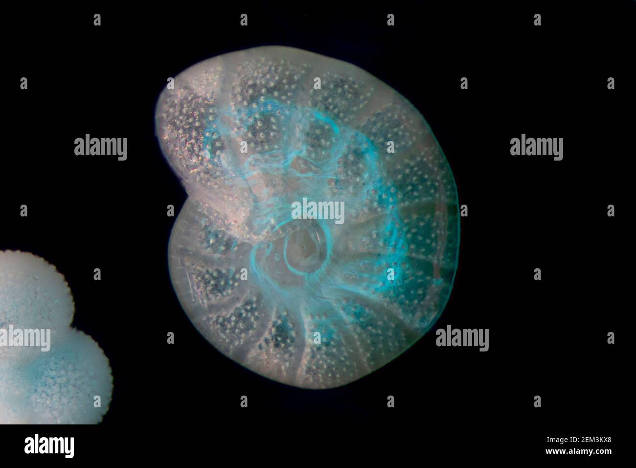 foraminiferans, forams (Foraminiferida), Recent planktonic foraminifera, dark field microscopic image, magnification x40 related to 35 mm Stock Photohttps://www.alamy.com/licenses-and-pricing/?v=1https://www.alamy.com/foraminiferans-forams-foraminiferida-recent-planktonic-foraminifera-dark-field-microscopic-image-magnification-x40-related-to-35-mm-image408213072.html
RM2EM3KX8–foraminiferans, forams (Foraminiferida), Recent planktonic foraminifera, dark field microscopic image, magnification x40 related to 35 mm
Search Results for Microscopic image Stock Photos and Images (11,192)
Page 1 of 112