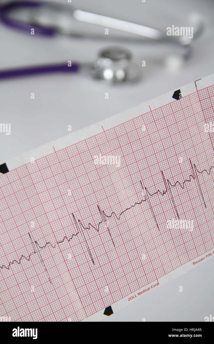
Atrial fibrillation ecg Stock Photos and Images
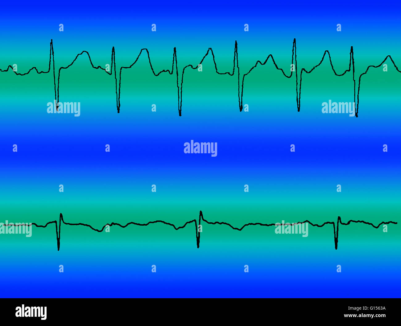 Composite image comparing an electrocardiogram (ECG) readout of atrial fibrillation (at top) and a normal heart beat (at bottom). Stock Photohttps://www.alamy.com/image-license-details/?v=1https://www.alamy.com/stock-photo-composite-image-comparing-an-electrocardiogram-ecg-readout-of-atrial-103991422.html
RMG1563A–Composite image comparing an electrocardiogram (ECG) readout of atrial fibrillation (at top) and a normal heart beat (at bottom).
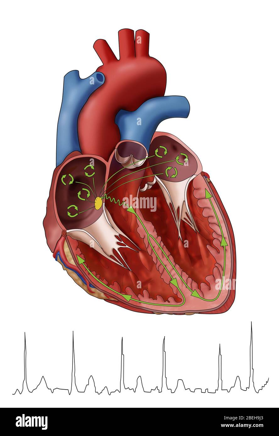 Atrial Fibrillation with EKG, Illustration Stock Photohttps://www.alamy.com/image-license-details/?v=1https://www.alamy.com/atrial-fibrillation-with-ekg-illustration-image353193291.html
RM2BEH9J3–Atrial Fibrillation with EKG, Illustration
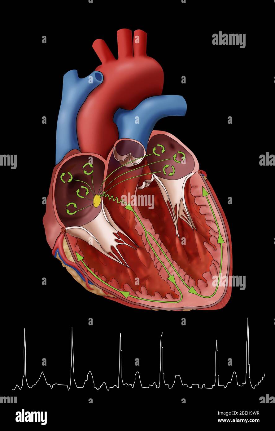 Atrial Fibrillation with EKG, Illustration Stock Photohttps://www.alamy.com/image-license-details/?v=1https://www.alamy.com/atrial-fibrillation-with-ekg-illustration-image353193507.html
RM2BEH9WR–Atrial Fibrillation with EKG, Illustration
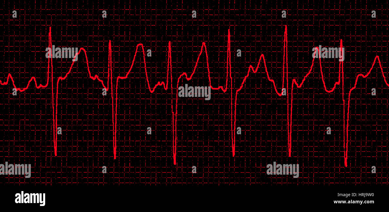 Atrial Fibrillation Stock Photohttps://www.alamy.com/image-license-details/?v=1https://www.alamy.com/stock-photo-atrial-fibrillation-135012556.html
RMHRJ9W0–Atrial Fibrillation
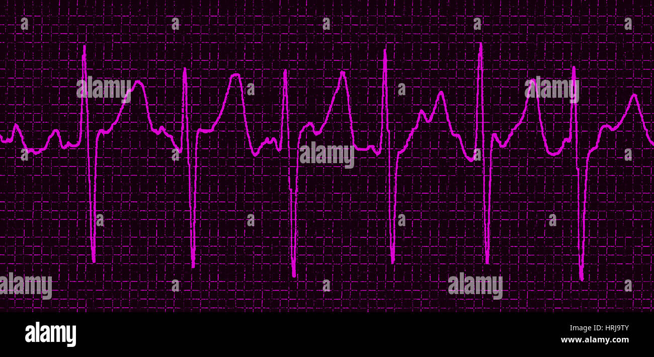 Atrial Fibrillation Stock Photohttps://www.alamy.com/image-license-details/?v=1https://www.alamy.com/stock-photo-atrial-fibrillation-135012555.html
RMHRJ9TY–Atrial Fibrillation
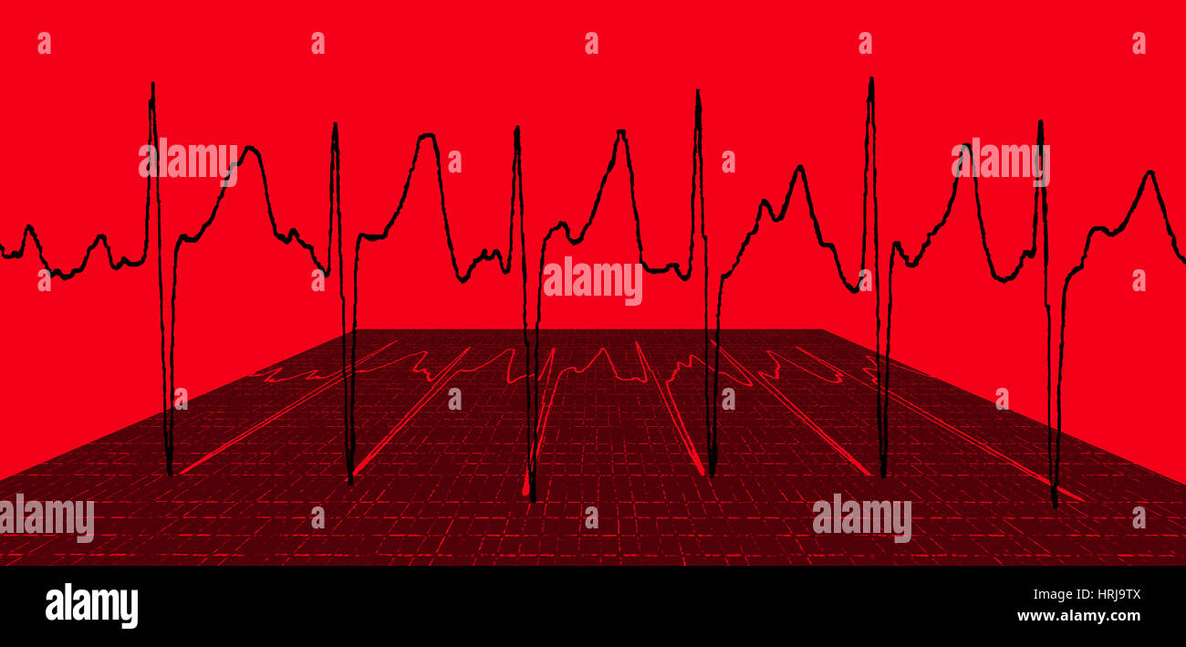 Atrial Fibrillation Stock Photohttps://www.alamy.com/image-license-details/?v=1https://www.alamy.com/stock-photo-atrial-fibrillation-135012554.html
RMHRJ9TX–Atrial Fibrillation
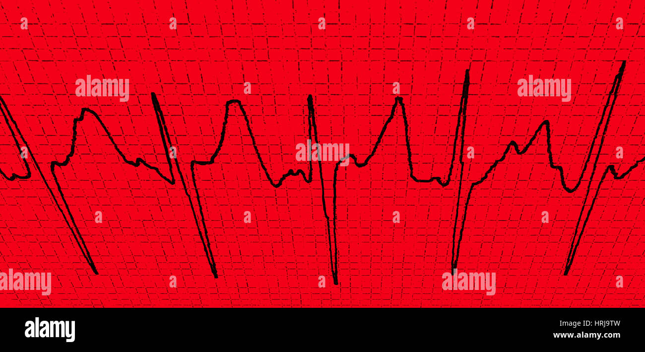 Atrial Fibrillation Stock Photohttps://www.alamy.com/image-license-details/?v=1https://www.alamy.com/stock-photo-atrial-fibrillation-135012553.html
RMHRJ9TW–Atrial Fibrillation
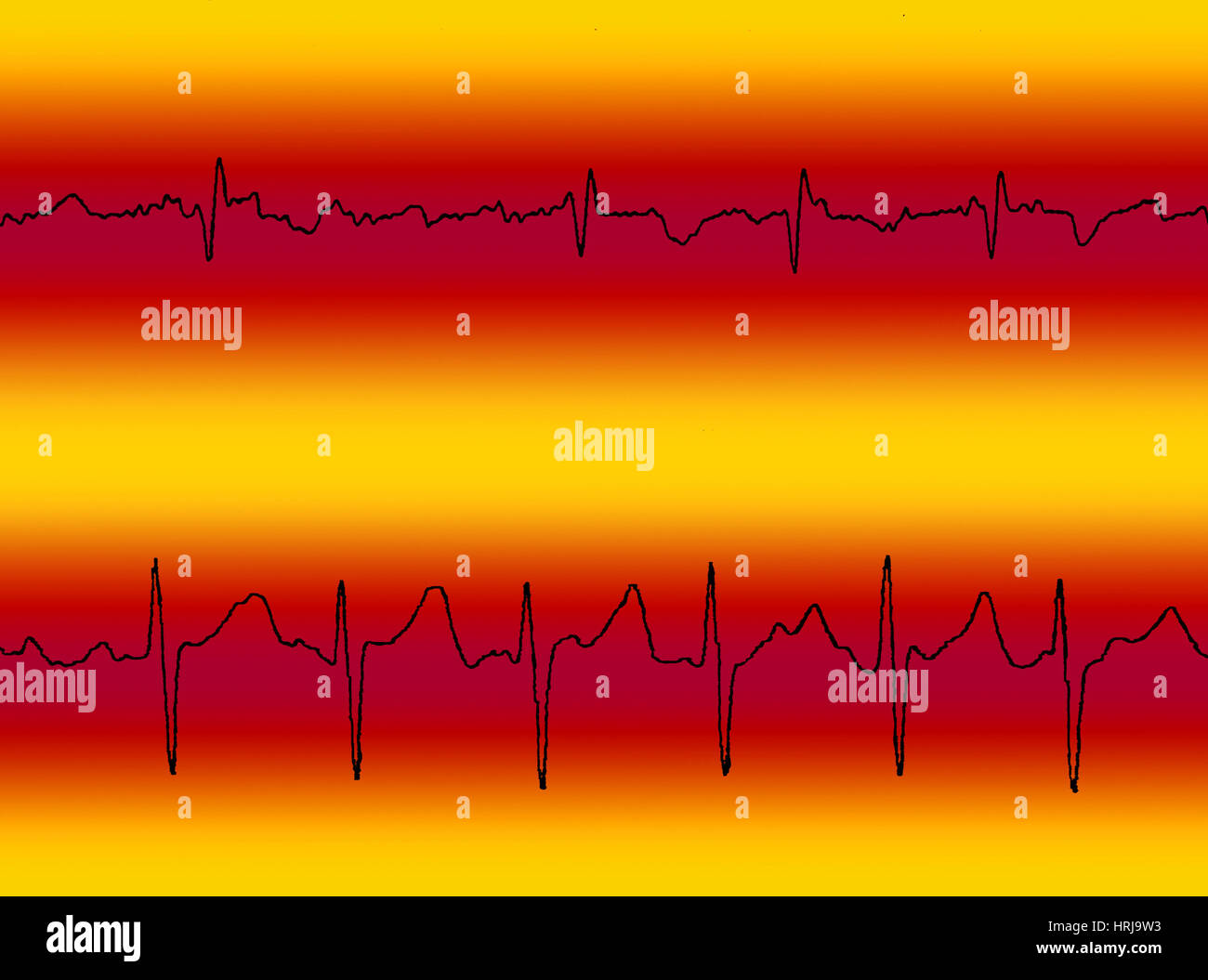 Atrial Flutter & Atrial Fibrillation Stock Photohttps://www.alamy.com/image-license-details/?v=1https://www.alamy.com/stock-photo-atrial-flutter-atrial-fibrillation-135012559.html
RMHRJ9W3–Atrial Flutter & Atrial Fibrillation
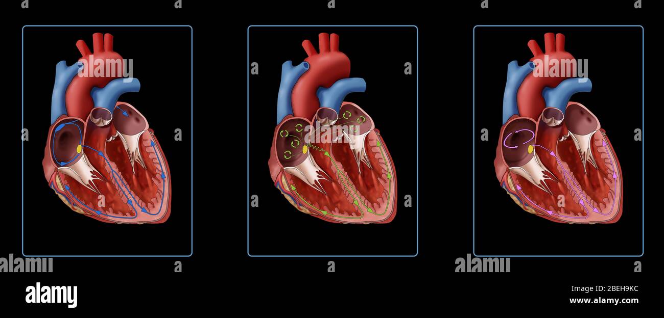 Illustration comparing a heart muscle with a normal heartbeat and irregular heartbeats. Showing a normal heart (left), heart with atrial fibrillation (center), and heart with atrial flutter (right). As shown in the illustration in atrial fibrillation (AFib), disorganized electrical signals (green circles) are originating from the SA Node (yellow dot). With atrial flutter, electrical signals travel around and around inside the atria (pink circle); these circling signals make the atria beat too fast. Stock Photohttps://www.alamy.com/image-license-details/?v=1https://www.alamy.com/illustration-comparing-a-heart-muscle-with-a-normal-heartbeat-and-irregular-heartbeats-showing-a-normal-heart-left-heart-with-atrial-fibrillation-center-and-heart-with-atrial-flutter-right-as-shown-in-the-illustration-in-atrial-fibrillation-afib-disorganized-electrical-signals-green-circles-are-originating-from-the-sa-node-yellow-dot-with-atrial-flutter-electrical-signals-travel-around-and-around-inside-the-atria-pink-circle-these-circling-signals-make-the-atria-beat-too-fast-image353193328.html
RM2BEH9KC–Illustration comparing a heart muscle with a normal heartbeat and irregular heartbeats. Showing a normal heart (left), heart with atrial fibrillation (center), and heart with atrial flutter (right). As shown in the illustration in atrial fibrillation (AFib), disorganized electrical signals (green circles) are originating from the SA Node (yellow dot). With atrial flutter, electrical signals travel around and around inside the atria (pink circle); these circling signals make the atria beat too fast.
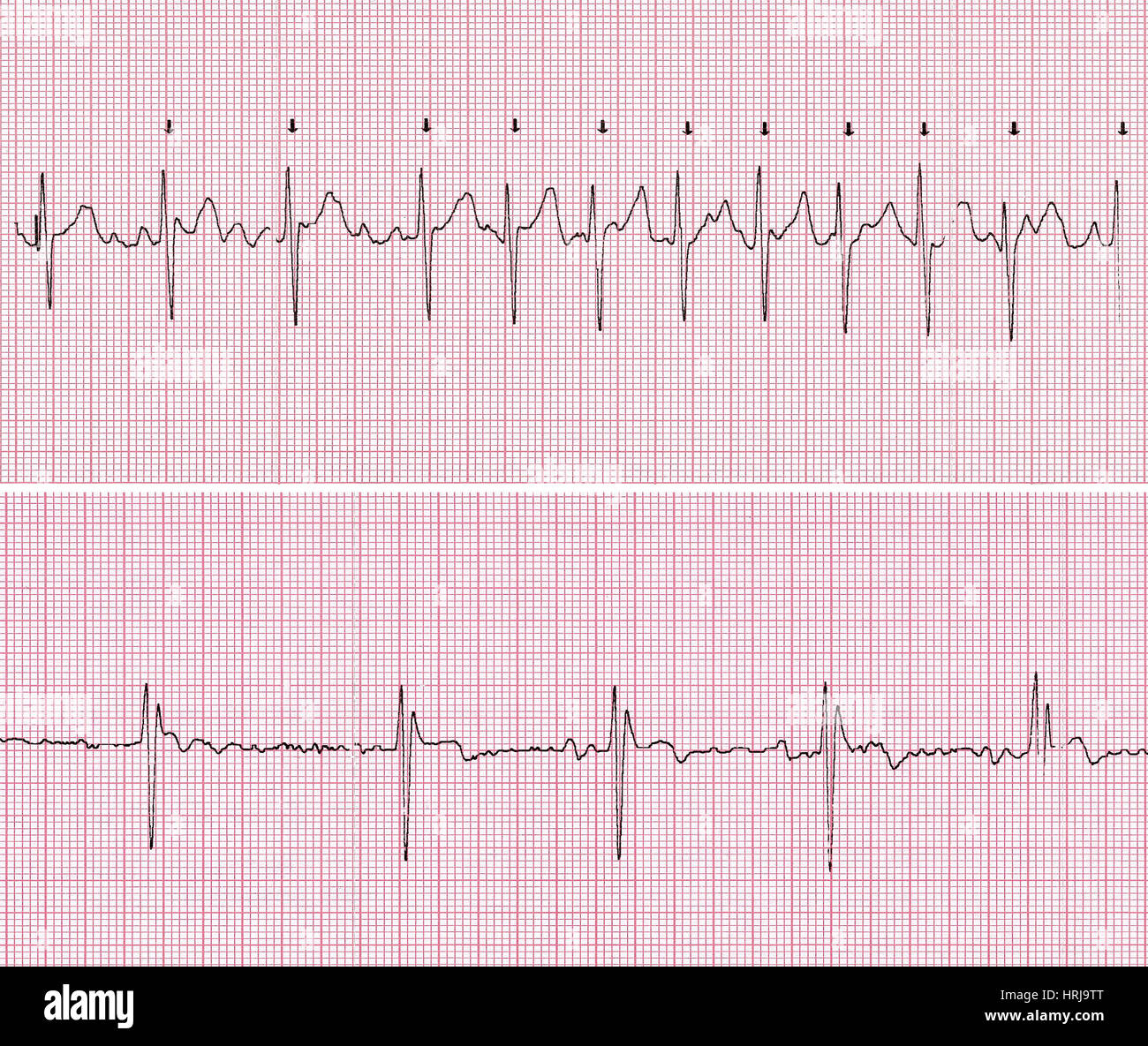 Cardioversion Stock Photohttps://www.alamy.com/image-license-details/?v=1https://www.alamy.com/stock-photo-cardioversion-135012552.html
RMHRJ9TT–Cardioversion
 Cardioversion, 1 of 2 Stock Photohttps://www.alamy.com/image-license-details/?v=1https://www.alamy.com/stock-photo-cardioversion-1-of-2-135012549.html
RMHRJ9TN–Cardioversion, 1 of 2
 Cardioversion, 2 of 2 Stock Photohttps://www.alamy.com/image-license-details/?v=1https://www.alamy.com/stock-photo-cardioversion-2-of-2-135012551.html
RMHRJ9TR–Cardioversion, 2 of 2
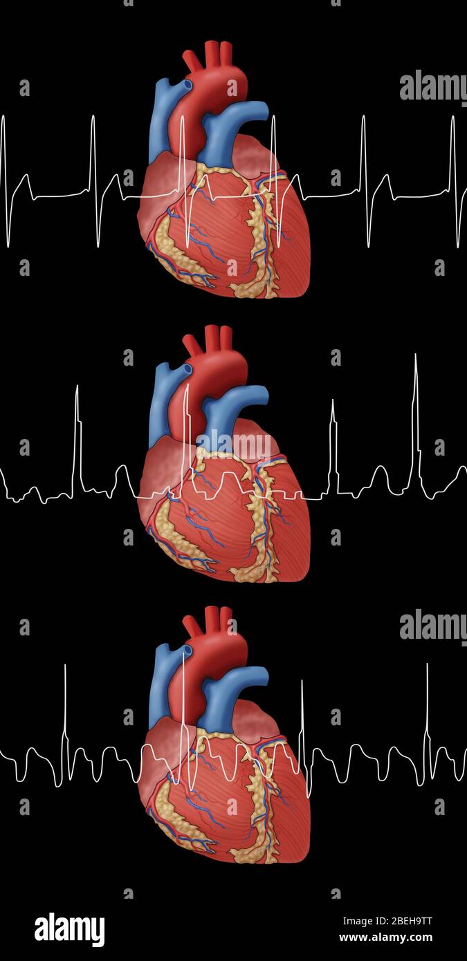 Heartbeat, AFib and Atrial Flutter, Illustration Stock Photohttps://www.alamy.com/image-license-details/?v=1https://www.alamy.com/heartbeat-afib-and-atrial-flutter-illustration-image353193480.html
RM2BEH9TT–Heartbeat, AFib and Atrial Flutter, Illustration
