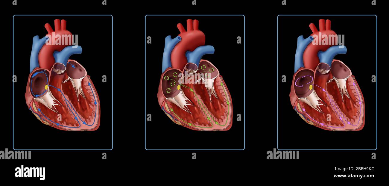
Illustration comparing a heart muscle with a normal heartbeat and irregular heartbeats. Showing a normal heart (left), heart with atrial fibrillation (center), and heart with atrial flutter (right). As shown in the illustration in atrial fibrillation (AFib), disorganized electrical signals (green circles) are originating from the SA Node (yellow dot). With atrial flutter, electrical signals travel around and around inside the atria (pink circle); these circling signals make the atria beat too fast.
RMID:Image ID:2BEH9KC
Image details
Contributor:
Science History ImagesImage ID:
2BEH9KCFile size:
66.2 MB (1,002.4 KB Compressed download)Releases:
Model - no | Property - noDo I need a release?Dimensions:
7500 x 3083 px | 63.5 x 26.1 cm | 25 x 10.3 inches | 300dpiDate taken:
8 March 2016Photographer:
Photo ResearchersMore information:
This image could have imperfections as it’s either historical or reportage.