MRI of Brain AVM
RMID:Image ID:HRH6N9
Image details
Contributor:
Science History Images / Alamy Stock PhotoImage ID:
HRH6N9File size:
37.1 MB (363.7 KB Compressed download)Releases:
Model - no | Property - noDo I need a release?Dimensions:
3600 x 3600 px | 30.5 x 30.5 cm | 12 x 12 inches | 300dpiPhotographer:
Medical Body ScansMore information:
This image could have imperfections as it’s either historical or reportage.
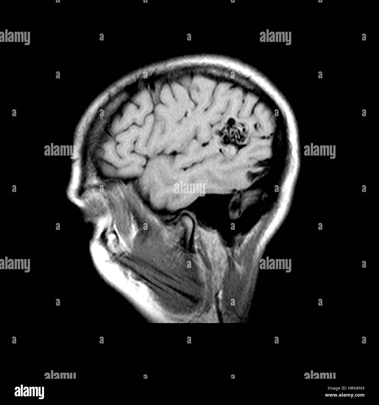
This sagittal (from the side) MRI image of the brain shows the typical appearance of a high-flow arterial venous malformation (AVM). This is depicted as the rounded area of black signal. This is located in the temporal-parietal region. This is a type of congenital brain vascular malformation that can result in serious disability or death. These can rupture (burst) and lead to bleeding inside the head.